



- The kidney concentrates urine by maintaining a gradient of increasing osmolality from the cortex to the medulla.
- This gradient is produced by the loops of Henle acting as countercurrent multipliers.
- The gradient is maintained by the vasa recta acting as countercurrent exchangers.
- A countercurrent system is one in which fluid flows in opposite directions through two closely placed parallel pathways.
- Both the loops of Henle and the vasa recta form countercurrent systems in the renal medulla.
- The loop of Henle acts as a countercurrent multiplier because:
- The thin descending limb is highly permeable to water through Aquaporin-1.
- The thick ascending limb actively transports Na⁺ and Cl⁻ into the interstitium.
- Tubular fluid continuously enters from the proximal tubule and leaves through the distal tubule.
- Initially, the descending limb, ascending limb, and medullary interstitium all have an osmolality of about 300 mOsm/kg H₂O.
- The thick ascending limb actively pumps about 100 mOsm/kg of Na⁺ and Cl⁻ into the medullary interstitium.
- This increases the interstitial osmolality to about 400 mOsm/kg H₂O.
- Water then leaves the thin descending limb until its fluid reaches the same osmolality as the surrounding interstitium.
- New tubular fluid with an osmolality of 300 mOsm/kg H₂O continuously enters from the proximal tubule.
- This allows more Na⁺ and Cl⁻ to be pumped into the interstitium.
- Hypotonic fluid moves into the distal tubule.
- Isotonic and then hypertonic fluid enters the thick ascending limb.
- This process repeats continuously.
- As a result, a gradual increase in osmolality develops from the top to the bottom of the loop of Henle.
- Juxtamedullary nephrons have longer loops of Henle than cortical nephrons.
- Their thin ascending limbs are relatively impermeable to water but permeable to Na⁺ and Cl⁻.
- Na⁺ and Cl⁻ passively diffuse into the medullary interstitium.
- This produces additional passive countercurrent multiplication.
- The longer the loop of Henle, the greater the osmolality that can be produced at the tip of the medulla.
- The high medullary osmolality is preserved by the vasa recta.
- The vasa recta act as countercurrent exchangers.
- Na⁺ and urea diffuse from blood leaving the medulla into blood entering the medulla.
- Water diffuses from the descending vessels into the ascending vessels.
- As a result, solutes remain within the medulla while water is carried away into the circulation.
- The vasa recta also remove the water reabsorbed from the collecting ducts.
- Countercurrent exchange is a passive process.
- It cannot maintain the medullary osmotic gradient if countercurrent multiplication by the loop of Henle stops.
- The countercurrent system spreads a large osmotic gradient over a long length of tubules (about 1 cm or more), rather than across a very thin cell layer.
- Countercurrent exchange also occurs in other parts of the body.
- For example, arteries and accompanying veins in the limbs exchange heat, helping conserve body heat, especially in mammals living in cold environments.
Figure: Figure 37–3, Figure 37–15, Figure 37–16
KEY CONCEPT
- The countercurrent mechanism allows the kidney to concentrate urine. The loop of Henle acts as a countercurrent multiplier by actively transporting Na⁺ and Cl⁻ while allowing water to leave the descending limb, creating a medullary osmotic gradient. The vasa recta act as countercurrent exchangers, preserving this gradient by retaining Na⁺ and urea in the medulla while removing reabsorbed water. Longer loops of Henle produce a greater medullary osmolality and increase the kidney’s ability to concentrate urine.
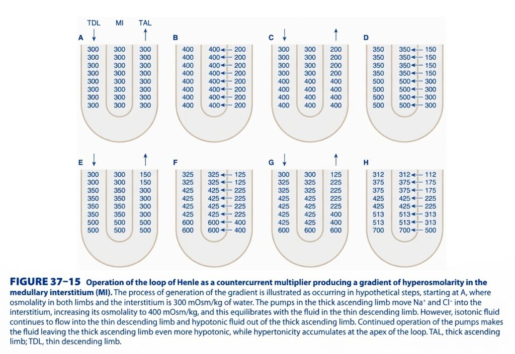
Operation of the Loop of Henle as a Countercurrent Multiplier (Figure 37-15)
Easiest & Most Conceptual Summary for self learners
This is one of the most important kidney physiology figures.
It explains how the Loop of Henle creates the high osmolarity (hyperosmotic medulla) that allows the kidney to produce concentrated urine.
⭐ One-Line Concept
The Thick Ascending Limb pumps salt out, the Descending Limb loses water, and continuous fluid flow multiplies a small difference into a large medullary osmotic gradient.
Before Understanding the Figure
Imagine a U-shaped tube buried inside a sponge.
- Left side = Thin Descending Limb (TDL)
- Middle = Medullary Interstitium (MI)
- Right side = Thick Ascending Limb (TAL)
Tubular Fluid
      ↓
 Thin Descending Limb
        │
        │
      Bottom
        │
        │
 Thick Ascending Limb
      ↑
Tubular Fluid Leaves
Very Important Properties
Descending Limb (TDL)
✅ Water can leave easily
❌ Salt cannot leave easily
Remember:
Descending = Water Leaves
Thick Ascending Limb (TAL)
❌ Water cannot leave
✅ Salt is actively pumped out
Remember:
Ascending = Salt Leaves
Main Characters
| Part | Main Job |
|---|---|
| Descending limb | Loses water |
| Ascending limb | Pumps NaCl |
| Interstitium | Becomes salty |
Now Let’s Understand Every Panel
A. Starting Situation
The figure begins with:
300
300
300
Everywhere is 300 mOsm/kg.
- Descending limb = 300
- Interstitium = 300
- Ascending limb = 300
Everything is equal.
Easy Concept
Imagine three glasses of water.
All contain exactly the same amount of salt.
No movement occurs.
B. Salt Pump Starts Working
Now look at Panel B.
The Thick Ascending Limb pumps NaCl into the interstitium.
The figure shows:
Interstitium
400Ascending limb
200What happened?
Salt moved:
Ascending Limb
      ↓
Interstitium
Result
Interstitium becomes:
400
Ascending limb becomes:
200
Why?
Because:
Salt leaves
Water cannot follow.
So the ascending limb becomes dilute.
Easy Concept
Imagine removing all the salt from soup,
but leaving the water.
The soup becomes dilute.
C. Descending Limb Responds
Now look at Panel C.
Descending limb becomes:
400Why?
The surrounding interstitium is now very salty.
Water leaves the descending limb.
Salt stays inside.
Therefore,
the descending limb becomes concentrated.
Easy Concept
Imagine grapes drying in the sun.
Water leaves.
Sugar stays.
The grapes become sweeter.
D. New Fluid Keeps Flowing
Fresh filtrate (300 mOsm) enters from above.
Older concentrated fluid moves downward.
Result
The bottom becomes:
500while the top remains closer to 350.
Why?
Continuous flow pushes concentrated fluid deeper into the medulla.
Easy Concept
Imagine adding fresh water to the top of a river.
Older water is pushed further downstream.
E. Ascending Limb Pumps Again
Now the TAL again pumps salt.
Notice:
Upper ascending limb becomes:
150Lower part remains more concentrated.
Result
Another 200 mOsm difference develops.
Easy Concept
The salt pump keeps working every moment.
F. Descending Limb Again Loses Water
Now the descending limb again equilibrates.
Numbers become:
325
425
600
Why?
Water continues leaving wherever the surrounding interstitium is saltier.
Easy Concept
Each time salt is pumped,
water follows in the descending limb.
G. Continuous Flow Again
Fresh fluid enters.
Old fluid moves down.
Gradient becomes larger.
Now bottom reaches:
600Easy Concept
The concentrated fluid is pushed deeper into the loop.
H. Final Gradient
Now the figure reaches:
Top
312Bottom
700Notice:
The medulla has become much more concentrated.
This is the Countercurrent Multiplier
A small difference
↓
Repeated again and again
↓
Becomes a huge gradient.
Why Is It Called “Countercurrent”?
Because fluid moves in opposite directions.
Descending Limb
        ↓↓↓↓

Ascending Limb
        ↑↑↑↑
Two opposite streams.
Why Is It Called “Multiplier”?
Initially,
only about 200 mOsm difference exists between:
Interstitium
and
Ascending limb.
But repeated cycles produce:
Top
300

↓

Bottom
700
In humans,
this eventually becomes:
≈ 1200–1400 mOsm/kg in the inner medulla.
Easy Concept
Think of climbing stairs.
Each step is only 20 cm.
But after many steps,
you reach the roof.
Small differences become a large overall gradient.
What Creates the Gradient?
Two things working together:
1. Salt Pump
Ascending Limb
      ↓
Pumps NaCl Out
2. Water Movement
Descending Limb
      ↓
Water Leaves
Together
Salt Out
+
Water Out
+
Continuous Flow
=
Large Osmotic Gradient
What Happens After This Gradient Is Created?
Later,
the Collecting Duct passes through this salty medulla.
When ADH is present,
water leaves the collecting duct.
The urine becomes concentrated.
Without this gradient,
ADH cannot concentrate urine effectively.
Complete Story of the Figure
Everything Starts at 300
        ↓
Ascending Limb Pumps NaCl
        ↓
Interstitium Becomes 400
Ascending Limb Becomes 200
        ↓
Descending Limb Loses Water
        ↓
Descending Limb Becomes 400
        ↓
Fresh Fluid Enters
Old Fluid Moves Down
        ↓
Salt Pump Works Again
        ↓
Water Leaves Again
        ↓
Repeated Hundreds of Times
        ↓
Top ≈300
Bottom ≈700
(≈1200 in Human Kidney)
        ↓
Hyperosmotic Medulla Created
        ↓
ADH Can Produce Concentrated Urine
Easy Waterfall Analogy
Imagine a mountain waterfall.
Every minute:
- New water enters from the top.
- Water keeps flowing downward.
- Workers keep throwing salt onto the ground beside the stream.
- The ground becomes saltier and saltier.
Eventually,
the bottom of the mountain becomes extremely salty.
The Loop of Henle works in the same way.
Easy Memory Trick
Remember: “SWF”
- S = Salt pumped out (Ascending limb)
- W = Water leaves (Descending limb)
- F = Flow multiplies the gradient
↓
Countercurrent Multiplier
Important Points from Figure 37-15
- The Loop of Henle functions as a countercurrent multiplier to generate a progressively hyperosmotic renal medulla.
- Initially, the fluid in the descending limb, interstitium, and ascending limb is approximately 300 mOsm/kg.
- The Thick Ascending Limb (TAL) actively pumps NaCl into the medullary interstitium but is impermeable to water, making the tubular fluid progressively dilute.
- The increased interstitial osmolarity causes water to leave the Thin Descending Limb (TDL), concentrating the tubular fluid in that segment.
- Continuous movement of fresh filtrate into the descending limb and concentrated fluid around the loop repeatedly re-establishes the transverse osmotic difference.
- Repeated cycles multiply a small horizontal osmotic difference (about 200 mOsm/kg) into a large vertical corticomedullary osmotic gradient.
- In the human kidney, this mechanism ultimately creates an interstitial osmolarity that may reach approximately 1200–1400 mOsm/kg in the deepest medulla.
- The hyperosmotic medulla is essential for water reabsorption from the collecting ducts in the presence of ADH, allowing the kidney to produce concentrated urine.
KEY CONCEPT (Figure 37-15)
Figure 37-15 demonstrates how the Loop of Henle acts as a countercurrent multiplier. The Thick Ascending Limb continuously pumps NaCl into the medullary interstitium without permitting water to follow, while the Thin Descending Limb, which is permeable to water but relatively impermeable to salt, loses water until it equilibrates with the increasingly hyperosmotic interstitium. Continuous tubular flow repeatedly re-establishes these small osmotic differences, progressively multiplying them into a large corticomedullary osmotic gradient. This hyperosmotic medulla provides the driving force for ADH-mediated water reabsorption in the collecting ducts, enabling the kidneys to produce concentrated urine.
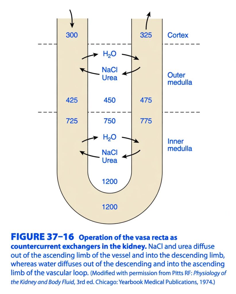
Figure 37-16: Operation of the Vasa Recta as Countercurrent Exchangers
Easiest & Most Conceptual Explanation for self learners
This figure explains how the vasa recta preserves (does not create) the high osmolarity of the renal medulla.
Loop of Henle = Creates the medullary osmotic gradient (Countercurrent Multiplier).
Vasa Recta = Preserves the gradient (Countercurrent Exchanger).
Golden Rule
The Loop of Henle makes the salty medulla.
The Vasa Recta protects the salty medulla.
First, What is the Vasa Recta?
The vasa recta are long, U-shaped capillaries that run parallel to the Loop of Henle.
Their job is not to create concentration.
Their job is to:
- supply oxygen and nutrients to the medulla,
- remove excess water,
- prevent the medullary salt from being washed away.
Why is the Vasa Recta Needed?
Imagine the medulla is like a deep salt lake.
The Loop of Henle spends a lot of energy making this lake salty.
If ordinary blood vessels flowed straight through:
- fresh blood would carry away all the salt,
- the kidney would lose its osmotic gradient,
- concentrated urine could never be formed.
So the kidney uses a special U-shaped blood vessel called the vasa recta.
Understand the Figure First
The figure shows one U-shaped blood vessel.
          Cortex
            │

Descending Vasa Recta
        ↓↓↓↓↓↓

Outer Medulla

Inner Medulla

Bottom (1200)

Ascending Vasa Recta
        ↑↑↑↑↑

Back to Cortex
Osmolarity in the Figure
Notice the numbers.
At the top
Blood enters:
300Normal plasma osmolarity.
Going Down
Blood becomes:
425

↓

725

↓

1200
Coming Up
Blood becomes
1200

↓

775

↓

475

↓

325
Eventually leaving the kidney at about:
325Almost the same as normal blood.
Why Does Blood Become More Concentrated While Descending?
Look at the arrows.
The figure shows:
Water
      →

NaCl
←

Urea
←
Meaning:
Water leaves the blood.
Salt enters the blood.
Urea enters the blood.
Step 1
Blood enters at
300The surrounding medulla is
450

↓

750

↓

1200
The medulla is much saltier.
What happens?
Water leaves blood.
Salt enters blood.
Urea enters blood.
Therefore,
blood osmolarity rises.
Easy Concept
Imagine walking into a desert.
You lose water.
Everything left behind becomes concentrated.
Exactly the same thing happens in descending vasa recta.
At the Bottom of the Loop
Now blood reaches
1200It is now almost equal to the surrounding medulla.
So there is almost no net movement.
Blood Turns Upward
Now blood starts ascending.
The surrounding medulla becomes progressively less concentrated.
What happens now?
Exactly the opposite.
Water enters blood.
Salt leaves blood.
Urea leaves blood.
The figure shows
H2O
←

NaCl
→

Urea
→
Easy Concept
The blood is now “too salty.”
It gives salt back to the medulla.
It receives water.
Blood Leaves the Kidney
Finally blood leaves as
325Almost normal plasma.
Why Doesn’t the Vasa Recta Wash Away the Salt?
This is the most important point.
When blood goes down,
it picks up salt.
When blood comes up,
it gives almost all that salt back.
So,
very little salt actually leaves the medulla.
Easy Analogy
Imagine carrying sand in a bucket.
You pick up sand going downhill.
You drop almost the same sand while coming uphill.
When you return,
almost no sand has been removed.
Exactly what the vasa recta does.
Why is it Called a Countercurrent Exchanger?
Because blood flows in opposite directions.
Descending
↓↓↓↓↓

Ascending
↑↑↑↑↑
Substances exchange between the two limbs.
Nothing is wasted.
Compare Loop of Henle and Vasa Recta
| Loop of Henle | Vasa Recta |
|---|---|
| Tubular fluid | Blood |
| Creates gradient | Preserves gradient |
| Countercurrent multiplier | Countercurrent exchanger |
| Pumps NaCl | Exchanges NaCl and water |
| Forms hyperosmotic medulla | Prevents washout of medulla |
Understanding Every Number in the Figure
Top
Blood entering
300Normal plasma.
Descending Limb
300

↓

425

↓

725

↓

1200
Reason:
- Water leaves
- Salt enters
- Urea enters
Blood becomes concentrated.
Bottom
1200Blood equals surrounding interstitium.
No major diffusion.
Ascending Limb
1200

↓

775

↓

475

↓

325
Reason:
- Water enters
- Salt leaves
- Urea leaves
Blood becomes dilute again.
What Happens to Water?
Descending
Water leaves blood.
Blood
↓

Water Out
Ascending
Water enters blood.
Blood
↑

Water In
What Happens to Salt?
Descending
Salt enters blood.
Interstitium

↓

Blood
Ascending
Salt leaves blood.
Blood

↓

Interstitium
What Happens to Urea?
Exactly like salt.
Descending
Urea enters blood.
Ascending
Urea returns to medulla.
One Complete Cycle
Blood Enters Cortex
300
      ↓
Water Leaves
NaCl Enters
Urea Enters
      ↓
Blood Becomes 425
      ↓
725
      ↓
1200
      ↓
Turns Upward
      ↓
Water Enters
NaCl Leaves
Urea Leaves
      ↓
775
      ↓
475
      ↓
325
      ↓
Returns to Circulation
Everyday Analogy
Imagine a sponge soaked with salty water.
A sponge must stay salty.
Now imagine a pipe passing through it.
If water flows quickly,
the sponge loses all its salt.
Instead,
the pipe is folded into a U.
As water goes down,
it absorbs salt.
As it comes back,
it releases the same salt.
The sponge remains salty.
That sponge is the renal medulla.
That pipe is the vasa recta.
High-Yield Exam Points
- Vasa recta act as countercurrent exchangers, not multipliers.
- They preserve the corticomedullary osmotic gradient created by the Loop of Henle.
- In the descending vasa recta, water diffuses out, while NaCl and urea diffuse into the blood, increasing blood osmolarity.
- At the hairpin turn, blood osmolarity approaches that of the surrounding deepest medulla (about 1200 mOsm/kg).
- In the ascending vasa recta, water diffuses into the blood, while NaCl and urea diffuse back into the medullary interstitium, decreasing blood osmolarity.
- Blood leaves the medulla only slightly more concentrated than when it entered (about 325 mOsm/kg), carrying away excess water but retaining most of the medullary solute.
- The slow blood flow through the vasa recta minimizes solute washout while still supplying oxygen and nutrients to the renal medulla.
KEY CONCEPT (Figure 37-16)
Figure 37-16 illustrates the vasa recta functioning as countercurrent exchangers. As blood descends into the increasingly hyperosmotic medulla, water leaves the blood while NaCl and urea enter, causing blood osmolarity to rise progressively. At the hairpin bend, the blood nearly equilibrates with the surrounding medulla. As blood ascends toward the cortex, the opposite exchange occurs: water enters the blood while NaCl and urea diffuse back into the medullary interstitium. Because nearly all of the solute gained during descent is returned during ascent, the vasa recta prevent washout of the medullary osmotic gradient, while simultaneously removing excess water reabsorbed from the nephron. This preservation of the hyperosmotic medulla is essential for ADH-dependent urine concentration.
ROLE OF UREA
- Urea helps create the osmotic gradient in the medullary pyramids.
- This osmotic gradient helps the kidneys produce concentrated urine.
- Urea moves through cell membranes by urea transporters.
- This transport occurs mainly by facilitated diffusion.
- The kidneys contain at least four UT-A urea transporters:
- UT-A1
- UT-A2
- UT-A3
- UT-A4
- UT-B is present in:
- Red blood cells (erythrocytes)
- Descending limbs of the vasa recta
- Urea transport in the collecting ducts is mainly carried out by UT-A1 and UT-A3.
- Both UT-A1 and UT-A3 are regulated by vasopressin (ADH).
- During antidiuresis (high vasopressin levels), more urea is deposited in the medullary interstitium.
- This increases the osmotic gradient in the medulla.
- As a result, the kidney can produce more concentrated urine.
- The amount of urea in the medullary interstitium depends on the amount of urea filtered by the kidneys.
- The amount of filtered urea depends on dietary protein intake.
- A high-protein diet increases urea production and improves the kidney’s ability to concentrate urine.
- A low-protein diet decreases urea production and reduces the kidney’s ability to concentrate urine.
KEY CONCEPT
- Urea is an important contributor to the medullary osmotic gradient that allows the kidneys to produce concentrated urine. Urea transport is mainly mediated by UT-A1 and UT-A3 in the collecting ducts under the influence of vasopressin. High vasopressin and a high-protein diet increase urea accumulation in the medulla, enhancing urine-concentrating ability, whereas a low-protein diet reduces this ability.
OSMOTIC DIURESIS
- Osmotic diuresis is an increase in urine volume caused by large amounts of unreabsorbed solutes in the renal tubules.
- Unreabsorbed solutes remain inside the tubules and attract water.
- As a result, water stays in the tubular fluid instead of being reabsorbed.
- These solutes also reduce the ability of the proximal tubule to reabsorb Na⁺.
- Normally, water reabsorption prevents a large Na⁺ concentration gradient from developing.
- When unreabsorbed solutes are present, less water is reabsorbed.
- The Na⁺ concentration in the tubular fluid falls.
- The maximum concentration gradient for Na⁺ reabsorption is reached.
- Further Na⁺ reabsorption from the proximal tubule is reduced.
- More Na⁺ remains inside the tubular fluid.
- Water stays with the Na⁺.
- As a result, a much larger volume of isotonic fluid enters the loop of Henle.
- Although the Na⁺ concentration is lower, the total amount of Na⁺ reaching the loop of Henle increases.
- In the loop of Henle, reabsorption of both water and Na⁺ decreases.
- This occurs because medullary hypertonicity decreases.
- The reduced medullary hypertonicity is mainly due to decreased reabsorption of Na⁺, K⁺, and Cl⁻ in the ascending limb.
- More fluid then reaches the distal tubule.
- Because the medullary osmotic gradient is reduced, less water is reabsorbed in the collecting ducts.
- The final result is:
- Marked increase in urine volume
- Increased excretion of Na⁺
- Increased excretion of other electrolytes
- Osmotic diuresis can be produced by substances that are filtered but not reabsorbed, such as mannitol and related polysaccharides.
- It can also occur when naturally occurring substances exceed the kidney’s reabsorptive capacity.
- In diabetes mellitus, high blood glucose increases the filtered glucose load.
- When the filtered glucose exceeds the transport maximum (TmG), glucose remains in the renal tubules.
- The retained glucose causes osmotic diuresis and produces polyuria.
- Large infusions of sodium chloride or urea can also cause osmotic diuresis.
- Osmotic diuresis is different from water diuresis.
- In water diuresis, water reabsorption in the proximal nephron remains normal.
- The maximum urine flow during water diuresis is about 16 mL/min.
- In osmotic diuresis, urine flow increases because water reabsorption decreases in the proximal tubule and loop of Henle.
- Therefore, much larger urine volumes can be produced.
- As solute excretion increases, urine osmolality gradually approaches that of plasma, even when vasopressin secretion is maximal.
- This happens because an increasing proportion of the urine consists of isotonic fluid from the proximal tubule.
- If osmotic diuresis occurs in diabetes insipidus, urine concentration also increases for the same reason.
Figure: Figure 37–17
KEY CONCEPT
- Osmotic diuresis occurs when unreabsorbed solutes remain in the renal tubules and hold water inside the tubular fluid. This decreases water and Na⁺ reabsorption, increases the amount of fluid reaching the distal nephron, and produces a large increase in urine volume and electrolyte excretion. Common causes include mannitol, severe hyperglycaemia in diabetes mellitus, and large amounts of sodium chloride or urea. Unlike water diuresis, osmotic diuresis begins with reduced water reabsorption in the proximal nephron and can produce much larger urine volumes.
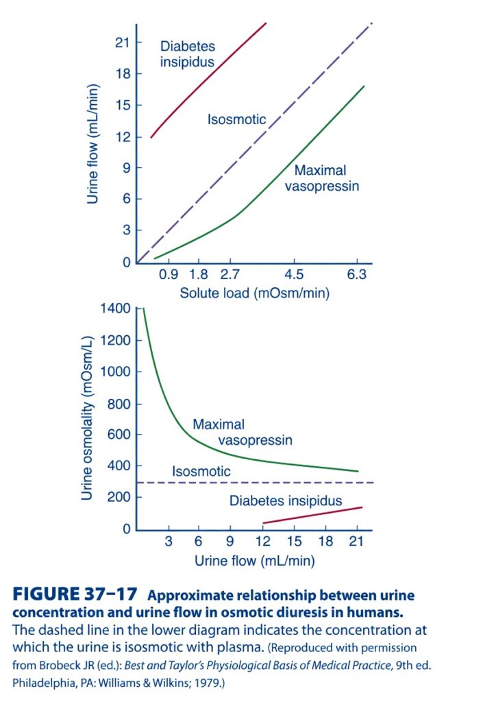
Figure 37-17 – Relationship Between Urine Flow and Urine Concentration During Osmotic Diuresis (Easy Conceptual Summary for self learners)
This figure explains how urine volume (urine flow) and urine concentration (urine osmolality) change under three different conditions:
- Maximum Vasopressin (ADH)
- Isosmotic urine
- Diabetes insipidus (No ADH action)
The figure has two graphs, and both explain the same physiology from different viewpoints.
Main Concept of the Figure
The kidney always tries to balance water and solute (osmoles).
Think of urine as:
Urine = Water + Dissolved Solutes (Na⁺, Urea, etc.)
The amount of urine produced depends mainly on:
- How much solute must be excreted
- How much water ADH allows the kidney to reabsorb
So,
- More ADH → More water reabsorbed → Less urine → More concentrated urine
- Less ADH → Less water reabsorbed → More urine → Dilute urine
TOP GRAPH
Axes
X-axis
Solute load (mOsm/min)
This means:
How many osmoles the kidney must excrete every minute.
More solute load means:
➡️ More waste must leave the body.
Y-axis
Urine flow (mL/min)
This means:
How much urine is produced every minute.
Three different lines are shown.
1. Green Line — Maximum Vasopressin (Maximum ADH)
This is the lowest curve.
What happens?
Even if solute load increases,
urine flow increases only a little.
Why?
ADH makes the collecting duct highly permeable to water.
Most water is reabsorbed back into the blood.
Only a small amount of water leaves as urine.
Example
Suppose the kidney must remove 100 osmoles.
With ADH,
the kidney packs all those osmoles into very little water.
Result:
✔ Small urine volume
✔ Highly concentrated urine
Easy Concept
Imagine putting 10 spoons of sugar into one small glass of water.
Very concentrated.
Very little water.
Exactly what ADH does.
Key Point
Maximum ADH
↓
Maximum water reabsorption
↓
Smallest urine volume
↓
Highest urine concentration
2. Purple Dashed Line — Isosmotic Urine
This is the middle straight line.
What does Isosmotic mean?
Urine has the same osmolality as plasma.
About
300 mOsm/L
What happens?
As solute load increases,
urine flow increases proportionally.
Why?
Every litre of urine always contains about the same concentration of solute.
Therefore,
if twice as much solute must be excreted,
twice as much urine must be produced.
Easy Concept
Imagine every bottle always contains:
300 grams of salt per litre.
If you must remove more salt,
you simply fill more bottles.
Key Point
Isosmotic urine has
constant concentration,
so urine volume rises directly with solute load.
3. Red Line — Diabetes Insipidus
This is the highest line.
What happens?
Even with small solute loads,
urine flow is already very high.
As solute load increases,
urine volume becomes even larger.
Why?
There is no effective ADH.
Collecting ducts cannot reabsorb water.
Large amounts of water are lost.
Example
Suppose the kidney must remove 100 osmoles.
Without ADH,
it cannot concentrate urine.
So,
it must use a huge amount of water.
Result:
Large volume of dilute urine.
Easy Concept
Imagine trying to dissolve one spoon of sugar in a whole bucket of water.
Very dilute.
Very large volume.
Key Point
Diabetes insipidus
↓
No ADH effect
↓
Water cannot be reabsorbed
↓
Very large urine volume
↓
Very dilute urine
Comparison of the Top Graph
| Condition | Urine Volume |
|---|---|
| Maximum ADH | Lowest |
| Isosmotic | Intermediate |
| Diabetes insipidus | Highest |
BOTTOM GRAPH
This graph looks at the relationship differently.
Instead of plotting solute load,
it compares:
Urine Flow
with
Urine Osmolality
Axes
X-axis
Urine flow
(mL/min)
Y-axis
Urine osmolality
(mOsm/L)
The dashed horizontal line represents:
Isosmotic urine
Approximately
300 mOsm/L
Green Curve — Maximum Vasopressin
What happens?
When urine flow is very low,
urine osmolality is extremely high.
As urine flow increases,
urine becomes less concentrated.
Why?
ADH allows maximum water reabsorption.
Small urine volume.
Same solute in less water.
Very concentrated urine.
As urine volume gradually increases,
more water stays in urine.
Therefore,
urine concentration falls.
Easy Concept
Imagine mixing one spoon of salt with:
100 mL water
Very concentrated.
Now mix the same salt with:
500 mL water
Less concentrated.
Key Point
Low urine flow
↓
High urine osmolality
Purple Dashed Line — Isosmotic Urine
This horizontal line stays constant.
Why?
Because urine concentration remains equal to plasma.
Around
300 mOsm/L
regardless of urine volume.
Key Point
Isosmotic urine always has the same concentration.
Red Line — Diabetes Insipidus
What happens?
Urine flow is always high,
but urine osmolality stays very low.
Why?
Without ADH,
water cannot be reabsorbed.
Urine contains lots of water.
Therefore,
it is always dilute.
Easy Concept
Think of adding one spoon of salt to a swimming pool.
Extremely dilute.
Key Point
Diabetes insipidus produces:
Large urine volume
Very low osmolality
How ADH Changes Urine
Maximum ADH
Water returns to blood.
↓
Urine becomes concentrated.
↓
Urine volume decreases.
No ADH
Water stays inside tubules.
↓
Urine becomes dilute.
↓
Urine volume increases.
Clinical Importance
Diabetes Insipidus
Problem:
ADH is absent or ineffective.
Patient develops:
- Massive polyuria
- Dilute urine
- Polydipsia
- Risk of dehydration
SIADH
Opposite condition.
Too much ADH.
Results:
- Very little urine
- Highly concentrated urine
Osmotic Diuresis
Seen in:
- Diabetes mellitus
- Mannitol therapy
Extra osmoles remain in tubules.
Water follows these osmoles.
Urine volume increases even if ADH is present.
Quick Comparison Table
| Condition | ADH | Urine Volume | Urine Osmolality |
|---|---|---|---|
| Maximum vasopressin | High | Very low | Very high |
| Isosmotic urine | Moderate | Moderate | ~300 mOsm/L |
| Diabetes insipidus | None | Very high | Very low |
Easy Memory Trick
Maximum ADH = “Save Water” 💧
- Small urine
- Concentrated urine
Isosmotic = “Same as Plasma” ⚖️
- About 300 mOsm/L
Diabetes Insipidus = “Dump Water” 🚿
- Huge urine volume
- Very dilute urine
Flow Diagram
More ADH
     ↓
More water reabsorbed
     ↓
Less urine produced
     ↓
Higher urine osmolality
No ADH
↓
Water stays in tubules
↓
More urine produced
↓
Lower urine osmolalityHigher Solute Load
↓
Kidney must excrete more osmoles
↓
Urine volume increases
↓
Increase depends on ADH levelKey Concept
This figure demonstrates how urine flow and urine concentration are determined by both the solute load and the action of vasopressin (ADH). In the presence of maximum ADH, the collecting ducts become highly permeable to water, allowing extensive water reabsorption. As a result, even when the solute load increases, urine volume remains low and urine becomes highly concentrated. During isosmotic urine formation, urine has approximately the same osmolality as plasma (about 300 mOsm/L), so urine volume increases directly with the amount of solute that must be excreted. In diabetes insipidus, where ADH is absent or ineffective, the collecting ducts cannot reabsorb water efficiently. Consequently, large volumes of dilute urine are produced, even with relatively small solute loads. Thus, ADH is the major hormone that determines whether the kidney conserves water by producing a small volume of concentrated urine or loses water by producing a large volume of dilute urine.
RELATION OF URINE CONCENTRATION TO GFR
- The concentration of urine depends partly on the glomerular filtration rate (GFR).
- When the flow of fluid through the loops of Henle decreases, the osmotic gradient in the medullary pyramids becomes greater.
- A stronger osmotic gradient allows the kidneys to produce more concentrated urine.
- During dehydration, GFR decreases.
- A lower GFR delivers less fluid to the countercurrent mechanism.
- As a result, the flow of fluid through the loops of Henle decreases.
- This increases the kidney’s ability to concentrate urine.
- Therefore, urine becomes more concentrated when GFR is low.
- When GFR is greatly reduced, urine can become highly concentrated even in the absence of vasopressin (ADH).
- If one renal artery is narrowed (constricted), the GFR decreases in that kidney.
- The kidney with the narrowed renal artery produces hypertonic (concentrated) urine.
- The opposite kidney, with a normal GFR, continues to produce hypotonic (dilute) urine.
KEY CONCEPT
- A decrease in GFR reduces the flow of fluid through the loops of Henle, which strengthens the medullary osmotic gradient and increases the kidney’s ability to concentrate urine. During dehydration or renal artery constriction, urine becomes more concentrated, and this can occur even without vasopressin.
“FREE WATER CLEARANCE”
- Free water clearance (CH₂O) is used to measure whether the kidneys are losing or conserving water.
- It is calculated by finding the difference between:
- Urine flow rate (V̇)
- Osmolar clearance (COsm)
- Formula: CH₂O = V̇ − COsm
- Since osmolar clearance (COsm) is calculated as: COsm = (UOsm × V̇) / POsm
- The complete formula becomes: CH₂O = V̇ − (UOsm × V̇) / POsm
Easy Concept of the Formula
- V̇ (Urine flow rate) = Total amount of urine produced each minute.
- UOsm (Urine osmolality) = Concentration of dissolved particles in urine.
- POsm (Plasma osmolality) = Concentration of dissolved particles in plasma.
- COsm (Osmolar clearance) = The amount of water needed to remove dissolved particles if the urine had the same concentration as plasma.
- CH₂O (Free water clearance) = Extra water that is either removed from the body or conserved by the kidneys.
- If CH₂O is positive (+):
- The urine is dilute (hypotonic).
- The kidneys are excreting extra free water.
- If CH₂O is negative (−):
- The urine is concentrated (hypertonic).
- The kidneys are conserving water.
- Using the values in Table 37–6:
- During maximal antidiuresis:
- CH₂O = −1.3 mL/min (−1.9 L/day)
- This means the kidneys are conserving water by producing concentrated urine.
- In the absence of vasopressin (ADH):
- CH₂O = +14.5 mL/min (20.9 L/day)
- This means the kidneys are excreting large amounts of free water by producing dilute urine.
- During maximal antidiuresis:
Table: Table 37–6
KEY CONCEPT
- Free water clearance (CH₂O) measures whether the kidneys are conserving or excreting water. A negative CH₂O means concentrated urine and water conservation, while a positive CH₂O means dilute urine and increased water excretion.
further simplified Formula
CH2O=V˙−POsmUOsm×V˙
Easiest Concept
Think of the formula in 2 simple steps.
Step 1: Start with all the urine produced
V˙
- V̇ = Total urine flow per minute
- This is the total amount of urine made by the kidneys.
Step 2: Remove the water that is needed to carry the dissolved particles
POsmUOsm×V˙
This part is called Osmolar Clearance (COsm).
It tells us:
“How much water is actually needed to remove all the dissolved solutes (osmoles) from the body?”
Step 3: What remains is “Free Water”
CH2O=Total Urine−Water needed for solutes
So,
Free Water Clearance = Total urine − Water required to carry dissolved particles
Meaning of Each Symbol
| Symbol | Easy Meaning |
|---|---|
| CH₂O | Free water clearance |
| V̇ | Total urine produced per minute |
| UOsm | Urine osmolality (concentration of urine) |
| POsm | Plasma osmolality (concentration of blood plasma) |
| COsm | Water needed to excrete the dissolved solutes |
How to Remember
If CH₂O is Positive (+)
- More water is excreted than needed.
- Urine is dilute (hypotonic).
- Kidney is losing free water.
Example:
- Urine = 10 mL/min
- Water needed for solutes = 6 mL/min
CH2O=10−6=+4
✅ 4 mL/min of free water is lost.
If CH₂O is Negative (−)
- Less water is excreted than needed.
- Urine is concentrated (hypertonic).
- Kidney is conserving water.
Example:
- Urine = 2 mL/min
- Water needed for solutes = 4 mL/min
CH2O=2−4=−2
✅ The kidney is retaining 2 mL/min of free water.
One-Line Memory Trick
Free Water Clearance = Total Urine − Water Needed for Solutes
- Positive CH₂O → Water is being excreted → Dilute urine
- Negative CH₂O → Water is being conserved → Concentrated urine
Made by Self learning CEO AND FOUNDER Dr sheen
