


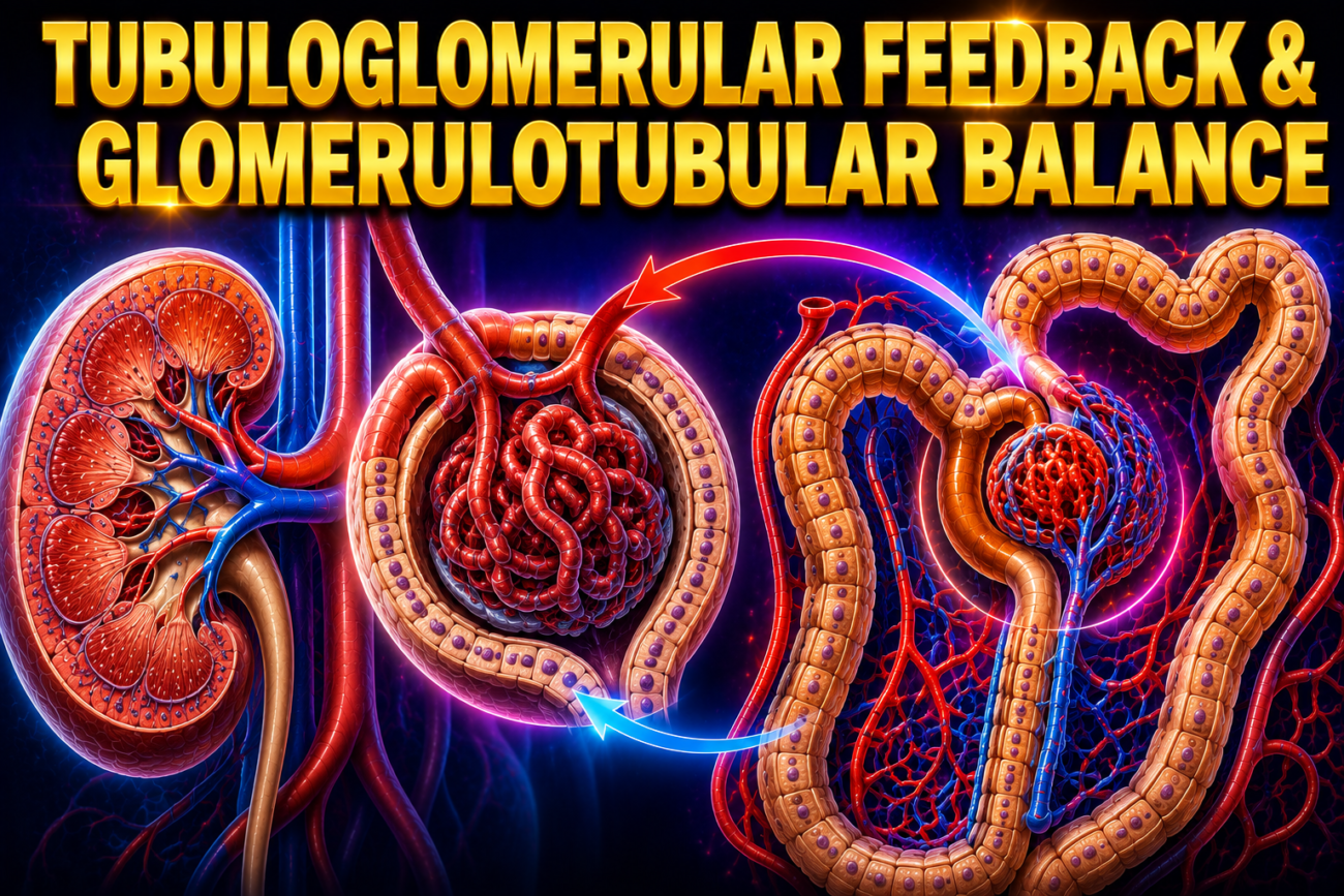
- Signals from the renal tubule can affect filtration in the glomerulus of the same nephron.
Tubuloglomerular Feedback
- When the flow through the ascending limb of the loop of Henle and the first part of the distal tubule increases, the GFR of the same nephron decreases.
- When the flow decreases, the GFR increases.
- This mechanism is called tubuloglomerular feedback.
- Its main function is to keep the amount of fluid reaching the distal tubule relatively constant.
- The macula densa acts as the sensor for this mechanism.
- The amount of fluid reaching the distal tubule depends on the amount of Na⁺ and Cl⁻ present in it.
- Na⁺ and Cl⁻ enter the macula densa cells through the Na–K–2Cl cotransporter in the apical membrane.
- Increased Na⁺ entry increases Na⁺/K⁺-ATPase activity.
- Increased ATP breakdown produces more adenosine.
- Adenosine is released from the basal membrane of the macula densa cells.
- Adenosine acts on A₁ receptors.
- This causes the macula densa to increase Ca²⁺ release to the vascular smooth muscle of the afferent arteriole.
- The afferent arteriole constricts (vasoconstriction).
- Afferent vasoconstriction decreases GFR.
- A similar mechanism may also decrease renin secretion from the nearby juxtaglomerular cells, but this is not yet fully confirmed.
Glomerulotubular Balance
- When GFR increases, reabsorption of solutes and water also increases, mainly in the proximal tubule.
- Because of this, the percentage of filtered solute that is reabsorbed remains nearly constant.
- This process is called glomerulotubular balance.
- It is especially important for Na⁺ reabsorption.
- Changes in Na⁺ reabsorption occur within seconds after a change in GFR.
- Therefore, an external hormonal factor is unlikely to be responsible.
- One important factor is the oncotic pressure in the peritubular capillaries.
- When GFR is high, the plasma leaving the glomerulus through the efferent arteriole has a higher oncotic pressure.
- This higher oncotic pressure increases Na⁺ reabsorption from the renal tubule.
- Other intrarenal mechanisms may also contribute, but they are not yet fully identified.
Figure: Figure 37–12
KEY CONCEPT
- Tubuloglomerular feedback is a local mechanism in which the macula densa senses Na⁺ and Cl⁻ delivery and adjusts GFR by changing afferent arteriole tone. Increased NaCl delivery causes adenosine release, afferent vasoconstriction, and decreased GFR. Glomerulotubular balance ensures that when GFR increases, proximal tubular reabsorption of Na⁺ and water also increases, keeping the percentage of reabsorption nearly constant.
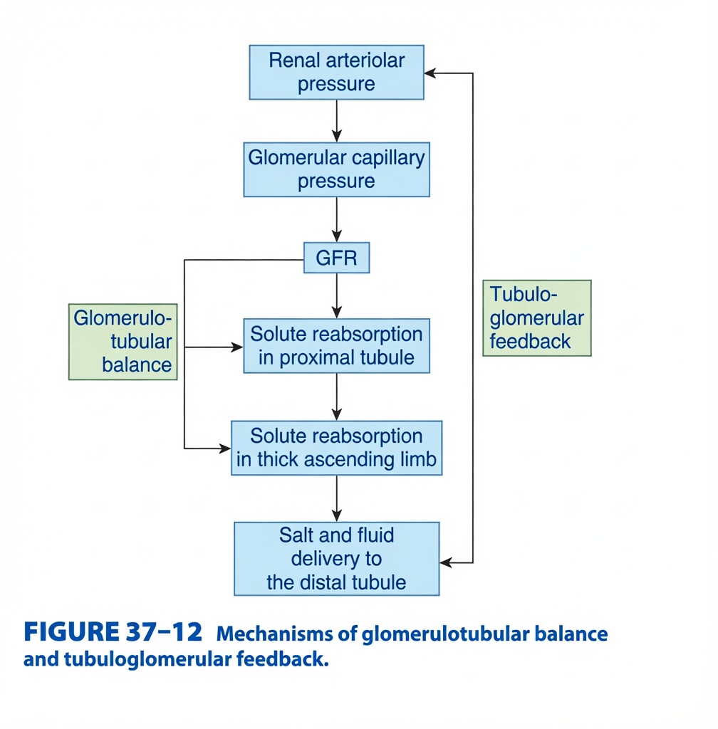
Mechanisms of Glomerulotubular Balance & Tubuloglomerular Feedback (Figure 37-12)
This figure explains how the kidney automatically keeps filtration and reabsorption balanced.
It shows two important kidney safety systems:
- Glomerulotubular Balance (GTB) → Tubules adjust reabsorption according to filtration.
- Tubuloglomerular Feedback (TGF) → Tubules adjust filtration according to salt delivery.
Together, they keep the kidney stable despite changes in blood pressure.
Main Concept
The kidney works like a smart water filtration plant.
- If more fluid is filtered, the tubules automatically reabsorb more (Glomerulotubular Balance).
- If too much salt reaches the distal tubule, the kidney automatically reduces filtration (Tubuloglomerular Feedback).
The Whole Story in One Flow
Renal Blood Pressure
        ↓
Glomerular Pressure
        ↓
GFR
        ↓
Tubules Reabsorb Salt & Water
        ↓
Amount Reaching Distal Tubule
        ↓
Kidney Checks the Amount
        ↓
Adjusts GFR if Needed
First Understand the Big Picture
Imagine your kidney is a water purification factory.
The factory has:
- Filter → Glomerulus
- Workers → Renal tubules
- Quality inspector → Macula densa
The filter sends water to the workers.
If the filter suddenly sends more water, the workers must work faster.
If the workers become overloaded,
the quality inspector slows down the filter.
This is exactly what happens in the kidney.
STEP 1 – Renal Arteriolar Pressure
The figure starts with:
Renal Arteriolar Pressure
This is simply the blood pressure entering the kidney.
If Blood Pressure Increases
↓
More blood reaches the glomerulus.
Easy Concept
Think of opening a tap wider.
More water enters the pipe.
STEP 2 – Glomerular Capillary Pressure
More blood pressure produces:
Higher Glomerular Capillary Pressure
This is the pressure inside the glomerular capillaries.
Easy Concept
Imagine pressing harder on a water filter.
More water passes through it.
STEP 3 – GFR (Glomerular Filtration Rate)
Higher glomerular pressure increases:
GFR
Meaning:
More plasma is filtered every minute.
Easy Concept
The filter now produces more filtrate.
STEP 4 – Proximal Tubule Reabsorption
The figure shows:
GFR
      ↓
Solute Reabsorption
in Proximal Tubule
Now the first tubule begins reabsorbing:
- Sodium
- Water
- Glucose
- Amino acids
What is Glomerulotubular Balance?
The green box on the left represents:
Glomerulotubular Balance (GTB)
Definition
When GFR increases, the proximal tubule automatically increases reabsorption by nearly the same proportion.
Easy Concept
Imagine:
Normally,
100 students enter a classroom.
One teacher teaches them.
Tomorrow,
120 students arrive.
The teacher works harder and teaches more students.
The teacher adjusts automatically.
The proximal tubule behaves exactly the same way.Why is GTB Important?
Without GTB,
an increase in GFR would cause:
Huge fluid loss in urine.
Instead,
the proximal tubule reabsorbs most of the extra filtrate.
Example
Normal:
100 mL filtered
↓
65 mL reabsorbed
Higher GFR:
120 mL filtered
↓
About 78 mL reabsorbed
The percentage remains nearly the same.
STEP 5 – Thick Ascending Limb
The figure continues:
Solute Reabsorption
in Thick Ascending Limb
This segment also reabsorbs sodium and chloride.
It continues the adjustment started by the proximal tubule.
Easy Concept
The second group of workers also works faster when more filtrate arrives.
STEP 6 – Salt & Fluid Reach Distal Tubule
Eventually,
the remaining filtrate reaches:
Distal Tubule
The figure labels:
Salt and Fluid Delivery to Distal Tubule
Why is this important?
The distal tubule acts like a monitoring station.
STEP 7 – Tubuloglomerular Feedback (TGF)
The green box on the right represents:
Tubuloglomerular Feedback
Definition
The distal tubule senses how much salt is arriving and sends signals back to the glomerulus to adjust GFR.
Easy Concept
Imagine a quality inspector.
If too many boxes arrive,
he calls the factory manager:
“Slow the machine!”
Who Detects the Salt?
A special group of cells called:
Macula Densa
These cells detect:
Mainly NaCl concentration.
Scenario 1 – GFR Too High
↑ GFR
      ↓
More NaCl reaches Macula Densa
The macula densa says:
“Too much salt is arriving.”
It releases signals that:
- Constrict the afferent arteriole
- Reduce glomerular pressure
- Decrease GFR
Flow
↑ GFR
      ↓
↑ NaCl at Macula Densa
      ↓
Afferent Arteriole Constricts
      ↓
↓ GFR
Easy Concept
Too many customers enter a shop.
The security guard closes one entrance.
Fewer customers enter.
Scenario 2 – GFR Too Low
Now imagine filtration becomes low.
↓ GFR
      ↓
Less NaCl reaches Macula Densa
The macula densa says:
“Too little salt is arriving.”
It responds by:
Flow
↓ GFR
      ↓
↓ NaCl at Macula Densa
      ↓
Renin Release
      ↓
↑ GFR
Easy Concept
Too few customers enter the shop.
The security guard opens the gate wider.
Difference Between GTB and TGF
| Glomerulotubular Balance (GTB) | Tubuloglomerular Feedback (TGF) |
|---|---|
| Occurs mainly in proximal tubule | Occurs at macula densa (distal tubule) |
| Adjusts reabsorption | Adjusts filtration (GFR) |
| Keeps filtered and reabsorbed amounts proportional | Prevents excessive or insufficient filtration |
| No neural control needed | Uses local signalling to afferent arteriole |
Complete Story of the Figure
Blood Pressure
      ↓
Renal Arteriolar Pressure
      ↓
Glomerular Pressure
      ↓
GFR
      ↓
────────────────────────────
Glomerulotubular Balance
(Proximal Tubule)
      ↓
More GFR
      ↓
More Reabsorption
────────────────────────────
Thick Ascending Limb
      ↓
Distal Tubule
      ↓
Macula Densa Measures NaCl
────────────────────────────
Too Much NaCl
      ↓
Tubuloglomerular Feedback
      ↓
↓ GFR
────────────────────────────
Too Little NaCl
      ↓
Renin Release
      ↓
↑ GFR
Easy Factory Analogy
Imagine a bottle-filling factory.
Stage 1
The filling machine (glomerulus) fills bottles.
↓
Stage 2
Workers (proximal tubule) pack bottles.
If more bottles arrive,
the workers pack more.
This is Glomerulotubular Balance.
↓
Stage 3
The quality inspector (macula densa) counts bottles.
If too many bottles arrive,
he slows the filling machine.
If too few arrive,
he speeds it up.
This is Tubuloglomerular Feedback.
Easy Memory Trick
GTB = “Tubules Keep Up”
↑ GFR
     ↓
↑ Reabsorption
Think:
“More Filter → More Reabsorb.”
TGF = “Tubules Talk Back”
Distal Tubule
      ↓
Talks to Glomerulus
      ↓
Adjusts GFR
Think:
“The tubule tells the filter what to do.”
Important Points from Figure 37-12
- Renal arteriolar pressure determines glomerular capillary pressure, which directly influences GFR.
- An increase in GFR increases the amount of filtrate entering the nephron.
- Glomerulotubular balance (GTB) ensures that the proximal tubule automatically reabsorbs a relatively constant fraction of the filtered sodium and water, preventing excessive fluid loss.
- The thick ascending limb also increases solute reabsorption when filtrate delivery increases.
- The amount of salt and fluid reaching the distal tubule is monitored by the macula densa.
- Tubuloglomerular feedback (TGF) adjusts GFR according to distal tubular NaCl delivery:
- High NaCl delivery → afferent arteriolar constriction → ↓ GFR.
- Low NaCl delivery → afferent arteriolar dilation and renin release → ↑ GFR.
- GTB primarily regulates tubular reabsorption, whereas TGF primarily regulates glomerular filtration, allowing the kidney to maintain stable filtration and fluid balance despite changes in blood pressure.
KEY CONCEPT (Figure 37-12)
Figure 37-12 illustrates two essential autoregulatory mechanisms of the kidney. Glomerulotubular balance enables the proximal tubule (and subsequently the thick ascending limb) to increase solute and water reabsorption in proportion to increases in GFR, maintaining a nearly constant fraction of filtrate reabsorbed. Tubuloglomerular feedback uses the macula densa in the distal tubule to monitor NaCl delivery and adjust glomerular filtration accordingly. When distal NaCl delivery is high, the afferent arteriole constricts to reduce GFR; when NaCl delivery is low, afferent dilation and renin release help increase GFR. Together, these mechanisms stabilize nephron function and protect the kidneys from large fluctuations in blood pressure and filtration.
WATER TRANSPORT
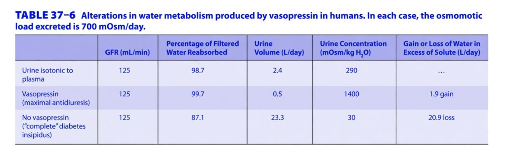
- Normally, about 180 L of fluid is filtered by the glomeruli each day.
- The average urine produced each day is only about 1 L.
- The same amount of solute can be excreted in different urine volumes.
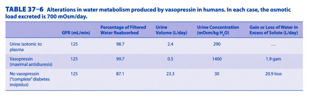
- It can be excreted in 500 mL of highly concentrated urine (1400 mOsm/kg).
- It can also be excreted in 23.3 L of very dilute urine (30 mOsm/kg).
- These observations show two important facts.
- First, at least 87% of the filtered water is reabsorbed, even when urine volume is as high as 23.3 L.
- Second, the amount of water reabsorbed can change without changing the total amount of solute excreted.
- When urine is concentrated, the kidneys retain more water than solute.
- When urine is dilute, the kidneys lose more water than solute.
- These mechanisms are very important for maintaining the normal osmolality of body fluids.
- Vasopressin is the main hormone that regulates water excretion.
- Vasopressin acts on the collecting ducts to control water output.
Table: Table 37–6
KEY CONCEPT
- About 180 L of fluid is filtered each day, but only about 1 L is excreted as urine because most filtered water is reabsorbed. The kidneys can produce either concentrated or dilute urine while excreting the same amount of solute. Vasopressin regulates water reabsorption in the collecting ducts, helping maintain normal body fluid osmolality.
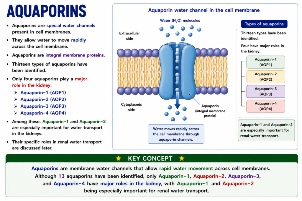
AQUAPORINS
- Aquaporins are special water channels present in cell membranes.
- They allow water to move rapidly across the cell membrane.
- Aquaporins are integral membrane proteins.
- Thirteen types of aquaporins have been identified.
- Only four aquaporins play a major role in the kidney:
- Aquaporin-1 (AQP1)
- Aquaporin-2 (AQP2)
- Aquaporin-3 (AQP3)
- Aquaporin-4 (AQP4)
- Among these, Aquaporin-1 and Aquaporin-2 are especially important for water transport in the kidneys.
- Their specific roles in renal water transport are discussed later.
KEY CONCEPT
- Aquaporins are membrane water channels that allow rapid water movement across cell membranes. Although 13 aquaporins have been identified, only Aquaporin-1, Aquaporin-2, Aquaporin-3, and Aquaporin-4 have major roles in the kidney, with Aquaporin-1 and Aquaporin-2 being especially important for renal water transport.
PROXIMAL TUBULE
- Many substances are actively transported from the fluid in the proximal tubule.
- Despite active transport, the tubular fluid remains almost iso-osmotic with plasma until the end of the proximal tubule.
- Aquaporin-1 (AQP1) is present in both the apical and basolateral membranes of proximal tubule cells.
- Aquaporin-1 allows water to move rapidly out of the tubule.
- Water follows the osmotic gradient created by active transport of solutes.
- As a result, the tubular fluid remains isotonic (iso-osmotic).
- At the end of the proximal tubule, the TF/P ratio of inulin is about 2.5–3.3.
- This indicates that about 60–70% of the filtered solutes have been reabsorbed.
- It also indicates that about 60–70% of the filtered water has been reabsorbed before the filtrate leaves the proximal tubule.
- When Aquaporin-1 was removed (knocked out) in mice, water permeability of the proximal tubule decreased by about 80%.
- When these mice became dehydrated, their urine osmolality did not increase above about 700 mOsm/kg, even though other renal aquaporins were present.
- In humans with mutations that eliminate Aquaporin-1 activity, the disturbance in water balance is less severe.
- However, these individuals have a reduced ability to respond to dehydration.
Figure: Figure 37–8, Figure 37–13
KEY CONCEPT
- The proximal tubule actively reabsorbs many solutes while Aquaporin-1 allows water to follow, keeping the tubular fluid iso-osmotic. By the end of the proximal tubule, about 60–70% of the filtered water and solutes have been reabsorbed. Loss of Aquaporin-1 greatly reduces water reabsorption and impairs the ability to concentrate urine during dehydration.
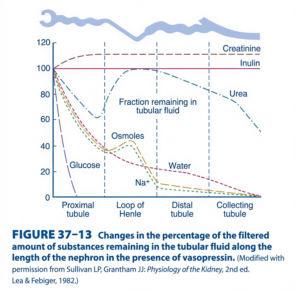
Changes in the Percentage of Filtered Substances Remaining in Tubular Fluid Along the Nephron (Figure 37.13) – Easy Conceptual Summary
This figure explains what happens to different filtered substances as they travel through the nephron in the presence of vasopressin (ADH).
Instead of showing concentration, this graph shows:
The percentage (%) of the originally filtered amount that remains in the tubular fluid as it moves from the proximal tubule → Loop of Henle → Distal tubule → Collecting duct.
The figure compares:
- Creatinine
- Inulin
- Urea
- Glucose
- Water
- Na⁺
- Osmoles
Basic Concept
Imagine that 100 molecules of each substance are filtered at the glomerulus.
At the start of the proximal tubule:
- Glucose = 100 molecules
- Water = 100 molecules
- Na⁺ = 100 molecules
- Urea = 100 molecules
As the filtrate travels through the nephron,
some substances are:
- Reabsorbed back into blood
- Secreted into the tubule
The graph simply shows:
How many of the original 100 molecules are still left inside the tubular fluid.
Understanding the Axes
X-axis
Shows different parts of the nephron:
- Proximal tubule
- Loop of Henle
- Distal tubule
- Collecting duct
Y-axis
Shows:
Percentage of filtered substance remaining in tubular fluid
100%
means
➡️ None has been reabsorbed.
0%
means
➡️ Completely removed from the tubular fluid.
Understanding Each Curve
1. Glucose Curve (Purple) – Falls Rapidly to Zero
This is the steepest downward curve.What happens?
At filtration:
100% glucose enters the nephron.
Within the early proximal tubule,
the curve reaches:
0%
Why?
Glucose is almost completely reabsorbed in the proximal tubule.
It is transported by:
- SGLT2 (early proximal tubule)
- SGLT1 (late proximal tubule)
Normally,
no glucose reaches the Loop of Henle.
Easy Concept
Imagine collecting 100 chocolates.
Before leaving the first room,
someone takes back all 100 chocolates.
Nothing is left.
Exactly the same happens with glucose.
Key Point
✔ 100% filtered glucose is normally reabsorbed in the proximal tubule.
2. Na⁺ Curve (Green Dashed Line)
What happens?
Na⁺ gradually decreases throughout the nephron.
By the end,
almost none remains.
Why?
Sodium is reabsorbed throughout the nephron.
Proximal tubule
About 65%
↓
Thick ascending limb
About 25%
↓
Distal tubule
About 5%
↓
Collecting duct
Small additional amount
Almost all sodium is reabsorbed.
Why does the curve rise slightly in the Loop of Henle?
Notice a small upward bend.
This occurs because:
The descending limb loses water but not sodium.
Water leaves.
Sodium remains.
Therefore,
the percentage of sodium remaining appears relatively higher for a short distance.
Afterwards,
the thick ascending limb actively reabsorbs sodium,
and the curve falls sharply again.
Easy Concept
Imagine removing water from soup.
The salt concentration temporarily increases,
even though you haven’t added more salt.
Key Point
Na⁺ is progressively reabsorbed throughout the nephron.
3. Water Curve (Red Dashed Line)
What happens?
Water gradually decreases.
Only a very small amount remains by the end.
Why?
Water is reabsorbed in:
Proximal tubule
About 65%
↓
Descending Loop
Large amount
↓
Collecting duct (because ADH is present)
Even more water is reabsorbed.
Therefore,
very little water remains in the urine.
Role of ADH
The graph specifically states:
“In the presence of vasopressin (ADH).”
ADH inserts:
Aquaporin-2 channels
into the collecting duct.
More water is reabsorbed.
Therefore,
the final water curve approaches zero.
Easy Concept
Think of squeezing water out of a sponge several times.
Each squeeze leaves less water behind.
Key Point
With ADH,
almost all filtered water is reabsorbed.
4. Osmoles Curve (Brown Line)
What happens?
Osmoles decrease,
rise slightly in the Loop of Henle,
then fall sharply.
Why?
The osmoles mainly include:
- Sodium
- Chloride
- Urea
The temporary rise occurs because:
Water leaves the descending limb faster than solutes.
Later,
active sodium reabsorption reduces osmoles.ey Point
Osmoles follow sodium and water handling.5. Urea Curve (Blue Dash-Dot Line)
This is one of the most interesting curves.hat happens?
Initially,
the percentage remaining decreases.
Then,
it rises in the Loop of Henle.
Later,
it gradually falls again.
Why?
Step 1
Some urea is reabsorbed in the proximal tubule.
Therefore,
the percentage decreases.
Step 2
Water leaves the descending limb.
Urea becomes concentrated.
ADH also promotes urea recycling in the medulla.
As urea is secreted into the thin loop of Henle,
the amount of urea in tubular fluid increases again.
This explains the rise.
Step 3
Later,
urea is reabsorbed again in the collecting duct,
so the curve falls.Easy Concept
Think of passengers leaving a bus,
then more passengers getting on,
then some leaving again.
The number changes in both directions.
Key Point
Urea undergoes both reabsorption and secretion (urea recycling).
6. Inulin Curve (Straight Horizontal Line)
What happens?
The line remains around 100% throughout.
Why?
Inulin is:
- Freely filtered.
- Neither reabsorbed.
- Nor secreted.
Therefore,
the amount inside the tubule does not change.
Easy Concept
Imagine 100 balls entering a tunnel.
No balls leave.
No new balls enter.
100 balls remain throughout.Key Point
Inulin is the ideal marker of GFR because it is neither reabsorbed nor secreted.7. Creatinine Curve (Brown Dashed Line Above Inulin)
What happens?
The curve rises slightly above 100%.
Why?
Creatinine is:
- Freely filtered.
- Slightly secreted by the proximal tubule.
Because additional creatinine is added into the tubular fluid,
more than the originally filtered amount is present.
Therefore,
the percentage becomes greater than 100%.
Easy Concept
Imagine starting with 100 marbles.
Along the way,
someone adds 10 more.
Now,
110 marbles remain.
Key Point
Creatinine is filtered and slightly secreted.omparison of All Curves
| Substance | Main Event | Final Amount Remaining |
|---|---|---|
| Glucose | Completely reabsorbed | ~0% |
| Water | Almost completely reabsorbed (ADH present) | Very little |
| Na⁺ | Progressively reabsorbed | Very little |
| Osmoles | Follow sodium and water | Very little |
| Urea | Reabsorbed + secreted (recycling) | Moderate amount |
| Inulin | Neither reabsorbed nor secreted | ~100% |
| Creatinine | Slight secretion | >100% |
Clinical Importance
Glucose in Urine
Occurs only when:
Transport maximum (Tm) is exceeded,
such as in diabetes mellitus.
Creatinine
Used clinically to estimate:
Glomerular Filtration Rate (GFR).
Because it is slightly secreted,
creatinine clearance slightly overestimates GFR.
Inulin
The most accurate marker for measuring GFR,
but rarely used clinically because it must be infused.
ADH
With ADH present:
- Water reabsorption increases.
- Urine volume decreases.
- Urine becomes concentrated.
Quick Memory Table
| Substance | Reabsorbed? | Secreted? |
|---|---|---|
| Glucose | ✔ Completely | ✘ No |
| Water | ✔ Almost completely | ✘ No |
| Na⁺ | ✔ Yes | ✘ No |
| Urea | ✔ Yes | ✔ Yes (recycling) |
| Inulin | ✘ No | ✘ No |
| Creatinine | ✘ No | ✔ Slightly |
Easy Memory Trick
Glucose = Gone 🍬
- Completely reabsorbed.
- Reaches zero.
Water = With ADH, Nearly Gone 💧
- Most reabsorbed.
Na⁺ = Saved 🧂
- Gradually reabsorbed.
Urea = Up and Down 🔄
- Reabsorbed.
- Secreted.
- Reabsorbed again.
Inulin = Innocent ⚖️
- Nothing happens.
- Perfect GFR marker.
Creatinine = Climbs 📈
- Slight secretion.
- Slightly above 100%.
Key Concept
This figure shows the percentage of the originally filtered amount of different substances remaining in the tubular fluid as filtrate passes through the nephron in the presence of vasopressin (ADH). Glucose is almost completely reabsorbed in the proximal tubule, so it falls rapidly to 0%. Water and sodium are progressively reabsorbed throughout the nephron, with ADH greatly increasing water reabsorption in the collecting duct, leaving very little water in the final urine. Osmoles generally follow the handling of sodium and water. Urea shows a unique pattern because it undergoes both reabsorption and secretion (urea recycling), causing its amount in tubular fluid to decrease, then increase, and finally decrease again. Inulin is filtered but neither reabsorbed nor secreted, so the filtered amount remaining stays essentially unchanged, making it the ideal marker for glomerular filtration rate (GFR). Creatinine is filtered and slightly secreted, so the amount remaining in the tubular fluid becomes slightly greater than the filtered amount, which is why creatinine clearance slightly overestimates GFR.
LOOP OF HENLE
- The loops of Henle of the juxtamedullary nephrons extend deep into the medullary pyramids.
- They then return to the cortex and drain into the distal convoluted tubules.
- All collecting ducts pass back through the medullary pyramids and open into the renal pelvis at the tips of the pyramids.
- The osmolality of the medullary interstitium gradually increases from the cortex toward the papilla.
- At the tip of the papilla, the osmolality can reach about 1200 mOsm/kg H₂O.
- This is about four times higher than the osmolality of plasma.
- The descending limb of the loop of Henle is permeable to water because it contains Aquaporin-1 in both the apical and basolateral membranes.
- The ascending limb is impermeable to water.
- In the thick ascending limb, Na⁺, K⁺, and Cl⁻ are transported out of the tubular fluid together.
- As water leaves the descending limb and enters the hypertonic medullary interstitium, the tubular fluid becomes hypertonic (more concentrated).
- In the ascending limb, Na⁺ and Cl⁻ leave the tubular fluid, but water cannot follow.
- As a result, the tubular fluid becomes progressively more dilute.
- By the time the fluid reaches the top of the ascending limb (the diluting segment), it is hypotonic compared with plasma.
- About 15% of the filtered water is reabsorbed while the filtrate passes through the loop of Henle.
- Therefore, only about 20% of the filtered water reaches the distal tubule.
- At this point, the TF/P ratio of inulin is about 5.
- In the thick ascending limb, one Na⁺, one K⁺, and two Cl⁻ ions are transported into the tubular cells by the Na⁺–K⁺–2Cl⁻ cotransporter.
- This transport is an example of secondary active transport.
- Na⁺ is then actively pumped from the tubular cells into the interstitium by the Na⁺/K⁺-ATPase in the basolateral membrane.
- This keeps the intracellular Na⁺ concentration low.
- The Na⁺–K⁺–2Cl⁻ cotransporter has 12 transmembrane domains with intracellular amino and carboxyl terminals.
- Similar transporters are also found in the salivary glands, gastrointestinal tract, and airways.
- K⁺ diffuses back into the tubular lumen and into the interstitium through ROMK and other K⁺ channels.
- Cl⁻ moves into the interstitium through ClC–Kb channels.
Figure: Figure 37–14
KEY CONCEPT
The loop of Henle creates concentrated and dilute tubular fluid by having different permeabilities in its two limbs. The descending limb is permeable to water because of Aquaporin-1, so water leaves and the tubular fluid becomes concentrated. The ascending limb is impermeable to water but actively reabsorbs Na⁺, K⁺, and Cl⁻ through the Na⁺–K⁺–2Cl⁻ cotransporter, making the tubular fluid dilute. About 15% of filtered water is reabsorbed in the loop of Henle, and only about 20% of filtered water reaches the distal tubule.
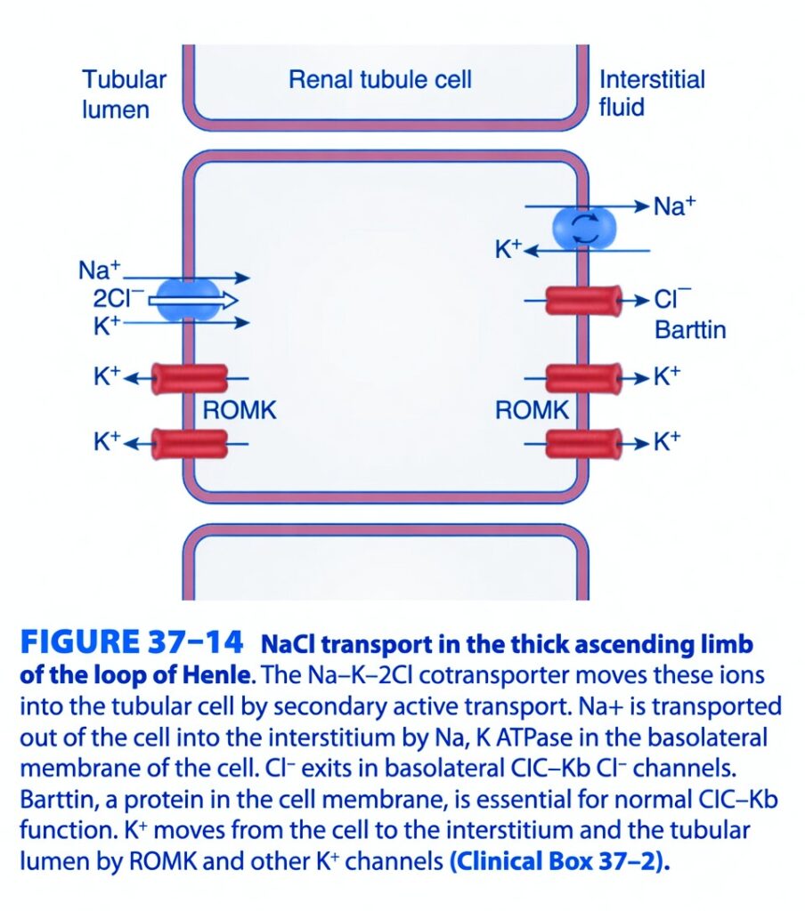
NaCl Transport in the Thick Ascending Limb of the Loop of Henle (Figure 37-14)
Easiest & Most Conceptual ANALYZE
This figure explains how the Thick Ascending Limb (TAL) reabsorbs sodium (Na⁺), potassium (K⁺), and chloride (Cl⁻) from the tubular fluid into the blood.
The main transporter is the Na⁺–K⁺–2Cl⁻ cotransporter (NKCC2).
Main Concept
The Thick Ascending Limb is the kidney’s “salt-pumping segment.”
It removes NaCl from the tubular fluid but does NOT allow water to follow.
Therefore:
- Tubular fluid becomes dilute
- Kidney medulla becomes concentrated
- This concentration gradient later helps ADH reabsorb water in the collecting duct.
Big Picture of the Figure
The figure shows one renal tubule cell.
There are two sides of this cell.
Tubular Lumen
      │
      │  (Apical membrane)
      │
[ Renal Tubule Cell ]
      │
      │ (Basolateral membrane)
      │
Interstitial Fluid (Blood Side)
Think of the cell as a warehouse.TEP 1 – Na⁺, K⁺, and Cl⁻ Enter the Cell
On the left (apical membrane) the figure shows:
Na⁺–K⁺–2Cl⁻ Cotransporter (NKCC2)
It transports:
1 Na⁺
1 K⁺
2 Cl⁻
All together into the cell.
Easy Concept
Imagine a school bus.
The bus only moves when it carries:
- 1 sodium
- 1 potassium
- 2 chlorides
Only when all passengers are present does the bus enter the cell.
Flow
Tubular Fluid
      ↓
Na⁺
K⁺
2Cl⁻
      ↓
NKCC2 Transporter
      ↓
Renal Tubule Cell
Why is this called Secondary Active Transport?
The transporter itself does not use ATP directly.
Instead,
it uses the sodium gradient created by the Na⁺/K⁺ ATPase on the opposite side.Easy Concept
Think of NKCC2 as a cart moving downhill.
It doesn’t have its own engine.
The downhill slope (Na⁺ gradient) provides the energy.
STEP 2 – Na⁺ Leaves the Cell
On the right side (basolateral membrane)
the figure shows:
Na⁺/K⁺ ATPase
This pump:
3 Na⁺ OUT
2 K⁺ IN
using ATP.
Why?
It keeps intracellular sodium low.
Because sodium stays low inside,
more sodium can enter from the lumen through NKCC2.
Easy Concept
Imagine workers continuously emptying boxes from a warehouse.
Since the warehouse never becomes full,
more boxes can keep arriving.
Flow
Cell
     ↓
Na⁺ Pump
     ↓
Interstitial Fluid
STEP 3 – Chloride Leaves the Cell
After chloride enters through NKCC2,
it leaves the cell through
ClC-Kb Chloride Channel
The figure labels:
Barttin
What is Barttin?
Barttin is not a transporter.
It is a helper protein.
Without Barttin,
the chloride channel cannot function normally.
Easy Concept
Think of Barttin as the electric switch.
Without the switch,
the door (chloride channel) cannot open.
Flow
Cell
     ↓
ClC-Kb Channel
     ↓
Interstitial Fluid
STEP 4 – Potassium (K⁺) Recycling
This is the most important concept in the figure.
The figure shows ROMK channels.What does ROMK do?
ROMK allows potassium to leave the cell.
Some potassium:
- Goes back into the lumen
- Some goes into the interstitial fluid
Why recycle potassium?
Remember,
NKCC2 requires potassium.
But tubular potassium concentration is low.
Therefore,
the cell recycles potassium back into the lumen.
Now NKCC2 always has enough potassium available.
Easy Concept
Imagine a school bus always needs one passenger (K⁺).
After arriving,
that passenger immediately gets off,
walks back,
and boards the next bus again.
The same potassium is used repeatedly.
Flow
Cell
      ↓
ROMK
      ↓
Tubular Lumen
      ↓
NKCC2 Uses It Again
Why Does Potassium Recycling Matter?
Because positive potassium ions move back into the lumen,
the lumen becomes positively charged.This positive charge pushes other positive ions between the cells into the blood.
Examples:
- Ca²⁺
- Mg²⁺
This is called
Paracellular Reabsorptionasy Concept
The positive charge acts like a gentle push,
forcing calcium and magnesium toward the blood.
Overall Transport Story
Tubular Fluid
      ↓
NKCC2
(Na⁺ + K⁺ + 2Cl⁻)
      ↓
Renal Tubule Cell
      ↓
────────────────────────
Na⁺
→ Na⁺/K⁺ ATPase
→ Blood
────────────────────────
Cl⁻
→ ClC-Kb Channel
(Barttin Required)
→ Blood
────────────────────────
K⁺
→ ROMK
→ Back to Lumen
(Recycling)
Why is the Thick Ascending Limb Called the Diluting Segment?
This segment:
✅ Reabsorbs large amounts of NaCl
❌ Does NOT allow water to leave.
Therefore,
Salt leaves.
Water stays.
The tubular fluid becomes more dilute.
Easy Concept
Imagine removing all the salt from soup,
but leaving all the water.
The soup becomes very dilute.
The TAL does exactly this.
Clinical Correlation
Loop Diuretics (e.g., Furosemide)
Loop diuretics block:
NKCC2
Result:
- ↓ Na⁺ reabsorption
- ↓ Cl⁻ reabsorption
- ↓ K⁺ recycling
- ↓ Medullary osmotic gradient
- ↑ Urine volume
- ↑ Na⁺, Cl⁻, K⁺, Ca²⁺, and Mg²⁺ excretion
Bartter Syndrome
A defect in:
- NKCC2
- ROMK
- ClC-Kb
- Barttin
produces impaired salt reabsorption,
similar to chronic loop diuretic use.
Complete Story of the Figure
Tubular Lumen
      ↓
Na⁺ + K⁺ + 2Cl⁻
      ↓
NKCC2
      ↓
Renal Tubule Cell
      ↓
────────────────────────
Na⁺
→ Na⁺/K⁺ ATPase
→ Blood
────────────────────────
Cl⁻
→ ClC-Kb Channel
(Barttin Required)
→ Blood
────────────────────────
K⁺
→ ROMK
→ Back Into Lumen
(Recycling)
────────────────────────
NaCl Reabsorbed
Water Stays
────────────────────────
Dilute Tubular Fluid
Concentrated Medulla
Easy Bus Analogy
Imagine a bus (NKCC2).
Every trip requires:
- 👤 1 Sodium
- 👤 1 Potassium
- 👤👤 2 Chlorides
The bus enters the warehouse (cell).
- Sodium exits to the blood through the Na⁺/K⁺ ATPase.
- Chloride exits through the ClC-Kb channel (helped by Barttin).
- Potassium quickly returns to the bus stop through ROMK so it can ride the next bus again.
This continuous cycling keeps the transport system running efficiently.
Easy Memory Trick
Remember: “NKCC–RBP”
- N = Na⁺
- K = K⁺
- CC = 2 Cl⁻
- R = ROMK recycles K⁺
- B = Barttin helps ClC-Kb
- P = Pump (Na⁺/K⁺ ATPase) sends Na⁺ to blood
Important Points from Figure 37-14
- The Thick Ascending Limb (TAL) reabsorbs NaCl without water, making it the diluting segment of the nephron.
- The apical Na⁺–K⁺–2Cl⁻ cotransporter (NKCC2) transports 1 Na⁺, 1 K⁺, and 2 Cl⁻ from the tubular lumen into the cell by secondary active transport.
- The Na⁺/K⁺ ATPase on the basolateral membrane uses ATP to pump Na⁺ into the interstitial fluid, maintaining the sodium gradient that drives NKCC2.
- Cl⁻ exits the cell through ClC-Kb chloride channels, which require Barttin for normal function.
- K⁺ leaves the cell through ROMK channels, with a portion recycled back into the tubular lumen to maintain NKCC2 activity.
- Potassium recycling creates a lumen-positive electrical potential, which promotes paracellular reabsorption of Ca²⁺ and Mg²⁺.
- Loop diuretics inhibit NKCC2, reducing NaCl reabsorption, collapsing the medullary osmotic gradient, and increasing urine output.
- Genetic defects affecting NKCC2, ROMK, ClC-Kb, or Barttin cause Bartter syndrome, producing impaired salt reabsorption and a loop diuretic–like phenotype.
KEY CONCEPT (Figure 37-14)
Figure 37-14 illustrates the mechanism of NaCl transport in the Thick Ascending Limb of the loop of Henle. The NKCC2 cotransporter brings Na⁺, K⁺, and 2 Cl⁻ into the tubular cell using the sodium gradient established by the Na⁺/K⁺ ATPase. Sodium is pumped into the interstitial fluid, chloride exits through ClC-Kb channels with the assistance of Barttin, and potassium is recycled into the tubular lumen through ROMK channels. Because this nephron segment is impermeable to water, NaCl is reabsorbed without water, diluting the tubular fluid while helping establish the hyperosmotic renal medulla that is essential for urine concentration.
DISTAL TUBULE
- The distal tubule, especially its first part, is a continuation of the thick ascending limb of the loop of Henle.
- It is relatively impermeable to water.
- Solutes continue to be reabsorbed without water.
- As a result, the tubular fluid becomes even more dilute.
COLLECTING DUCTS
- The collecting ducts have two parts:
- Cortical collecting duct
- Medullary collecting duct
- The volume and osmolality of the tubular fluid in the collecting ducts depend on the amount of vasopressin (ADH).
- Vasopressin is an antidiuretic hormone released from the posterior pituitary gland.
- Vasopressin increases the permeability of the collecting ducts to water.
- Aquaporin-2 is the key water channel responsible for the action of vasopressin.
- Unlike other aquaporins, Aquaporin-2 is stored inside vesicles in the cytoplasm of principal cells.
- Vasopressin rapidly inserts these vesicles into the apical membrane of the principal cells.
- This effect is mediated through:
- V₂ receptor
- cAMP
- Protein kinase A (PKA)
- Cytoskeletal proteins, including dynein, dynactin, myosin-1, and actin filaments, help move these vesicles to the cell membrane.
- When enough vasopressin is present (maximum antidiuresis), water leaves the hypotonic fluid in the cortical collecting duct and enters the cortical interstitium.
- The tubular fluid becomes isotonic.
- About 10% of the filtered water is reabsorbed in the cortical collecting duct.
- The isotonic fluid then enters the medullary collecting duct.
- At this point, the TF/P ratio of inulin is about 20.
- An additional 4.7% or more of the filtered water is reabsorbed into the hypertonic medullary interstitium.
- This produces highly concentrated urine.
- The TF/P ratio of inulin increases to more than 300.
- In humans, urine osmolality can reach about 1400 mOsm/kg H₂O.
- This is about five times greater than the osmolality of plasma.
- About 99.7% of the filtered water is reabsorbed.
- Some animals can concentrate urine even more:
- Dogs: about 2500 mOsm/kg
- Laboratory rats: about 3200 mOsm/kg
- Desert rodents: up to 5000 mOsm/kg
- When vasopressin is absent, the collecting ducts become relatively impermeable to water.
- The tubular fluid remains hypotonic.
- Large amounts of dilute urine pass into the renal pelvis.
- In humans, urine osmolality may fall to about 30 mOsm/kg H₂O.
- Even without vasopressin, about 2% of the filtered water is still reabsorbed along with salt.
- However, as much as 13% of the filtered water may be excreted.
- Urine flow may increase to 15 mL/min or more.
Table: Table 37–6
KEY CONCEPT
- The distal tubule is relatively impermeable to water and continues to dilute the tubular fluid. In the collecting ducts, vasopressin (ADH) acts through Aquaporin-2 to increase water reabsorption. With maximum ADH, about 99.7% of filtered water is reabsorbed, producing highly concentrated urine (up to 1400 mOsm/kg in humans). Without ADH, the collecting ducts remain relatively impermeable to water, producing large volumes of dilute urine.
GENETIC MUTATIONS IN RENAL TRANSPORTERS
- Mutations in genes that code for renal sodium transporters, proteins, and channels can cause specific kidney diseases.
- Examples include:
- Bartter syndrome
- Liddle syndrome
- Dent disease
- Polycystic kidney disease
- Many different genetic mutations have been identified.
- Bartter syndrome is a rare disorder caused by defective transport in the thick ascending limb of the loop of Henle.
- It is characterized by:
- Chronic loss of Na⁺ in the urine
- Hypovolemia
- Increased renin secretion
- Increased aldosterone secretion
- No hypertension
- Hyperkalemia
- Alkalosis
- Bartter syndrome can result from loss-of-function mutations in any of these proteins:
- Na⁺–K⁺–2Cl⁻ cotransporter
- ROMK K⁺ channel
- ClC–Kb Cl⁻ channel
- Barttin protein
- Barttin is an integral membrane protein required for the normal function of ClC–Kb Cl⁻ channels.
- The stria vascularis of the inner ear maintains the high K⁺ concentration needed for normal hearing.
- It contains both ClC–Kb and ClC–Ka Cl⁻ channels.
- Patients with Bartter syndrome caused by ClC–Kb mutations usually do not develop deafness because ClC–Ka channels can compensate.
- Both ClC–Kb and ClC–Ka channels require barttin for normal function.
- Therefore, patients with Bartter syndrome caused by barttin mutations also develop deafness.
- Polycystin-1 (PKD-1) and Polycystin-2 (PKD-2) are proteins involved in kidney function.
- PKD-1 appears to act as a Ca²⁺ receptor that activates an ion channel associated with PKD-2.
- The normal function of this ion channel is not fully known.
- Mutations in PKD-1 or PKD-2 cause autosomal dominant polycystic kidney disease.
- In this disease, normal kidney tissue is gradually replaced by fluid-filled cysts.
- Progressive cyst formation eventually leads to complete renal failure.
CLINICAL CONCEPT CHECK 37–2
Question:
A 42-year-old man presents with hematuria, abdominal pain, hypertension (160/110 mmHg), flank tenderness, and bilateral renal cysts on ultrasound. What is the most likely diagnosis?
Answer:
- Autosomal dominant polycystic kidney disease (ADPKD).
Why?
- Hematuria is common due to bleeding from renal cysts.
- Abdominal and flank pain occurs because the kidneys become enlarged with multiple cysts.
- Hypertension is a common early feature.
- Bilateral renal cysts on ultrasound are the characteristic finding.
- The disease is caused by mutations in PKD-1 or PKD-2.
- It progressively destroys normal kidney tissue and may lead to chronic kidney failure.
KEY CONCEPT
- Genetic mutations in renal transporters and channels cause several kidney disorders. Bartter syndrome results from defects in transport proteins of the thick ascending limb, while mutations in PKD-1 or PKD-2 cause autosomal dominant polycystic kidney disease, characterized by bilateral renal cysts, hematuria, hypertension, progressive loss of kidney tissue, and eventual renal failure.
MADE BY SELF LEARNING DR SHEEN
