


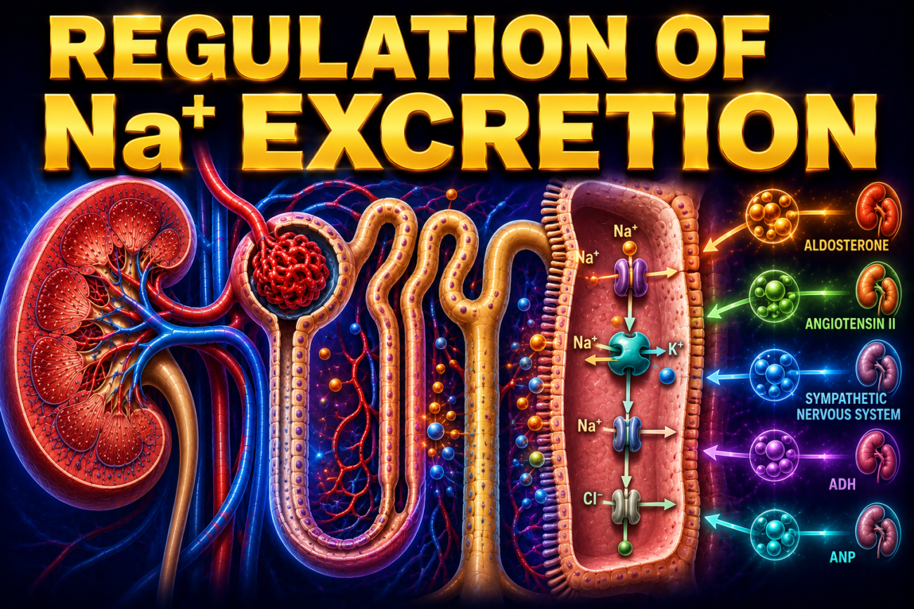
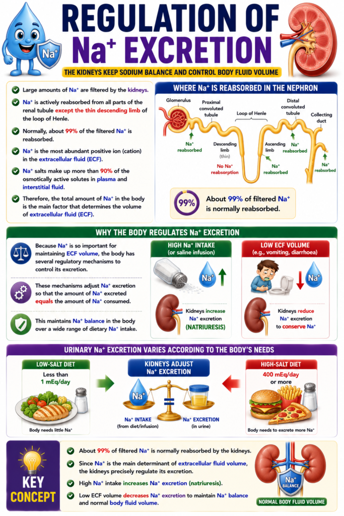
- Large amounts of Na⁺ are filtered by the kidneys.
- Na⁺ is actively reabsorbed from all parts of the renal tubule except the thin descending limb of the loop of Henle.
- Normally, about 99% of the filtered Na⁺ is reabsorbed.
- Na⁺ is the most abundant positive ion (cation) in the extracellular fluid (ECF).
- Na⁺ salts make up more than 90% of the osmotically active solutes in plasma and interstitial fluid.
- Therefore, the total amount of Na⁺ in the body is the main factor that determines the volume of extracellular fluid (ECF).
- Because Na⁺ is so important for maintaining ECF volume, the body has several regulatory mechanisms to control its excretion.
- These mechanisms adjust Na⁺ excretion so that the amount of Na⁺ excreted equals the amount of Na⁺ consumed.
- This maintains Na⁺ balance in the body over a wide range of dietary Na⁺ intake.
- When Na⁺ intake is high or saline is infused, the kidneys increase Na⁺ excretion (natriuresis).
- When ECF volume decreases, such as after vomiting or diarrhoea, the kidneys reduce Na⁺ excretion to conserve Na⁺.
- Urinary Na⁺ excretion varies according to the body’s needs:
- Less than 1 mEq/day on a low-salt diet.
- 400 mEq/day or more on a high-salt diet.
KEY CONCEPT
- About 99% of filtered Na⁺ is normally reabsorbed by the kidneys. Since Na⁺ is the main determinant of extracellular fluid volume, the kidneys precisely regulate its excretion. High Na⁺ intake increases Na⁺ excretion (natriuresis), while low ECF volume decreases Na⁺ excretion to maintain Na⁺ balance and normal body fluid volume.
MECHANISMS
- Changes in Na⁺ excretion occur by two main mechanisms:
- Changes in glomerular filtration rate (GFR).
- Changes in tubular reabsorption of Na⁺.
- Most regulation of Na⁺ reabsorption occurs in the small amount (about 3%) of filtered Na⁺ that reaches the collecting ducts.
- Factors that change GFR, including tubuloglomerular feedback, also affect Na⁺ excretion.
- Several factors regulate Na⁺ reabsorption in the renal tubules.
- Aldosterone and other adrenocortical hormones increase Na⁺ reabsorption.
- Atrial natriuretic peptide (ANP) and other natriuretic hormones decrease Na⁺ reabsorption and increase Na⁺ excretion.
- The rate of H⁺ secretion also influences Na⁺ reabsorption.
- The rate of K⁺ secretion also influences Na⁺ reabsorption.
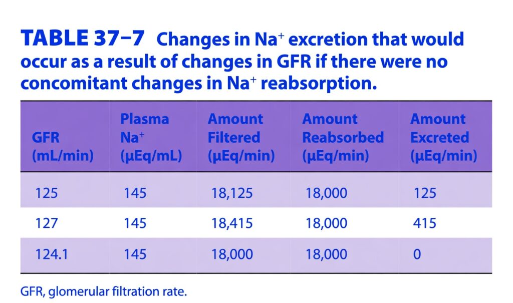
Table: Table 37–7
KEY CONCEPT
- Na⁺ excretion is regulated by changes in GFR and by changes in tubular Na⁺ reabsorption, mainly in the collecting ducts. Aldosterone increases Na⁺ reabsorption, whereas ANP increases Na⁺ excretion. H⁺ and K⁺ secretion also influence Na⁺ reabsorption.
EFFECTS OF ADRENOCORTICAL STEROIDS
- Adrenal mineralocorticoids, such as aldosterone, increase Na⁺ reabsorption by the renal tubules.
- This increase in Na⁺ reabsorption occurs together with increased secretion of K⁺ and H⁺.
- Aldosterone also promotes the reabsorption of Cl⁻ along with Na⁺.
- When mineralocorticoids are given to adrenalectomized animals, their effects on Na⁺ reabsorption do not appear immediately.
- There is a delay of about 10–30 minutes before Na⁺ reabsorption increases.
- This delay occurs because the steroids must first act on DNA to produce new proteins.
- Mineralocorticoids may also produce rapid effects through the cell membrane.
- However, these rapid effects do not significantly change Na⁺ excretion in the whole animal.
- Mineralocorticoids mainly act on the collecting ducts of the nephron.
- They increase the number of active epithelial sodium channels (ENaCs) in the collecting ducts.
- More active ENaCs allow more Na⁺ to enter the tubular cells, increasing Na⁺ reabsorption.
- In Liddle syndrome, mutations occur in the genes that code for the β subunit and, less commonly, the γ subunit of ENaC.
- These mutations keep the ENaC channels continuously active.
- As a result, excessive Na⁺ is reabsorbed by the kidneys.
- This causes Na⁺ retention and hypertension (high blood pressure).
Figure: Figure 37–18
KEY CONCEPT
- Aldosterone increases Na⁺ reabsorption and Cl⁻ reabsorption while promoting K⁺ and H⁺ secretion, mainly by increasing the number of active ENaC channels in the collecting ducts. In Liddle syndrome, continuously active ENaC channels cause excessive Na⁺ retention and hypertension.
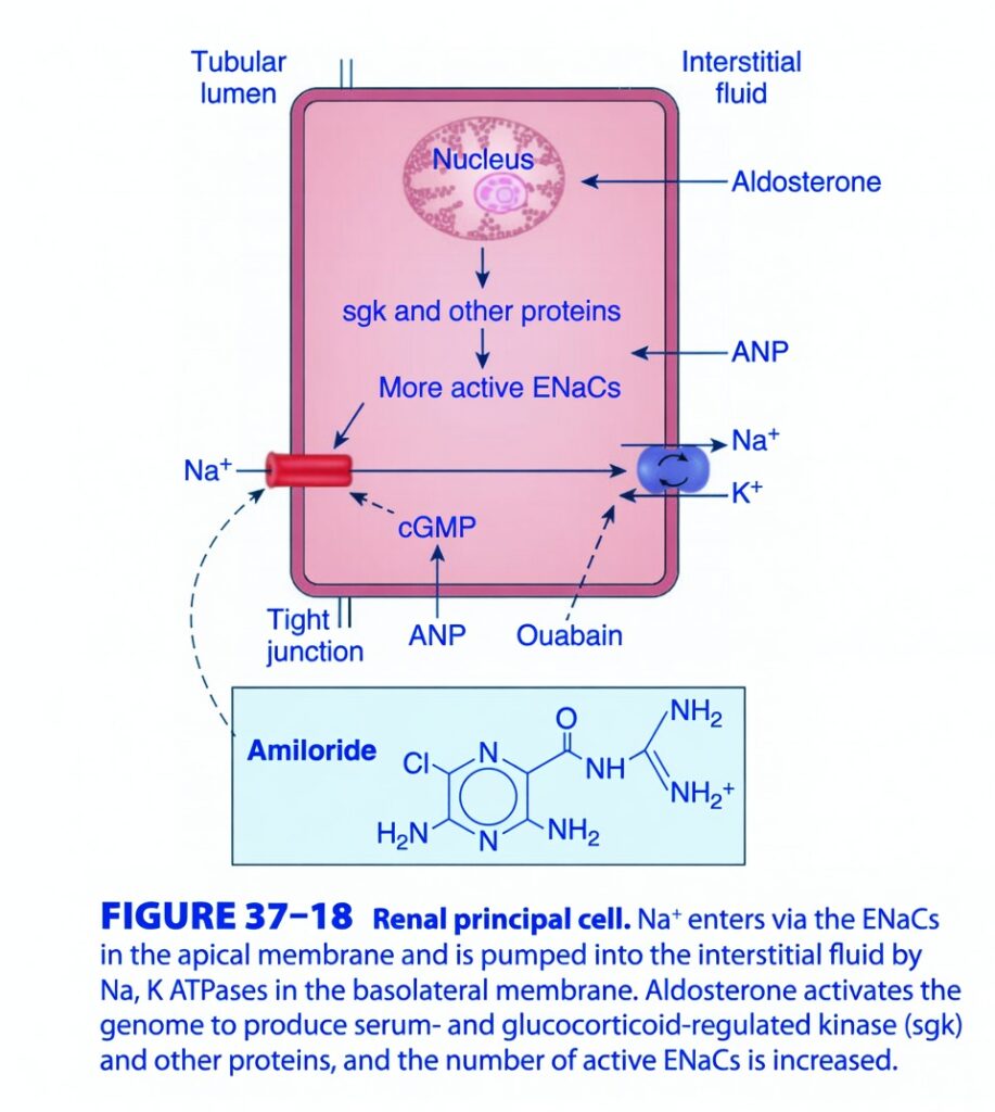
Figure 37-18: Renal Principal Cell (Collecting Duct)
Easiest & Most Conceptual Explanation for SELF LEARNERS
This figure explains how the principal cell of the late distal tubule and collecting duct controls sodium (Na⁺), potassium (K⁺), and water balance, mainly under the influence of Aldosterone and ANP.
Think of the Principal Cell as the kidney’s “Salt-Control Room.”
It decides:
- How much Na⁺ to save
- How much K⁺ to remove
- Indirectly, how much water the body keeps
⭐ One-Line Concept
Na⁺ enters the principal cell from the urine through ENaC, then is pumped into the blood by the Na⁺/K⁺ pump. Aldosterone speeds up this process, while ANP slows it down.
First Understand the Cell
The picture shows one principal cell.
Urine (Tubular lumen)
        │
        │
   [ ENaC Channel ]
        │
   PRINCIPAL CELL
        │
 [Na+/K+ ATPase Pump]
        │
Blood (Interstitial fluid)
The cell has two sides:
Left Side
Faces the tubular lumen (urine).
Right Side
Faces the interstitial fluid (blood).
Step 1: Sodium Enters the Cell
Look at the left membrane.
The red channel is called:
ENaC
(Epithelial Sodium Channel)
Urine
   │
 Na+
   ↓
 ENaC
   ↓
Cell
What happens?
Na⁺ moves from urine into the cell.
Easy Analogy
Imagine ENaC is the front door.
Na⁺ enters the house through the front door.Step 2: Sodium Leaves the Cell
Now look at the right membrane.
There is a blue pump.
This is:
Na⁺/K⁺ ATPase
It uses ATP.
Cell
   │
3 Na+
   ↓
Blood
At the same time
Blood
   │
2 K+
   ↓
Cell
Easy Concept
The pump pushes Na⁺ into the blood.
So the body keeps sodium.
Complete Sodium Journey
Urine
   │
 ENaC
   │
Principal Cell
   │
Na+/K+ Pump
   │
Blood
This is the main pathway of sodium reabsorption.
What Happens to Potassium?
The pump brings K⁺ inside the cell.
Blood
   │
2 K+
   ↓
Cell
Later,
K⁺ leaves into the urine through potassium channels (not emphasized in this figure).
So:
- Na⁺ is reabsorbed
- K⁺ is secreted
Aldosterone (The Sodium-Saving Hormone)
Look at the arrow entering from the blood side.
It shows:
Aldosterone
Aldosterone enters the cell and reaches the nucleus.
Inside the nucleus
It increases production of:
SGK
+
Other proteins
These proteins increase:
More Active ENaCsWhat does this mean?
More ENaC channels are available.
More Na⁺ enters the cell.
More Na⁺ is pumped into blood.
Therefore,
the body saves more sodium.
Easy Analogy
Imagine a supermarket opens more checkout counters.
More customers can enter.
Similarly,
more ENaC channels allow more Na⁺ to enter.
Effects of Aldosterone
↑ ENaC channels
↓
↑ Na⁺ reabsorption
↓
↑ Water reabsorption (water follows sodium)
↓
↑ Blood volume
↓
↑ Blood pressure
Also,
↑ K⁺ secretion into urine.
ANP (Atrial Natriuretic Peptide)
Look at the right side.
ANP acts on the cell.
Inside the cell it produces:
cGMPWhat does ANP do?
It opposes aldosterone.
Meaning:
Less ENaC activity
↓
Less Na⁺ reabsorption
↓
More Na⁺ remains in urine.
Easy Concept
Aldosterone says:
“Save salt.”
ANP says:Amiloride
Look at the blue box.
This drug blocks:
ENaC
Amiloride
      ↓
Blocks ENaC
Result
Na⁺ cannot enter the cell.
So:
Less sodium is reabsorbed.
More sodium is excreted.
Because Na⁺ entry is reduced, less K⁺ is secreted, making amiloride a potassium-sparing diuretic.
Easy Memory
Amiloride
↓
Blocks the front door (ENaC)
↓
Na⁺ stays in urine.
Ouabain
Look near the Na⁺/K⁺ pump.
Ouabain blocks:
Na⁺/K⁺ ATPase
If the pump stops:
Na⁺ cannot move into blood.
So sodium reabsorption decreases.
Tight Junction
Between neighboring cells is the:
Tight Junction
It seals the cells together.
So sodium must pass through the cell, not between cells.
Entire Sodium Story
Na+ in Urine
      ↓
ENaC
      ↓
Principal Cell
      ↓
Na+/K+ Pump
      ↓
Blood
Hormone Story
Aldosterone
Aldosterone
      ↓
Nucleus
      ↓
SGK Proteins
      ↓
More ENaC
      ↓
More Na+ Reabsorbed
ANP
ANP
      ↓
cGMP
      ↓
Less ENaC Activity
      ↓
Less Na+ Reabsorbed
Drug Story
Amiloride
Blocks ENaC
        ↓
Less Na+ Entry
Ouabain
Blocks Na+/K+ Pump
        ↓
Less Na+ Leaves Cell
Everyday Analogy
Imagine a warehouse.
- ENaC = Front gate where packages (Na⁺) enter.
- Na⁺/K⁺ pump = Delivery truck sending packages to the city (blood).
- Aldosterone = Manager who opens more front gates and hires more workers.
- ANP = Inspector who tells workers to slow down and send fewer packages.
- Amiloride = Lock placed on the front gate.
- Ouabain = Stops the delivery truck.
High-Yield Exam Points
- Principal cells are located in the late distal tubule and collecting duct.
- ENaC on the apical (luminal) membrane allows Na⁺ to enter from the tubular lumen.
- Na⁺/K⁺ ATPase on the basolateral membrane pumps Na⁺ into the interstitial fluid and brings K⁺ into the cell.
- The net effect is Na⁺ reabsorption and K⁺ secretion.
- Aldosterone enters the cell, binds intracellular receptors, and stimulates gene transcription (including SGK), increasing the number and activity of ENaC channels and enhancing Na⁺ reabsorption.
- Increased Na⁺ reabsorption also promotes water retention, increasing extracellular fluid volume and blood pressure.
- ANP activates cGMP, reducing ENaC activity and promoting natriuresis (Na⁺ excretion).
- Amiloride blocks ENaC and is a potassium-sparing diuretic.
- Ouabain inhibits the Na⁺/K⁺ ATPase, reducing Na⁺ transport across the principal cell.
KEY CONCEPT (Figure 37-18)
Figure 37-18 illustrates the transport mechanisms of the renal principal cell. Sodium enters the cell from the tubular lumen through epithelial sodium channels (ENaC) in the apical membrane and is actively pumped into the interstitial fluid by the Na⁺/K⁺ ATPase in the basolateral membrane. This process results in Na⁺ reabsorption, while K⁺ brought into the cell by the pump is available for secretion into the tubular lumen. Aldosterone enhances Na⁺ reabsorption by stimulating the synthesis of SGK and other proteins, increasing the number and activity of ENaC channels. In contrast, ANP reduces Na⁺ reabsorption through cGMP-mediated inhibition. Pharmacologically, amiloride blocks ENaC, whereas ouabain inhibits the Na⁺/K⁺ ATPase. Together, these mechanisms regulate extracellular fluid volume, potassium balance, and arterial blood pressure.
OTHER HUMORAL EFFECTS
- A low-salt diet increases aldosterone secretion.
- Higher aldosterone levels gradually reduce Na⁺ excretion.
- Several other hormones and chemical messengers also regulate Na⁺ reabsorption.
- Prostaglandin E₂ (PGE₂) increases Na⁺ excretion (natriuresis).
- PGE₂ may increase Na⁺ excretion by:
- Inhibiting the Na⁺, K⁺-ATPase pump.
- Increasing intracellular Ca²⁺, which reduces Na⁺ transport through ENaC channels.
- Endothelin and interleukin-1 (IL-1) also increase Na⁺ excretion.
- They probably do this by increasing the production of PGE₂.
- Atrial natriuretic peptide (ANP) and related hormones increase the level of cyclic GMP (cGMP) inside cells.
- Increased cGMP inhibits Na⁺ transport through ENaC channels.
- Another natriuretic hormone, thought to be naturally produced ouabain, inhibits the Na⁺, K⁺-ATPase pump.
- This inhibition also increases Na⁺ excretion.
- Angiotensin II increases the reabsorption of Na⁺ and HCO₃⁻ in the proximal tubules.
- The kidneys contain a large amount of angiotensin-converting enzyme (ACE).
- The kidneys convert about 20% of circulating angiotensin I into angiotensin II.
- The kidneys also produce angiotensin I locally.
- Long-term exposure to high levels of mineralocorticoids does not usually cause oedema in healthy individuals.
- This is because the kidneys eventually escape from the effects of mineralocorticoids.
- This is called the escape phenomenon.
- The escape phenomenon may occur because of increased secretion of ANP.
- The escape phenomenon is reduced or absent in:
- Nephrosis
- Cirrhosis
- Heart failure
- In these conditions, the kidneys continue to retain Na⁺ when mineralocorticoid levels are high.
- Continued Na⁺ retention causes oedema.
KEY CONCEPT
- Many hormones regulate Na⁺ excretion. Aldosterone and angiotensin II increase Na⁺ reabsorption, while PGE₂, endothelin, IL-1, ANP, and endogenous ouabain increase Na⁺ excretion. Normally, the kidneys escape the long-term effects of mineralocorticoids, but this escape is impaired in nephrosis, cirrhosis, and heart failure, leading to persistent Na⁺ retention and oedema.
REGULATION OF WATER EXCRETION
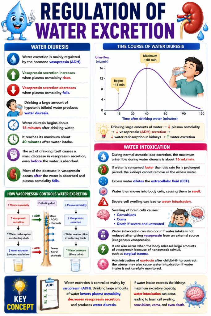
WATER DIURESIS
- Water excretion is mainly regulated by the hormone vasopressin (ADH).
- Vasopressin secretion increases when plasma osmolality rises.
- Vasopressin secretion decreases when plasma osmolality falls.
- Drinking a large amount of hypotonic (dilute) water produces water diuresis.
- Water diuresis begins about 15 minutes after drinking water.
- It reaches its maximum about 40 minutes after water intake.
- The act of drinking itself causes a small decrease in vasopressin secretion, even before the water is absorbed.
- Most of the decrease in vasopressin occurs after the water is absorbed and plasma osmolality falls.
WATER INTOXICATION
- During normal osmotic load excretion, the maximum urine flow during water diuresis is about 16 mL/min.
- If water is consumed faster than this rate for a prolonged period, the kidneys cannot remove all the excess water.
- Excess water dilutes the extracellular fluid (ECF).
- Water then moves into body cells, causing them to swell.
- Severe cell swelling can lead to water intoxication.
- Swelling of brain cells causes:
- Convulsions
- Coma
- Death if severe and untreated
- Water intoxication can also occur if water intake is not reduced after giving vasopressin from an external source (exogenous vasopressin).
- It can also occur when the body releases large amounts of vasopressin because of nonosmotic stimuli, such as surgical trauma.
- Administration of oxytocin after childbirth to contract the uterus may also cause water intoxication if water intake is not carefully monitored.
KEY CONCEPT
- Water excretion is controlled mainly by vasopressin (ADH). Drinking large amounts of water lowers plasma osmolality, decreases vasopressin secretion, and produces water diuresis. If water intake exceeds the kidneys’ maximum excretory capacity, water intoxication can occur, leading to brain cell swelling, convulsions, coma, and even death.
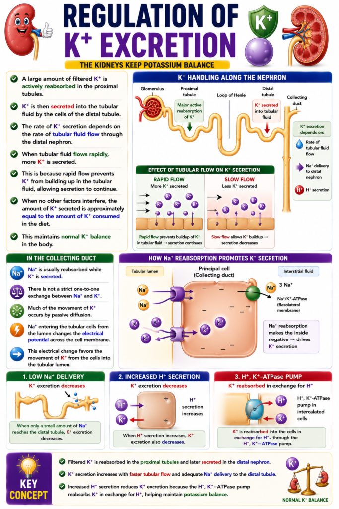
REGULATION OF K⁺ EXCRETION
- A large amount of filtered K⁺ is actively reabsorbed in the proximal tubules.
- K⁺ is then secreted into the tubular fluid by the cells of the distal tubule.
- The rate of K⁺ secretion depends on the rate of tubular fluid flow through the distal nephron.
- When tubular fluid flows rapidly, more K⁺ is secreted.
- This is because rapid flow prevents K⁺ from building up in the tubular fluid, allowing secretion to continue.
- When no other factors interfere, the amount of K⁺ secreted is approximately equal to the amount of K⁺ consumed in the diet.
- This maintains normal K⁺ balance in the body.
- In the collecting ducts, Na⁺ is usually reabsorbed while K⁺ is secreted.
- There is not a strict one-to-one exchange between Na⁺ and K⁺.
- Much of the movement of K⁺ occurs by passive diffusion.
- Na⁺ entering the tubular cells from the lumen changes the electrical potential across the cell membrane.
- This electrical change favors the movement of K⁺ from the cells into the tubular lumen.
- K⁺ excretion decreases when only a small amount of Na⁺ reaches the distal tubule.
- When H⁺ secretion increases, K⁺ excretion also decreases.
- This occurs because K⁺ is reabsorbed into the collecting duct cells in exchange for H⁺ through the H⁺, K⁺-ATPase pump.
KEY CONCEPT
- Filtered K⁺ is reabsorbed in the proximal tubules and later secreted in the distal nephron. K⁺ secretion increases with faster tubular flow and adequate Na⁺ delivery to the distal tubule. Increased H⁺ secretion reduces K⁺ excretion because the H⁺, K⁺-ATPase pump reabsorbs K⁺ in exchange for H⁺, helping maintain potassium balance.
MADE BY SELF LEARNING CEO AND FOUNDER DR SHEEN
