


After studying this topic, students will be able to:
- Explain postsynaptic inhibition and IPSP.
- Describe presynaptic inhibition and the role of GABA and chloride ions.
- Explain the time course of EPSP and IPSP.
Introduction
Neuronal inhibition is an important process in which synaptic activity reduces the transmission of nerve signals. In postsynaptic inhibition, inhibitory synapses increase the movement of chloride ions into the neuron and/or potassium ions out of the neuron, making the inside of the neuron more negative. This increased negativity is called hyperpolarization and produces an inhibitory postsynaptic potential (IPSP).
Inhibition can also occur at the presynaptic terminal before the nerve signal reaches the synapse. In most cases, GABA opens anion channels and allows chloride ions to enter the presynaptic terminal, reducing the excitatory effect of sodium ions and inhibiting synaptic transmission. This type of inhibition is common in sensory pathways and helps reduce sideways spread and mixing of sensory signals.
Postsynaptic potentials also have a specific time course. Excitatory synapses increase sodium permeability for about 1–2 milliseconds, producing an EPSP, which gradually declines over about 15 milliseconds. Inhibitory synapses similarly increase potassium and/or chloride permeability for 1–2 milliseconds, producing an IPSP, which also fades in about 15 milliseconds. Some transmitters, especially certain neuropeptides, can produce excitation or inhibition for much longer periods.
ELECTRICAL EVENTS DURING NEURONAL INHIBITION
- Inhibitory synapses mainly open chloride (Cl⁻) channels.
- This allows chloride ions to move more easily across the postsynaptic membrane.
- The Nernst potential for chloride ions is about −70 mV.
- The normal resting neuronal membrane potential is about −65 mV.
- Because −70 mV is more negative than −65 mV, opening chloride channels allows negatively charged Cl⁻ ions to move into the neuron.
- As Cl⁻ enters the neuron, the inside becomes more negative, moving toward −70 mV.
- Opening potassium (K⁺) channels allows positively charged potassium ions to move out of the neuron.
- Loss of positive K⁺ ions also makes the inside of the neuron more negative.
- Therefore:
- Cl⁻ influx → more negative inside
- K⁺ efflux → more negative inside
- This increase in negativity inside the neuron is called hyperpolarization.
- The neuron becomes inhibited because its membrane potential becomes more negative than the normal resting level.
- This increase in negativity beyond the resting membrane potential is called an inhibitory postsynaptic potential (IPSP).
- Fig. 46.11C shows this effect.
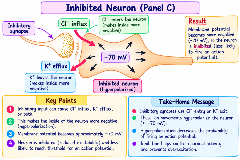
- Activation of inhibitory synapses causes:
- Cl⁻ influx into the cell
- and/or K⁺ efflux out of the cell
- The membrane potential changes from:
- −65 mV → −70 mV
- Difference:
- −70 − (−65) = −5 mV
- Therefore, the neuron develops an IPSP of −5 mV.
- This inhibits transmission of the nerve signal through the synapse.
KEY CONCEPT
Inhibitory synapse opens Cl⁻ and/or K⁺ channels → Cl⁻ enters and/or K⁺ leaves → inside becomes more negative → hyperpolarization occurs → IPSP develops → nerve signal transmission is inhibited.
Easiest example:
−65 mV → −70 mV = 5 mV more negative → IPSP = −5 mV → neuron is inhibited.
Short Conceptual Story – Neuronal Inhibition
Think of a neuron resting at −65 mV.
An inhibitory synapse becomes active and opens Cl⁻ channels.
Now Cl⁻ moves from outside → inside the neuron. Because Cl⁻ is negative, the inside becomes more negative.
At the same time, K⁺ channels may open.
Now K⁺ moves from inside → outside the neuron. Positive charge leaves, so the inside again becomes more negative.
So:
Cl⁻ enters + K⁺ leaves → neuron becomes more negative → −65 mV moves toward −70 mV.
This extra negativity is called hyperpolarization.
The change from −65 mV → −70 mV = −5 mV IPSP.
Memory Line
Cl⁻ IN + K⁺ OUT → negativity increases → hyperpolarization → IPSP → neuron inhibited.
Presynaptic Inhibition
- In addition to postsynaptic inhibition, inhibition can also occur at the presynaptic terminal.
- This happens before the nerve signal reaches the synapse.
- Presynaptic inhibition occurs when an inhibitory substance is released onto the outside of the presynaptic nerve fiber.
- This happens before the presynaptic nerve ending terminates on the postsynaptic neuron.
- In most cases, the inhibitory transmitter is GABA.
- GABA opens anion channels.
- This allows large numbers of chloride ions (Cl⁻) to enter the terminal fiber.
- These chloride ions carry negative charges.
- Their negative charges reduce synaptic transmission.
- They do this by cancelling much of the excitatory effect of the positively charged sodium ions (Na⁺).
- Sodium ions also enter the terminal fiber when an action potential arrives.
- Therefore:
- GABA opens anion channels
- Cl⁻ enters the presynaptic terminal
- Cl⁻ reduces the excitatory effect of Na⁺
- Synaptic transmission is inhibited
- Presynaptic inhibition occurs in many sensory pathways of the nervous system.
- Adjacent sensory nerve fibers can inhibit one another.
- This reduces the sideways spread of signals.
- It also reduces the mixing of signals in sensory tracts.
KEY CONCEPT
GABA released → anion channels open → Cl⁻ enters presynaptic terminal → excitatory effect of Na⁺ is reduced → synaptic transmission is inhibited.
Easiest example:
One sensory nerve fiber can inhibit a nearby sensory nerve fiber → this reduces sideways spread and mixing of sensory signals.
Presynaptic Inhibition — Short Conceptual Story
Imagine a nerve signal is traveling toward the presynaptic terminal.
Before the signal can reach the synapse, another inhibitory nerve releases GABA onto the presynaptic nerve fiber.
GABA opens anion channels → Cl⁻ enters the presynaptic terminal.
Now the action potential arrives and Na⁺ also enters the terminal. But the negative charge of Cl⁻ cancels much of the excitatory effect of the positive Na⁺.
So the presynaptic signal becomes weaker before it reaches the postsynaptic neuron. This is called presynaptic inhibition.
In sensory pathways, neighboring sensory fibers can inhibit each other in this way, which reduces sideways spread and mixing of sensory signals.
Easy Memory Flow
GABA released → anion channels open → Cl⁻ enters presynaptic terminal → Na⁺ excitatory effect reduced → synaptic transmission inhibited.
Remember:
Presynaptic inhibition = inhibition happens BEFORE the signal reaches the synapse.
Time Course of Postsynaptic Potentials
- When an excitatory synapse excites the anterior motor neuron, the neuronal membrane becomes highly permeable to sodium ions (Na⁺) for 1–2 milliseconds.
- During this short time, enough Na⁺ enters the postsynaptic motor neuron.
- This increases the intracellular potential by a few millivolts.
- This produces an EPSP, shown by the blue and green curves in Fig. 46.12.
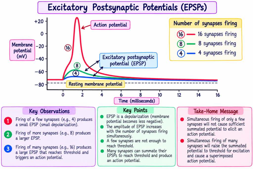
- The EPSP then slowly decreases over the next 15 milliseconds.
- During this time, the extra positive charges leak out of the excited neuron.
- The membrane then returns toward its normal resting membrane potential.
- The opposite effect occurs with an IPSP.
- An inhibitory synapse increases membrane permeability to potassium ions (K⁺), chloride ions (Cl⁻), or both for 1–2 milliseconds.
- This makes the intracellular potential more negative than normal.
- This produces an IPSP.
- The IPSP also gradually disappears in about 15 milliseconds.
- Some other transmitter substances can excite or inhibit the postsynaptic neuron for much longer periods.
- Their effects may last for hundreds of milliseconds, seconds, minutes, or even hours.
- This is especially true for some neuropeptide transmitters.
KEY CONCEPT
Excitatory synapse → Na⁺ permeability increases for 1–2 ms → Na⁺ enters → EPSP develops → EPSP fades over about 15 ms.
Inhibitory synapse → K⁺ and/or Cl⁻ permeability increases for 1–2 ms → membrane becomes more negative → IPSP develops → IPSP fades over about 15 ms.
Easiest example:
EPSP = brief excitation, then gradual return toward resting level.
IPSP = brief inhibition, then gradual return toward resting level.
Physiological Importance
- Postsynaptic inhibition makes the inside of the neuron more negative than normal.
- Chloride influx (Cl⁻) and potassium efflux (K⁺) increase intracellular negativity.
- This produces hyperpolarization and an inhibitory postsynaptic potential (IPSP).
- The IPSP inhibits transmission of the nerve signal through the synapse.
- Presynaptic inhibition reduces the nerve signal before it reaches the postsynaptic neuron.
- GABA opens anion channels and allows chloride ions to enter the presynaptic terminal.
- The negative chloride ions reduce much of the excitatory effect of sodium ions entering during the action potential.
- Therefore, synaptic transmission is reduced.
- Presynaptic inhibition is important in many sensory pathways.
- Adjacent sensory nerve fibers can inhibit one another.
- This helps minimize sideways spread of sensory signals.
- It also reduces mixing of signals in sensory tracts.
- The time course of postsynaptic potentials allows excitation and inhibition to occur for a limited period.
- An EPSP develops after increased sodium permeability and then gradually declines as excess positive charges leave the neuron.
- An IPSP develops after increased potassium and/or chloride permeability and also gradually disappears.
- Some transmitter substances, especially certain neuropeptides, can produce excitation or inhibition for much longer periods.
Key Physiological Concept
Neuronal inhibition controls nerve signal transmission by making the neuron more negative or by reducing excitation before the signal reaches the synapse.
Postsynaptic inhibition → hyperpolarization → IPSP → signal inhibited
Presynaptic inhibition → GABA → Cl⁻ enters → Na⁺ excitatory effect reduced → synaptic transmission reduced
Sensory pathways → neighboring fibers inhibit each other → less sideways spread and less signal mixing
High-Yield Points
- Inhibitory synapses mainly open chloride (Cl⁻) channels.
- The Nernst potential for chloride is about −70 mV, compared with the normal resting neuronal membrane potential of −65 mV.
- Opening chloride channels allows Cl⁻ to enter the neuron, making the inside more negative.
- Opening potassium channels allows K⁺ to leave the neuron, also making the inside more negative.
- Cl⁻ influx + K⁺ efflux → increased intracellular negativity → hyperpolarization.
- Increased negativity beyond the resting membrane potential is called an inhibitory postsynaptic potential (IPSP).
- In Fig. 46.11C, the membrane potential changes from −65 mV to −70 mV, producing an IPSP of −5 mV.
- An IPSP inhibits transmission of the nerve signal through the synapse.
- Presynaptic inhibition occurs at the presynaptic terminal before the signal reaches the synapse.
- In most cases, the inhibitory transmitter is GABA.
- GABA opens anion channels, allowing Cl⁻ to enter the presynaptic terminal.
- The negative charge of Cl⁻ reduces much of the excitatory effect of Na⁺ entering when an action potential arrives.
- Therefore, synaptic transmission is inhibited.
- Presynaptic inhibition occurs in many sensory pathways.
- Adjacent sensory nerve fibers may inhibit one another.
- This helps minimize sideways spread and mixing of signals in sensory tracts.
- During excitation, the neuronal membrane becomes highly permeable to Na⁺ for 1–2 milliseconds.
- Na⁺ enters the postsynaptic neuron and produces an EPSP.
- The EPSP then gradually decreases over about 15 milliseconds as excess positive charges leak out.
- Fig. 46.12 shows the EPSP by the blue and green curves.
- During inhibition, membrane permeability to K⁺ and/or Cl⁻ increases for 1–2 milliseconds.
- This makes the intracellular potential more negative and produces an IPSP.
- The IPSP also fades in about 15 milliseconds.
- Some transmitters can produce excitation or inhibition for hundreds of milliseconds, seconds, minutes, or hours.
- This prolonged effect is especially true for some neuropeptide transmitters.
Quick Memory Line
Postsynaptic inhibition → Cl⁻ in / K⁺ out → hyperpolarization → IPSP
Presynaptic inhibition → GABA → Cl⁻ in → Na⁺ effect reduced → transmission inhibited
EPSP/IPSP → develop rapidly → usually fade in about 15 ms
Reference
Guyton and Hall Textbook of Medical Physiology, 15th Edition
Chapter 46: Organization of the Nervous System
Topics: Electrical Events During Neuronal Inhibition, Presynaptic Inhibition, and Time Course of Postsynaptic Potentials
Figures: Fig. 46.11C and Fig. 46.12
