



LEARNING OBJECTIVES
After studying this topic, students will be able to:
- Describe the synthesis, transport, release, and prolonged actions of neuropeptides.
- Explain how neuropeptides and small-molecule transmitters may coexist and be co-released from the same neuron.
- Describe the resting membrane potential and ionic concentration differences across the neuronal somal membrane.
- Explain the Nernst potentials of Na⁺, K⁺, and Cl⁻ and their directions of movement.
- Explain how synaptic excitation produces an EPSP and summation.
- Describe the threshold and generation of an action potential at the axon initial segment.
INTRODUCTION
Neuronal communication depends on chemical transmitters and electrical changes in neurons. Neuropeptides are slowly acting transmitters that are formed in the neuronal cell body, transported to nerve terminals, and can produce prolonged effects. Neuropeptides and small-molecule transmitters may also be released from the same neuron. Neuronal excitation depends on changes in membrane potential, ion concentration gradients, excitatory postsynaptic potentials (EPSPs), summation of incoming signals, and generation of an action potential at the initial segment of the axon.
- Neuropeptides are made in a different way from small-molecule transmitters.
- Their actions are usually slow and are also different in other ways.
- Neuropeptides are not made in the cytosol of presynaptic terminals.
- Instead, they are made as parts of large protein molecules by ribosomes in the neuronal cell body.
- These large protein molecules then enter the endoplasmic reticulum of the cell body.
- After this, they move into the Golgi apparatus.
- In the Golgi apparatus, two important changes occur:
- The neuropeptide-forming protein is enzymatically split into smaller fragments.
- Some fragments become the neuropeptide itself or its precursor.
- The Golgi apparatus then packages the neuropeptide into very small transmitter vesicles.
- These vesicles are released into the cytoplasm.
- The transmitter vesicles are then transported from the cell body to the tips of the nerve fibers.
- This transport occurs by axonal streaming of the axon cytoplasm.
- The transport is slow, only a few centimeters per day.
- At the neuronal terminal, an action potential causes these vesicles to release their transmitter.
- This release occurs in the same general way as with small-molecule transmitters.
- However, after release, the neuropeptide vesicle is autolyzed and is not reused.
- Because neuropeptide formation is a slow and laborious process, much smaller amounts of neuropeptides are usually released compared with small-molecule transmitters.
- This smaller amount is partly compensated because neuropeptides are generally 1000 times or more potent than small-molecule transmitters.
- Another important feature is that neuropeptides usually produce much longer-lasting actions.
- Their prolonged actions may include:
- Prolonged closure of calcium channels
- Prolonged changes in the metabolic machinery of cells
- Prolonged activation or deactivation of specific genes in the cell nucleus
- Prolonged changes in the number of excitatory or inhibitory receptors
- Some neuropeptide effects may last for days.
- Other effects may last for months or even years.
KEY CONCEPT
Neuronal cell body → ribosomes make large protein → endoplasmic reticulum → Golgi apparatus → neuropeptide formed and packed into vesicles → slow axonal transport → nerve terminal → action potential releases neuropeptide → strong and prolonged effect
SHORT CONCEPTUAL STOREY – NEUROPEPTIDES
The ribosomes in the cell body make a large protein.
This protein goes to the endoplasmic reticulum, then to the Golgi apparatus.
The Golgi cuts the large protein into a neuropeptide or its precursor and packs it into vesicles.
These vesicles then travel slowly down the axon, only a few centimeters per day, until they reach the nerve terminal.
When an action potential arrives, the neuropeptide is released.
After release, the vesicle is destroyed and not reused.
Because making neuropeptides is slow, only a small amount is released—but they are about 1000 times or more potent and their effects can last from days to months or even years.
Memory line:
Made slowly → travels slowly → released in small amount → acts very strongly → lasts very long.
Easy example:
A small amount of neuropeptide can still produce a very strong and long-lasting effect because neuropeptides are much more potent than small-molecule transmitters.
Neuropeptide and Small-Molecule Transmitters May Coexist in the Same Neurons
- Slowly acting neuropeptide transmitters and rapidly acting small-molecule transmitters are often stored and released from the same neurons.
- In some cases, two or more transmitters are stored together in the same synaptic vesicles.
- When an action potential reaches the presynaptic terminal, these transmitters are released together (Fig. 46.9A).
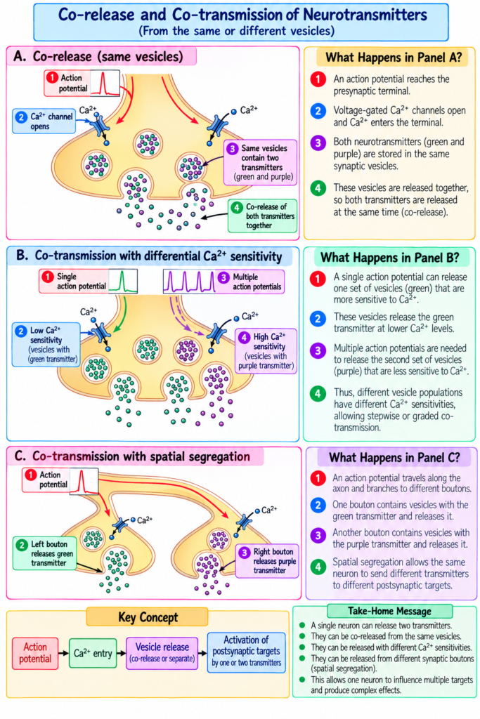
- In other cases, the transmitters are stored in different groups of synaptic vesicles within the same neuron.
- These different transmitters can still work together to produce co-transmission of signals to the postsynaptic neuron.
- Their release may also be controlled differently because of:
- Different sensitivities to calcium ions (Fig. 46.9B).
- Separation of vesicles on different boutons (Fig. 46.9C).
- Co-release of transmitters and co-transmission of signals have important functional effects.
- Each transmitter released from the same presynaptic neuron has its own specific receptors.
- Each transmitter may produce either:
- Inhibitory effects, or
- Excitatory effects
on the postsynaptic target.
- Different neurons may release different combinations of:
- Fast-acting transmitters → directly activate postsynaptic receptors.
- Slow-acting transmitters → require activation of second-messenger cascades and changes in postsynaptic gene expression.
- An example of co-release of two small-molecule transmitters is found in the raphe nucleus in the brain stem.
- Neurons of the raphe nucleus provide innervation to several brain regions.
- These neurons can co-release serotonin and glutamate.
- They play an important role in the sleep–wake cycle (see Chapters 59 and 60).
KEY CONCEPT
Same neuron → may contain fast + slow transmitters → action potential reaches presynaptic terminal → transmitters may be released together → each acts on its own receptor → postsynaptic neuron may receive excitatory or inhibitory effects
EASIEST EXAMPLE
Raphe nucleus neuron → releases serotonin + glutamate together → signals reach different brain regions → help in the sleep–wake cycle.
ELECTRICAL EVENTS DURING NEURONAL EXCITATION
- Electrical events during neuronal excitation have been studied especially in the large motor neurons of the anterior horns of the spinal cord.
- Therefore, the events described here mainly apply to these neurons.
- Except for some quantitative differences, the same basic events also occur in most other neurons of the nervous system.
- Resting Membrane Potential of the Neuronal Soma
- Fig. 46.10 shows the soma of a spinal motor neuron.
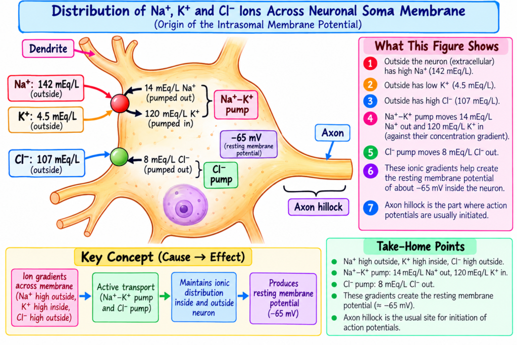
- Its resting membrane potential is about −65 mV.
- This resting membrane potential is less negative than that of:
- Large peripheral nerve fibers
- Skeletal muscle fibers
- This lower resting voltage is important because it allows both positive and negative control of neuronal excitability.
- A less negative membrane potential makes the neuron more excitable.
- A more negative membrane potential makes the neuron less excitable.
- This mechanism forms the basis of the two main functional states of a neuron:
- Excitation
- Inhibition
KEY CONCEPT
Resting membrane potential ≈ −65 mV
Less negative membrane potential → neuron becomes more excitable → excitation
More negative membrane potential → neuron becomes less excitable → inhibition
EASIEST EXAMPLE
If the neuronal membrane becomes less negative than −65 mV, the neuron becomes easier to excite.
If the membrane becomes more negative than −65 mV, the neuron becomes harder to excite.
Concentration Differences of Ions Across the Neuronal Somal Membrane
- Fig. 46.10 shows the concentration differences of the three most important ions across the neuronal somal membrane:
- Sodium (Na⁺)
- Potassium (K⁺)
- Chloride (Cl⁻)
- Sodium concentration
- Sodium concentration is high outside the neuron in extracellular fluid = 142 mEq/L.
- Sodium concentration is low inside the neuron = 14 mEq/L.
- This difference is maintained by the strong sodium-potassium pump.
- The pump continuously moves sodium out of the neuron.
- Potassium concentration
- Potassium concentration is high inside the neuronal soma = 120 mEq/L.
- Potassium concentration is low outside the neuron = 4.5 mEq/L.
- The sodium-potassium pump moves potassium into the neuron.
- Chloride concentration
- Chloride concentration is high outside the neuron.
- Chloride concentration is low inside the neuron.
- The membrane may be somewhat permeable to chloride ions.
- There may also be a weak chloride pump.
- However, the main reason for low chloride inside the neuron is the −65 mV negative voltage inside the neuron.
- This negative voltage repels negatively charged chloride ions.
- Therefore, chloride ions are pushed outward through channels until their concentration becomes much lower inside than outside.
- As described in Chapters 4 and 5, an electrical potential across a cell membrane can oppose the movement of ions.
- If the electrical potential has the correct polarity and magnitude, it can exactly oppose the movement of a particular ion.
- The potential that exactly opposes the movement of an ion is called the Nernst potential.
- Now the Nernst potentials for sodium, potassium, and chloride can be calculated.
Nernst Equation
EMF means the Nernst potential in millivolts inside the membrane.
For positive ions, the sign is negative (−).
For negative ions, the sign is positive (+).
Now the Nernst potentials for sodium, potassium, and chloride can be calculated.
Sodium (Na⁺)
- From Fig. 46.10:
- Outside = 142 mEq/L
- Inside = 14 mEq/L
{Nernst potential for Na⁺} = +61 { mV}
- Therefore, +61 mV would exactly oppose sodium movement through sodium channels.
- However, the actual membrane potential is −65 mV, not +61 mV.
- Therefore, sodium ions that leak into the neuron are immediately pumped back outside by the sodium pump.
- This helps maintain the −65 mV negative potential inside the neuron.
Potassium (K⁺)
- Potassium concentration:
- Inside = 120 mEq/L
- Outside = 4.5 mEq/L
{Nernst potential for K⁺} = -86 { mV}
- The calculated potassium Nernst potential is −86 mV.
- This is more negative than the actual membrane potential of −65 mV.
- Because potassium concentration is high inside the neuron, potassium ions have a net tendency to diffuse outside.
- This outward movement is opposed by continuous pumping of potassium back into the neuron.
Chloride (Cl⁻)
- Chloride concentration:
- Outside = 107 mEq/L
{Nernst potential for Cl⁻} = -70 { mV}
- The chloride Nernst potential is −70 mV.
- This is only slightly more negative than the actual membrane potential of −65 mV.
- Therefore, chloride ions tend to leak slightly into the neuron.
- The small amount that enters is moved back outside by an active chloride pump.
- These three Nernst potentials and the direction of ion movement are important for understanding neuronal excitation and inhibition when synapses activate or inactivate ion channels.
KEY CONCEPT
Na⁺:
142 outside → 14 inside → tends to enter → Na⁺ pump moves it back outside
K⁺:
120 inside → 4.5 outside → tends to leave → pump moves it back inside
Cl⁻:
107 outside → 8 inside → slight tendency to enter → chloride pump moves it back outside
EASIEST EXAMPLE
Think of the neuronal membrane as keeping each ion at a different concentration:
- Na⁺ mostly outside
- K⁺ mostly inside
- Cl⁻ mostly outside
The Nernst potential is the electrical voltage that exactly opposes the movement of each ion.
Uniform Distribution of Electrical Potential Inside the Neuronal Soma
- The fluid inside the neuronal soma is a highly conductive electrolytic solution.
- The neuronal soma has a relatively large diameter of about 10–80 micrometers.
- There is almost no resistance to the flow of electric current from one part of the inside of the soma to another.
- Therefore, if the electrical potential changes in one part of the soma, almost the same change occurs at all other points inside the soma.
- This uniform change occurs as long as the neuron is not transmitting an action potential.
- This principle is important because it helps in the summation of signals entering the neuron from multiple sources.
KEY CONCEPT
Highly conductive fluid + very low internal resistance → change in potential at one point → almost equal change throughout the soma → helps in summation of multiple incoming signals
EASIEST EXAMPLE
If an incoming signal changes the electrical potential in one part of the neuronal soma, almost the same electrical change spreads throughout the soma, helping the neuron combine signals coming from different sources.
Effect of Synaptic Excitation on the Postsynaptic Membrane—Excitatory Postsynaptic Potential
- Fig. 46.11A shows a resting neuron with an unexcited presynaptic terminal on its surface.
- The resting membrane potential throughout the soma is −65 mV.
- Fig. 46.11B shows that the presynaptic terminal releases an excitatory transmitter into the synaptic cleft.
- The transmitter acts on an excitatory receptor of the postsynaptic membrane.
- This increases the membrane permeability to Na⁺.
- Because:
- Na⁺ concentration is much higher outside the neuron, and
- The inside of the neuron is strongly negative,
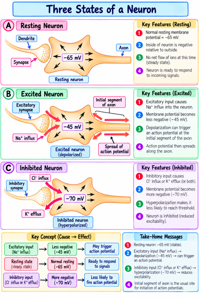
Na⁺ rapidly diffuses into the neuron.
- The entry of positively charged Na⁺ reduces some of the negative charge inside the membrane.
- Therefore, in Fig. 46.11B, the membrane potential changes from:
−65 mV → −45 mV
- This means the membrane potential becomes 20 mV more positive.
- This change toward a less negative membrane potential is called an excitatory postsynaptic potential (EPSP).
- In this example:
EPSP = +20 mV
- If the EPSP becomes sufficiently positive, it can produce an action potential in the postsynaptic neuron.
- Therefore, the postsynaptic neuron becomes excited.
- One presynaptic terminal alone normally cannot change the membrane potential from −65 mV to −45 mV.
- A change this large usually requires simultaneous or rapidly successive discharge of many presynaptic terminals.
- In a usual anterior motor neuron, about 40–80 terminals are required.
- The combined effect of many terminals is called summation.
EPSP SHORT CONCEPTUAL STOREY
A neuron is resting at −65 mV.
An excitatory transmitter is released from the presynaptic terminal and opens the way for Na⁺ to enter the postsynaptic neuron.
Because positive Na⁺ enters, the inside becomes less negative:
−65 mV → −45 mV
This +20 mV positive shift is called an EPSP.
One terminal is usually not enough, so about 40–80 excitatory terminals fire together or rapidly one after another.
Their effects add together (summation).
If the EPSP becomes large enough → action potential is produced → neuron is excited.
KEY CONCEPT
Excitatory transmitter released → excitatory receptor activated → Na⁺ permeability increases → Na⁺ enters neuron → membrane becomes less negative → EPSP develops → if sufficiently large, action potential occurs
EASIEST EXAMPLE
Resting potential = −65 mV
Many excitatory terminals discharge together → Na⁺ enters → membrane potential rises to −45 mV
Change = +20 mV → EPSP → neuron moves toward excitation.
Generation of Action Potentials in the Initial Segment of the Axon Leaving the Neuron—Threshold for Excitation
- When the EPSP becomes sufficiently positive, an action potential is generated.
- The action potential does not begin near the excitatory synapses.
- Instead, it begins at the initial segment of the axon, where the axon leaves the neuronal soma.
- The soma has relatively few voltage-gated Na⁺ channels.
- Therefore, it is difficult for an EPSP to open enough Na⁺ channels in the soma to produce an action potential.
- In contrast, the axon initial segment has about 7 times more voltage-gated Na⁺ channels than the soma.
- Therefore, an action potential can be produced much more easily at the initial segment.
- The EPSP needed to produce an action potential is:
- Axon initial segment: about +10 to +20 mV
- Soma: about +30 to +40 mV or more
- Once the action potential starts, it travels forward along the axon.
- It usually also travels backward over the soma.
- Sometimes it also travels backward into the dendrites.
- However, it does not enter all dendrites because dendrites, like the soma, have very few voltage-gated Na⁺ channels.
- Therefore, dendrites often cannot generate action potentials.
- In Fig. 46.11B, the threshold for neuronal excitation is about −45 mV.
- The normal resting membrane potential is −65 mV.
- Therefore:
−65 mV → −45 mV = +20 mV EPSP
- So, in this example, an EPSP of +20 mV reaches the threshold and triggers an action potential.
KEY CONCEPT
EPSP becomes sufficiently positive → initial segment reaches threshold → many voltage-gated Na⁺ channels open → action potential begins → travels along the axon
SHORT CONCEPTUAL STORY
The neuron is resting at −65 mV.
Excitatory signals produce an EPSP, making the membrane more positive.
When the potential reaches about −45 mV, the threshold is reached.
But the action potential does not start in the soma.
It starts at the axon initial segment because this area has about 7 times more voltage-gated Na⁺ channels.
So remember:
−65 mV → EPSP +20 mV → −45 mV threshold → axon initial segment fires → action potential travels along the axon.
PHYSIOLOGICAL IMPORTANCE
- Neuropeptides are important because they produce slow and prolonged effects compared with small-molecule transmitters.
- Their synthesis in the neuronal cell body, processing in the endoplasmic reticulum and Golgi apparatus, and slow transport to nerve terminals allow them to act as powerful chemical messengers.
- Although neuropeptides are released in smaller quantities, they are generally 1000 or more times as potent as small-molecule transmitters.
- Neuropeptides can produce prolonged effects by causing:
- Prolonged closure of calcium channels
- Long-lasting changes in cellular metabolism
- Activation or deactivation of specific genes
- Changes in the number of excitatory or inhibitory receptors
- These effects may last for days, months, or even years.
- Neuropeptides and small-molecule transmitters may be present in the same neuron and may be released together.
- Co-release allows one presynaptic neuron to produce different effects on the postsynaptic neuron through different receptors.
- Different transmitters from the same neuron may produce either excitatory or inhibitory effects.
- Fast-acting transmitters can directly activate postsynaptic receptors.
- Slow-acting transmitters can act through second-messenger cascades and changes in gene expression.
- Neurons of the raphe nucleus can co-release serotonin and glutamate and are important in the sleep–wake cycle.
- The resting membrane potential of the neuronal soma is about −65 mV, which allows both excitation and inhibition of the neuron.
- A less negative membrane potential makes the neuron more excitable.
- A more negative membrane potential makes the neuron less excitable.
- The concentration differences of Na⁺, K⁺, and Cl⁻ across the neuronal membrane are important for normal neuronal electrical activity.
- The sodium-potassium pump maintains:
- High Na⁺ outside
- High K⁺ inside
- The negative potential inside the neuron helps keep Cl⁻ concentration low inside the neuron.
- The Nernst potential is important because it represents the electrical potential that can exactly oppose the movement of a particular ion.
- The Nernst potentials of Na⁺, K⁺, and Cl⁻ help explain the direction in which these ions tend to move.
- These ion movements are important for understanding neuronal excitation and inhibition.
- The highly conductive fluid inside the neuronal soma allows a change in electrical potential at one point to produce an almost equal change throughout the soma.
- This property is important for summation of signals coming from multiple sources.
- An excitatory transmitter increases membrane permeability to Na⁺.
- Na⁺ enters the postsynaptic neuron and makes the membrane potential less negative.
- This positive change in membrane potential produces an excitatory postsynaptic potential (EPSP).
- If the EPSP becomes sufficiently positive, it can produce an action potential.
- Many presynaptic terminals may need to discharge together or rapidly one after another to produce a large enough EPSP.
- This process is called summation.
- The axon initial segment is especially important because it has about seven times more voltage-gated Na⁺ channels than the soma.
- Therefore, the initial segment can generate an action potential more easily than the soma.
- In the example given, the neuron reaches its excitation threshold at about −45 mV, corresponding to an EPSP of +20 mV from the resting level of −65 mV.
- Once generated, the action potential travels along the axon and may also travel backward over the soma and into some dendrites.
CLINICAL – PHYSIOLOGICAL IMPORTANCE
- Understanding neuropeptide action is important because their effects can be very prolonged, lasting from days to months or years.
- Changes produced by neuropeptides in:
- Calcium channels
- Cellular metabolism
- Gene activity
- Excitatory and inhibitory receptor numbers
can produce long-lasting changes in neuronal function.
- Co-release of different transmitters is important because the same neuron can produce both excitatory and inhibitory influences on its postsynaptic targets.
- The serotonin and glutamate co-release from raphe nucleus neurons is functionally important in the sleep–wake cycle.
- Changes in the neuronal membrane potential directly affect how easily a neuron can be excited:
- Less negative → more excitable
- More negative → less excitable
- Proper Na⁺, K⁺, and Cl⁻ concentration gradients are important for maintaining normal neuronal excitation and inhibition.
- The balance between ionic concentration gradients and membrane electrical potential determines the direction in which ions move across the neuronal membrane.
- Disturbance of the electrical conditions that control Na⁺, K⁺, or Cl⁻ movement would alter neuronal excitability.
- EPSP formation and summation are important because a neuron usually requires the combined effect of many excitatory inputs before reaching threshold.
- The axon initial segment is the main site where an action potential is initiated because of its high concentration of voltage-gated Na⁺ channels.
- The difference between the resting membrane potential (−65 mV) and threshold (about −45 mV) shows how much excitation is needed before the neuron fires.
KEY CLINICAL-PHYSIOLOGICAL CONCEPT
Neurotransmitter release → ion-channel/receptor effects → change in membrane potential → EPSP or inhibition → summation → threshold at axon initial segment → action potential
The whole process determines whether a neuron will remain quiet, become excited, or become inhibited.
HIGH-YIELD POINTS
- Neuropeptides are synthesized in the neuronal cell body, not in the presynaptic terminal.
- They are formed as parts of large protein molecules by ribosomes.
- They pass through the endoplasmic reticulum and then the Golgi apparatus.
- In the Golgi apparatus:
- The protein is split into smaller fragments.
- Neuropeptides are packed into transmitter vesicles.
- These vesicles move slowly toward nerve terminals by axonal streaming, only a few centimeters per day.
- Neuropeptide vesicles release their transmitter in response to action potentials.
- After release, the vesicle is autolyzed and not reused.
- Neuropeptides are released in smaller amounts than small-molecule transmitters.
- They are generally 1000 or more times more potent than small-molecule transmitters.
- Their effects are often long-lasting and may continue for days, months, or years.
- Neuropeptides can cause prolonged:
- Closure of calcium channels
- Changes in cell metabolism
- Changes in gene activation or deactivation
- Changes in excitatory or inhibitory receptor numbers
- Neuropeptides and small-molecule transmitters may coexist in the same neuron.
- They may be:
- Stored in the same synaptic vesicles, or
- Stored in different vesicle populations.
- Their release may differ because of:
- Different calcium sensitivities
- Different locations on separate boutons
- Different transmitters released from the same neuron can act on different receptors.
- They may produce either excitatory or inhibitory effects.
- Fast-acting transmitters directly activate postsynaptic receptors.
- Slow-acting transmitters act through second-messenger pathways and changes in gene expression.
- Raphe nucleus neurons can co-release serotonin and glutamate.
- These neurons are involved in the sleep–wake cycle.
- The resting membrane potential of the neuronal soma is about −65 mV.
- A less negative membrane potential makes the neuron more excitable.
- A more negative membrane potential makes the neuron less excitable.
- Important ion concentrations:
- Na⁺: high outside, low inside
- K⁺: high inside, low outside
- Cl⁻: high outside, low inside
- The Na⁺-K⁺ pump moves:
- Na⁺ out
- K⁺ in
- The negative voltage inside the neuron helps keep Cl⁻ outside.
- The Nernst potential is the voltage that exactly opposes movement of a particular ion.
- Nernst potentials in the provided text:
- Na⁺ = +61 mV
- K⁺ = −86 mV
- Cl⁻ = −70 mV
- Na⁺ tends to move into the neuron.
- K⁺ tends to move out of the neuron.
- Cl⁻ has a slight tendency to move into the neuron.
- The intracellular fluid of the neuronal soma is highly conductive.
- Electrical potential changes spread almost equally throughout the soma.
- This property is important for summation of incoming signals.
- An excitatory transmitter increases membrane permeability to Na⁺.
- Na⁺ enters the neuron and makes the membrane potential less negative.
- This change is called an excitatory postsynaptic potential (EPSP).
- Example:
- Resting potential = −65 mV
- Excited level = −45 mV
- EPSP = +20 mV
- A single presynaptic terminal usually cannot produce such a large EPSP.
- About 40–80 terminals may need to discharge together or rapidly one after another.
- Their combined effect is called summation.
- The action potential usually begins at the initial segment of the axon.
- The initial segment has about 7 times more voltage-gated Na⁺ channels than the soma.
- Therefore, it reaches threshold more easily.
- EPSP required at the initial segment:
- About +10 to +20 mV
- EPSP required at the soma:
- About +30 to +40 mV or more
- In Fig. 46.11B, the threshold for excitation is about −45 mV.
- This corresponds to an EPSP of +20 mV from the resting potential of −65 mV.
- Once generated, the action potential travels along the axon and may also travel backward over the soma and into some dendrites.
QUICK REVISION
Neuropeptide: slow synthesis → slow transport → small amount → very potent → long-lasting effect
Resting neuron: −65 mV
Na⁺: mainly outside
K⁺: mainly inside
Cl⁻: mainly outside
Excitation: Na⁺ enters → membrane becomes less negative → EPSP develops
Summation: many excitatory inputs combine
Threshold: about −45 mV
Main firing site: axon initial segment because it has many more voltage-gated Na⁺ channels
- REFERENCE
- Guyton and Hall Textbook of Medical Physiology, 15th Edition
