


- About 93% of the metabolically active thyroid hormones secreted are thyroxine (T₄), while 7% are triiodothyronine (T₃).
- Almost all T₄ is eventually converted into T₃ in the tissues, so both hormones are functionally important.
- T₃ and T₄ have qualitatively similar functions, but they differ in the speed and strength of their actions.
- T₃ is about 4 times more potent than T₄, but it is present in much smaller amounts in blood and remains there for a shorter time.
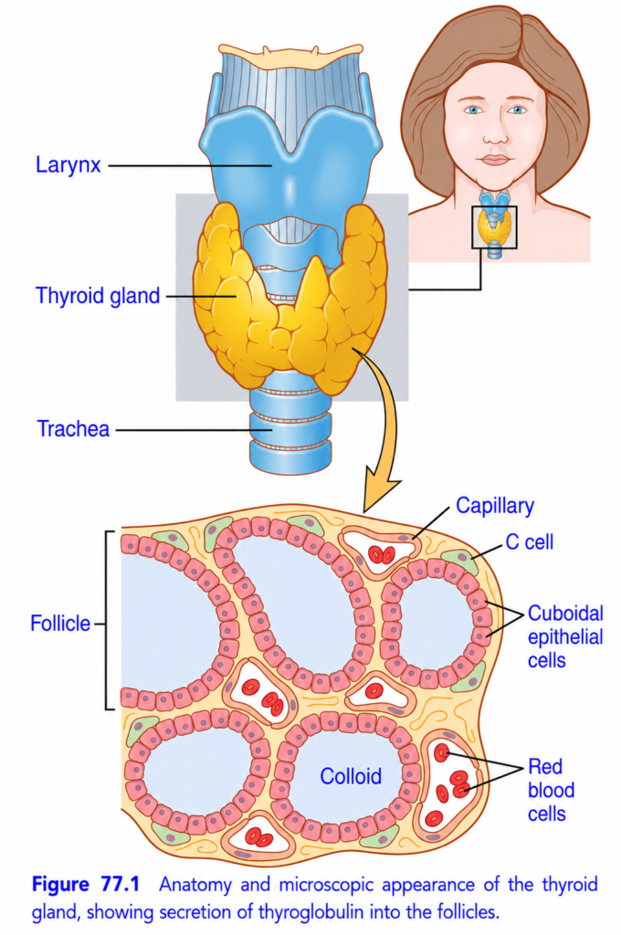
- The thyroid gland consists of many closed follicles, about 100–300 μm in diameter (Fig. 77.1).
- These follicles are filled with colloid and lined by cuboidal epithelial cells that secrete into the follicle.
- The main component of colloid is the large glycoprotein thyroglobulin, which contains the thyroid hormones.
- After thyroid hormone secretion enters the follicles, it must be absorbed back through the follicular epithelium into the blood before it can act in the body.
- The thyroid has a very high blood flow, about 5 times the weight of the gland each minute.
- This blood supply is among the greatest in the body, possibly exceeded only by the adrenal cortex.
- The thyroid also contains C cells, which secrete calcitonin.
- Calcitonin contributes to the regulation of plasma calcium ion concentration.
KEY CONCEPT
- T₄ = 93%, T₃ = 7% of thyroid secretion.
- T₄ → tissues → T₃, and T₃ is ~4× more potent.
- Thyroid follicle → colloid → thyroglobulin → thyroid hormones → blood.
- C cells → calcitonin → regulation of plasma Ca²⁺.
CONCEPTUAL EXAMPLES
- T₄: More abundant and lasts longer, but is less potent.
- T₃: Less abundant and shorter-lasting, but about 4× more potent.
- Thyroid follicle: Hormones enter colloid first, then are absorbed into blood.
- C cell: Produces calcitonin for plasma calcium regulation.
IODINE IS REQUIRED FOR THYROXINE FORMATION
- Normal thyroxine formation requires about 50 mg of ingested iodine per year, or about 1 mg/week.
- To prevent iodine deficiency, common table salt is iodized with about 1 part sodium iodide per 100,000 parts sodium chloride.
- Ingested iodides are absorbed from the gastrointestinal tract into the blood, similarly to chlorides.
- Most iodide is rapidly excreted by the kidneys, but about one-fifth is selectively taken up by thyroid cells and used to make thyroid hormones.
- The first step in thyroid hormone formation is transporting iodide from the blood into thyroid cells and follicles (Fig. 77.2).
- The thyroid cell’s basal membrane contains a sodium-iodide symporter.
- This symporter moves 1 iodide ion together with 2 sodium ions into the thyroid cell.
- The energy for this process comes indirectly from the Na⁺-K⁺ ATPase pump.
- Na⁺-K⁺ ATPase pumps sodium out of the cell, creating a low intracellular sodium concentration.
- This sodium gradient allows sodium to move back into the cell and helps bring iodide into the cell.
- Concentrating iodide inside the thyroid cell is called iodide trapping.
- Normally, the iodide pump concentrates iodide to about 30 times its blood concentration.
- When the thyroid is maximally active, this can increase to about 250 times the blood concentration.
- TSH is the most important factor controlling iodide trapping.
- TSH stimulates the iodide pump, whereas hypophysectomy greatly decreases its activity.
- Iodide then moves across the apical membrane from the thyroid cell into the follicle through pendrin, a chloride-iodide counter-transporter.
- Thyroid epithelial cells also secrete thyroglobulin into the follicle.
- Thyroglobulin contains tyrosine amino acids, to which iodine will bind for thyroid hormone formation.
KEY CONCEPT
- Iodine → essential for thyroxine formation.
- Blood iodide → sodium-iodide symporter → thyroid cell → iodide trapping → follicle.
- 1 I⁻ + 2 Na⁺ enter the cell through the symporter.
- Na⁺-K⁺ ATPase → creates Na⁺ gradient → drives iodide uptake.
- TSH ↑ iodide trapping.
- Normal concentration: ~30× blood → maximum activity: ~250× blood.
- Pendrin ( anion or transport exchange protein ) → moves iodide into follicle.
- Thyroglobulin + tyrosine → provides the site for iodine binding.
CONCEPTUAL EXAMPLES
- Low iodine intake → inadequate iodine available for thyroxine formation.
- TSH stimulation → increased iodide trapping by thyroid cells.
- Active thyroid → iodide concentration can reach ~250× blood level.
- Pendrin → moves trapped iodide from thyroid cell into the follicle.
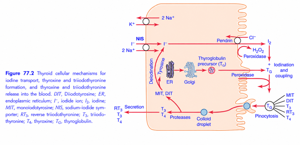
This Guyton figure shows almost the entire process of thyroid hormone formation inside one thyroid follicular cell.
The easiest way to understand it is as a factory story.
🧠 BIG PICTURE
The thyroid cell takes:
Iodide (I⁻) + Tyrosine → T₃ and T₄
Then it stores these hormones in thyroglobulin (Tg) inside the colloid and later releases them into the blood.
The whole pathway:
I⁻ enters cell → I⁻ enters colloid → I₂ → iodination of tyrosine → MIT + DIT → coupling → T₃ + T₄ → stored in Tg → endocytosis → digestion → T₃/T₄ released into blood
🏭 THINK OF THE THYROID AS A FACTORY
Raw material:
Iodide (I⁻)
Building material:
Tyrosine
Storage protein:
Thyroglobulin (Tg)
Factory enzyme:
Thyroid peroxidase (TPO)
Final products:
T₃ + T₄
Now let’s follow every arrow.
1️⃣ IODIDE COMES FROM THE BLOOD 🩸
On the left side of the cell is the blood/interstitial side.
The thyroid needs:
Iodide = I⁻
This is the raw material for thyroid hormone.
But iodide cannot simply enter efficiently by itself.
So the cell uses:
NIS
Sodium-Iodide Symporter
NIS brings:
2 Na⁺ + I⁻
into the thyroid cell.
Think:
NIS = iodine entrance door 🚪
2️⃣ WHY DOES NIS WORK?
The sodium gradient provides the energy.
The Na⁺/K⁺ ATPase maintains low intracellular Na⁺ by pumping:
3 Na⁺ OUT
and:
2 K⁺ IN
This Na⁺ gradient helps NIS bring iodide into the cell.
Simple:
Na⁺/K⁺ pump creates gradient → NIS uses gradient → I⁻ enters
🧠 HIGH-YIELD:
NIS = brings I⁻ INTO thyroid cell
Don’t confuse it with pendrin.
3️⃣ IODIDE MOVES TO THE COLLOID SIDE
Once inside the cell, iodide must get to the apical side facing the colloid.
There is a transporter called:
Pendrin
Pendrin helps move:
I⁻ → colloid
It is shown exchanging iodide with chloride.
So:
NIS = blood → cell
Pendrin = cell → colloid
This distinction is VERY important.
4️⃣ IODIDE MUST BECOME IODINE
Here’s a major step.
The iodide entering the colloid is:
I⁻
But it needs to be oxidized into an active form of iodine.
This is done by:
Thyroid peroxidase (TPO)
with:
H₂O₂
Think:
TPO = iodine activation machine ⚙️
So:
I⁻ → activated iodine
The figure represents this as:
I⁻ → I₂
5️⃣ THYROGLOBULIN = THE GIANT STORAGE SCAFFOLD 🏗️
Meanwhile, inside the thyroid cell, the rough ER and Golgi make:
Thyroglobulin (Tg)
Tg is a huge protein containing many:
Tyrosine residues
Think:
Thyroglobulin = giant protein scaffold carrying tyrosine building sites.
It is secreted into the colloid.
6️⃣ IODINE ATTACHES TO TYROSINE 🧩
Now activated iodine meets tyrosine residues on thyroglobulin.
TPO helps attach iodine to tyrosine.
This is called:
Iodination
Two important products are formed:
MIT
= Monoiodotyrosine
DIT
= Diiodotyrosine
Think:
Tyrosine + 1 iodine → MIT
Tyrosine + 2 iodines → DIT
⭐ EASY MEMORY
MIT = 1 iodine
DIT = 2 iodines
The letters are basically telling you:
Mono = 1
Di = 2
7️⃣ NOW COMES THE MAGIC: COUPLING 🔗
The iodinated tyrosines are coupled together.
DIT + DIT → T₄
Two DIT molecules combine:
T₄ = Thyroxine
And:
MIT + DIT → T₃
One MIT + one DIT:
T₃ = Triiodothyronine
So memorize:
DIT + DIT = T₄
MIT + DIT = T₃
🔥 This is one of the highest-yield parts of the figure.
8️⃣ WHERE ARE T₃ AND T₄ STORED?
They are still attached to:
Thyroglobulin
inside the:
Colloid
So the thyroid doesn’t immediately release all the hormone.
It stores a large amount as:
Tg + MIT + DIT + T₃ + T₄
Think:
Colloid = thyroid hormone warehouse 📦
9️⃣ WHEN THE BODY NEEDS THYROID HORMONE…
The thyroid cell takes the thyroglobulin-containing colloid back into the cell.
This process is:
Pinocytosis
Think:
Pinocytosis = cell “drinks” a little droplet 🥤 of colloid.
The figure shows a:
Colloid droplet
being brought into the cell.
🔟 PROTEASES CUT THYROGLOBULIN ✂️
Inside the cell, enzymes called:
Proteases
break down thyroglobulin.
This releases:
T₃
and
T₄
Now they are free and ready for secretion.
1️⃣1️⃣ T₃ AND T₄ ENTER THE BLOOD 🩸
The final arrow on the left shows:
T₃ + T₄ → blood
These are the main thyroid hormones released.
The figure also shows:
RT₃
Reverse T₃
but the major secreted thyroid hormones are:
T₄ and T₃
1️⃣2️⃣ WHAT HAPPENS TO MIT AND DIT?
Very important!
When thyroglobulin is broken down, MIT and DIT are also recovered.
They undergo:
Deiodination
The iodine is removed and recycled.
So:
MIT/DIT → iodine recycled → reused
Think:
The thyroid doesn’t waste iodine. ♻️
🧠 NOW LET’S FOLLOW THE ENTIRE FIGURE
STEP 1 — IODIDE ENTRY
Blood I⁻
↓ NIS
Thyroid cell
STEP 2 — IODIDE EXIT TO COLLOID
I⁻
↓ Pendrin
Colloid
STEP 3 — ACTIVATION
I⁻
↓ TPO + H₂O₂
Iodine
STEP 4 — IODINATION
Tyrosine on Tg + iodine
↓
MIT + DIT
STEP 5 — COUPLING
MIT + DIT → T₃
DIT + DIT → T₄
STEP 6 — STORAGE
T₃/T₄ attached to Tg
↓
Colloid warehouse
STEP 7 — RETRIEVAL
Pinocytosis
↓
Colloid droplet enters cell
STEP 8 — BREAKDOWN
Proteases
↓
T₃ + T₄ released from Tg
STEP 9 — SECRETION
T₃ + T₄ → BLOOD
🔥 THE MOST IMPORTANT TRANSPORTERS
This is where exam questions love to attack.
NIS
I⁻ INTO thyroid cell
Pendrin
I⁻ OUT toward colloid
Na⁺/K⁺ ATPase
Maintains Na⁺ gradient that supports NIS.
🧪 THE MOST IMPORTANT ENZYME
Thyroid peroxidase (TPO)
TPO is involved in:
1. Oxidation of iodide
2. Iodination
3. Coupling
So think:
TPO = thyroid hormone assembly enzyme
🎯 THE THREE BIG REACTIONS
Memorize these:
① Iodination
Tyrosine + iodine → MIT/DIT
② Coupling
MIT + DIT → T₃
DIT + DIT → T₄
③ Release
Tg → T₃ + T₄ → blood
🧠 WHAT DOES THE ER AND GOLGI DO?
Look in the middle of the cell.
ER → Golgi → thyroglobulin
The ER and Golgi manufacture/process:
Thyroglobulin
Then Tg is secreted toward the colloid.
So:
ER + Golgi = Tg manufacturing/processing department
📦 WHAT EXACTLY IS THYROGLOBULIN?
Don’t think of Tg as the thyroid hormone itself.
Thyroglobulin = huge storage protein/scaffold
It contains tyrosine residues where iodine is attached.
Eventually:
Tg + iodinated tyrosines → stored T₃/T₄
⚠️ VERY IMPORTANT: T₃ vs T₄
T₃
MIT + DIT
= 3 iodines
T₄
DIT + DIT
= 4 iodines
Therefore:
T₃ has 3 iodines; T₄ has 4 iodines.
🧩 STORY
Imagine the thyroid is a factory.
Iodide = raw iodine 🧱
NIS = entrance gate 🚪
Pendrin = delivery truck 🚚
TPO = chemical worker ⚙️
Thyroglobulin = giant assembly platform 🏗️
Tyrosine = building sites
MIT = tyrosine with 1 iodine
DIT = tyrosine with 2 iodines
MIT + DIT = T₃
DIT + DIT = T₄
Colloid = warehouse 📦
Pinocytosis = warehouse pickup 🚚
Proteases = scissors ✂️
T₃/T₄ = finished products
Blood = delivery highway 🩸
🚨 COMMON EXAM TRAPS
❌ NIS moves iodine into colloid
✅ NIS moves iodide into thyroid cell.
❌ Pendrin brings iodide from blood
✅ Pendrin moves iodide toward colloid.
❌ TPO makes thyroglobulin
✅ ER/Golgi make thyroglobulin.
❌ MIT + MIT = T₃
✅ MIT + DIT = T₃
❌ MIT + DIT = T₄
✅ DIT + DIT = T₄
❌ T₃/T₄ are stored freely in colloid
✅ They are stored attached to thyroglobulin.
❌ Proteases make T₃/T₄
✅ Proteases release T₃/T₄ from thyroglobulin.
🏆 10-SECOND FINAL REVISION
I⁻ → NIS → thyroid cell → Pendrin → colloid → TPO → iodine → Tg tyrosine → MIT/DIT → coupling → T₃/T₄ → colloid storage → pinocytosis → proteases → blood
And memorize the GOLDEN FOUR:
NIS = I⁻ IN
TPO = IODINATION + COUPLING
MIT + DIT = T₃
DIT + DIT = T₄
.THYROGLOBULIN AND FORMATION OF THYROXINE AND TRIIODOTHYRONINE
- Thyroid cells are protein-secreting glandular cells (Fig. 77.2).
- Their endoplasmic reticulum and Golgi apparatus make and secrete a large glycoprotein called thyroglobulin (Tg) into the follicles.
- Thyroglobulin has a molecular weight of about 335,000.
- Each thyroglobulin molecule contains about 70 tyrosine amino acids.
- These tyrosine molecules combine with iodine to form thyroid hormones.
- Therefore, T₄ and T₃ are formed within the thyroglobulin molecule and remain attached to it while being synthesized and stored in the follicular colloid.
- The first essential step is converting iodide (I⁻) into an oxidized form of iodine that can combine with tyrosine.
- This oxidation is performed by thyroid peroxidase (TPO) with hydrogen peroxide.
- TPO is located on or attached to the apical membrane of the thyroid cell.
- This places oxidized iodine exactly where thyroglobulin enters the follicular colloid.
- If the peroxidase system is blocked or genetically absent, thyroid hormone formation falls to zero.
- The binding of iodine to thyroglobulin is called organification.
- TPO makes this iodine-binding process occur very rapidly, within seconds to minutes.
- Iodine binds to about one-sixth of the tyrosine amino acids in thyroglobulin.
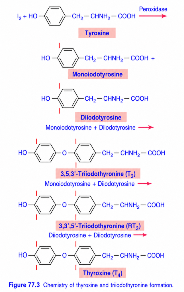
- Tyrosine is first iodinated to monoiodotyrosine (MIT) and then to diiodotyrosine (DIT).
- These iodinated tyrosines then couple with each other.
- 2 DIT molecules → Thyroxine (T₄).
- 1 DIT + 1 MIT → Triiodothyronine (T₃).
- T₃ represents about one-fifteenth of the final thyroid hormones.
- A small amount of reverse T₃ (rT₃) is also formed, but it has little functional significance in humans.
- The thyroid can store large amounts of thyroid hormone, which is unusual among endocrine glands.
- After synthesis, each thyroglobulin molecule can contain up to 30 T₄ molecules and a few T₃ molecules.
- These hormones are stored in the follicles in an amount sufficient for about 2–3 months of normal body needs.
- Therefore, even if thyroid hormone synthesis stops, deficiency effects may not appear for several months.
KEY CONCEPT
- Thyroglobulin + tyrosine + iodine → thyroid hormones.
- I⁻ → oxidized iodine → tyrosine iodination → MIT/DIT → coupling → T₃/T₄.
- 2 DIT → T₄
- DIT + MIT → T₃
- TPO + H₂O₂ → iodine oxidation and rapid organification.
- Thyroglobulin stores T₃/T₄ for ~2–3 months.
CONCEPTUAL EXAMPLES
Storage: Thyroglobulin can hold up to 30 T₄ molecules, allowing hormone supply for 2–3 months.
Thyroglobulin: Provides tyrosine sites where iodine attaches and thyroid hormones are formed.
T₄ formation: DIT + DIT → T₄.
T₃ formation: DIT + MIT → T₃.
TPO blocked: Iodide cannot be effectively oxidized → thyroid hormone formation falls to zero.
RELEASE OF THYROXINE AND TRIIODOTHYRONINE FROM THE THYROID GLAND
- Most thyroglobulin does not enter the blood; instead, T₄ and T₃ are separated from thyroglobulin and released.
- The thyroid cell forms pseudopods around small portions of colloid, creating pinocytic vesicles that enter the cell.
- Lysosomes fuse with these vesicles and release digestive enzymes.
- Proteases digest thyroglobulin and free T₄ and T₃.
- T₄ and T₃ then pass through the base of the thyroid cell into nearby capillaries and enter the blood.
- Some thyroglobulin enters thyroid cells by endocytosis after binding to megalin.
- The megalin–thyroglobulin complex crosses the cell by transcytosis and reaches the basolateral membrane.
- Part of megalin remains attached to thyroglobulin and is released into the capillary blood.
- About three-fourths of iodinated tyrosine in thyroglobulin does not become thyroid hormone.
- It remains as MIT and DIT.
- During thyroglobulin digestion, MIT and DIT are also released.
- They are not secreted into the blood.
- A deiodinase enzyme removes their iodine, allowing almost all of the iodine to be recycled inside the thyroid for new hormone formation.
- Congenital absence of deiodinase can cause iodine deficiency because iodine recycling fails.
- About 93% of thyroid hormone released is T₄, while 7% is T₃.
- During the following few days, about half of T₄ is slowly converted into T₃.
- Therefore, the hormone ultimately delivered to and used by tissues is mainly T₃, about 35 μg/day.
TRANSPORT OF THYROXINE AND TRIIODOTHYRONINE TO TISSUES
- After entering blood, more than 99% of T₄ and T₃ bind to plasma proteins made by the liver.
- They mainly bind to thyroxine-binding globulin, and less to thyroxine-binding prealbumin and albumin.
- Because these proteins bind thyroid hormones strongly, especially T₄, the hormones are released slowly to tissues.
- About half of blood T₄ is released to tissues every ~6 days.
- About half of T₃ is released every ~1 day, because T₃ binds less strongly to plasma proteins.
- Inside tissue cells, T₄ and T₃ again bind to intracellular proteins.
- T₄ binds more strongly, so both hormones are stored inside target cells and used slowly over days or weeks.
- Thyroid hormones have a slow onset and long duration of action.
- After a large dose of T₄, metabolic effects are usually not noticeable for 2–3 days.
- T₄ activity gradually increases and reaches maximum after 10–12 days (Fig. 77.4).
- T₄ then decreases with a half-life of about 15 days.
- Some T₄ activity can continue for 6–8 weeks.
- T₃ acts about 4 times faster than T₄.
- T₃ can begin acting within 6–12 hours and reaches maximum cellular activity within 2–3 days.
- Much of the slow onset and long action is due to binding of thyroid hormones to plasma and tissue proteins and their slow release.
- Part of the delay also results from how these hormones act inside cells.
KEY CONCEPT
Thyroglobulin → digestion → free T₄/T₃ → blood → plasma proteins → tissue cells → intracellular proteins → slow release and action
- T₄: 93% released → ~50% converted to T₃
- T₃: 7% released → main active hormone used by tissues
- T₄ release to tissues: ~6 days
- T₃ release to tissues: ~1 day
- T₄ onset: 2–3 days → peak: 10–12 days → half-life: ~15 days
- T₃ onset: 6–12 hours → peak: 2–3 days
CONCEPTUAL EXAMPLES
- Thyroglobulin digestion: Lysosomes → proteases → T₄/T₃ released → blood.
- Iodine recycling: MIT/DIT → deiodinase → iodine recovered → reused for thyroid hormone formation.
- T₄: Strong protein binding → slow release → slow, prolonged action.
- T₃: Weaker protein binding → faster release → faster action.
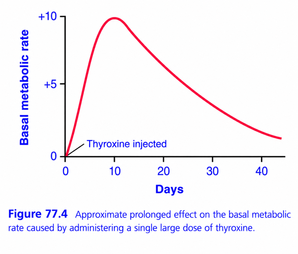
THYROXINE → BASAL METABOLIC RATE
🎯 BIG IDEA
One large dose of thyroxine (T4) → metabolism rises strongly → then slowly returns toward normal.
Think:
Thyroxine = body’s metabolic accelerator 🔥
📊 Every part of the graph
⬆️ Y-axis = Basal metabolic rate
How fast the body’s basic energy use is running.
- 0 = baseline
- +5 = moderately increased
- +10 = very high
👉 Higher = body is burning energy faster.
➡️ X-axis = Days
Shows what happens after the single thyroxine injection.
0 → 10 → 20 → 30 → 40+ days
💉 At Day 0 = thyroxine injected
The large dose is given once.
Immediately afterward:
Thyroxine ↑ → metabolic rate ↑
🔴 THE RED CURVE
Phase 1: Rapid rise 🚀
From Day 0 → ~10 days
BMR rises dramatically.
It reaches approximately:
+10
🔥 Maximum metabolic effect
Phase 2: Slow decline 📉
After ~10 days:
The effect gradually decreases:
+10 → +7 → +5 → +3 → +1
But notice:
👉 It takes many weeks to disappear.
⭐ WHY DOES IT LAST SO LONG?
Thyroxine has a long-lasting effect.
Even though only one large dose was given, T4 remains in the body and continues influencing metabolism for a prolonged period.
🧠 THE GRAPH IN ONE LINE
One large T4 dose 💉 → BMR rises slowly/strongly → peaks around 10 days → gradually falls over several weeks.
🔥 Memory trick:
T4 = “slow but long-lasting accelerator.”
Rise ↑ → Peak ~10 days → Slow fall ↓
⭐ Exam concept:
A single large dose of thyroxine produces a prolonged increase in basal metabolic rate, with the maximal effect occurring after about 10 days and gradually declining over subsequent weeks.
