


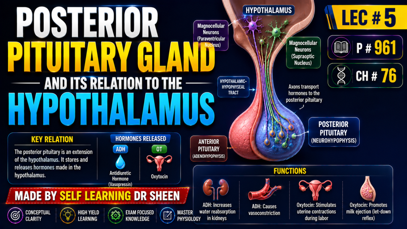
- The posterior pituitary (neurohypophysis) is mainly made of glial-like cells called pituicytes.
- Pituicytes do not secrete hormones; they mainly support nerve fibers and nerve endings coming from the supraoptic and paraventricular nuclei of the hypothalamus (Fig. 76.11).
- These nerve fibers travel through the pituitary stalk to the posterior pituitary.
- Their nerve endings contain many secretory granules and lie next to capillaries.
- They release two posterior pituitary hormones:
- Antidiuretic hormone (ADH/vasopressin)
- Oxytocin
- If the pituitary stalk is cut while the hypothalamus remains intact, posterior pituitary hormones continue to be released normally after a temporary decrease lasting a few days.
- This occurs because the hormones are released from the cut ends of the nerve fibers within the hypothalamus, rather than from the posterior pituitary nerve endings.
- The hormones are originally made in the cell bodies of the supraoptic and paraventricular nuclei.
- They travel down the nerve fibers together with carrier proteins called neurophysins, taking several days to reach the posterior pituitary.
- ADH is produced mainly by the supraoptic nuclei.
- Oxytocin is produced mainly by the paraventricular nuclei.
- Each nucleus can also produce about one-sixth as much of the other hormone.
- When nerve impulses travel down these fibers, the hormone is released from secretory granules by exocytosis.
- The hormone then enters nearby capillaries.
- Hormone and neurophysin are released together, but they separate almost immediately because they are only loosely attached.
- Neurophysin has no known function after leaving the nerve endings.
KEY CONCEPT
Hypothalamic neurons make ADH and oxytocin → hormones travel down nerve fibers → posterior pituitary nerve endings release them into blood.
CONCEPTUAL EXAMPLES
- Supraoptic nucleus → mainly ADH → posterior pituitary → blood.
- Paraventricular nucleus → mainly oxytocin → posterior pituitary → blood.
- Pituitary stalk cut → hormones can still be released from hypothalamic nerve fibers.
- Nerve impulse → exocytosis → hormone enters nearby capillary.
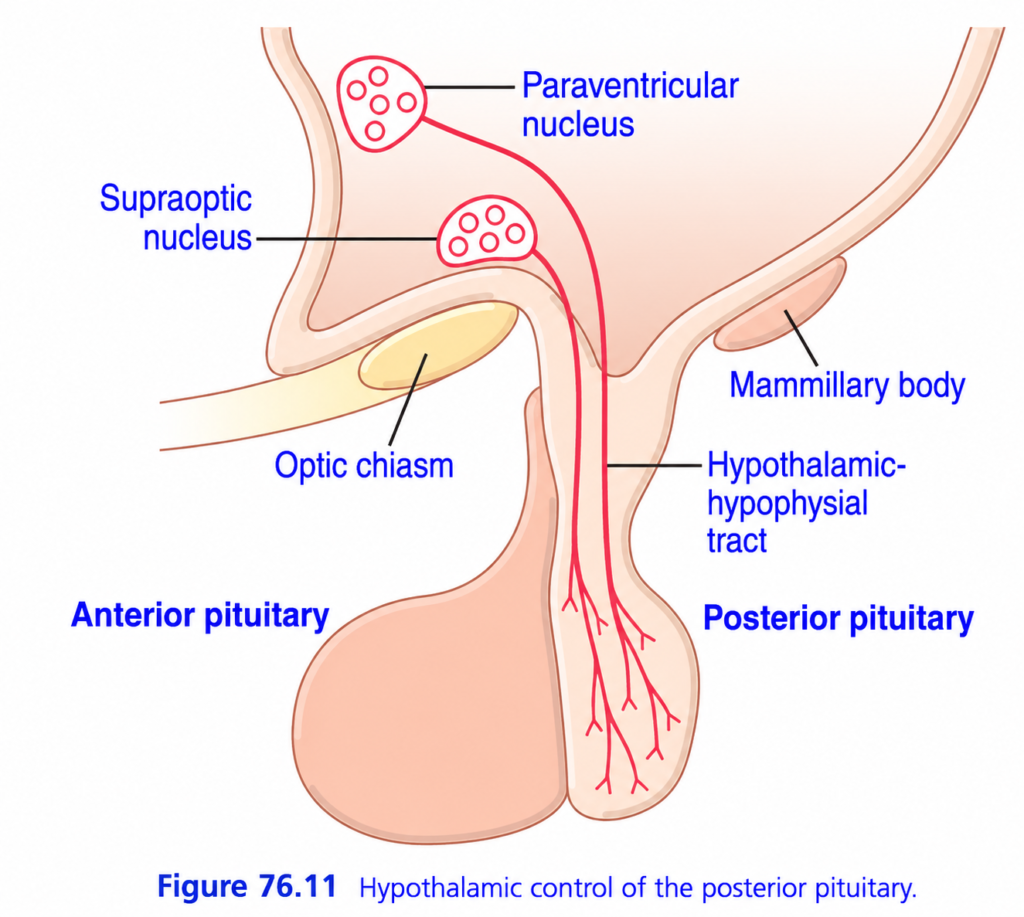
This figure is much easier than it looks.
🧠 BIG IDEA
This picture shows how the hypothalamus controls the posterior pituitary.
The key concept:
The posterior pituitary is basically an extension of the hypothalamus.
The hypothalamus makes the hormones, and nerve fibers carry them down to the posterior pituitary, where they are stored and released into blood.
⭐ THE WHOLE FIGURE IN ONE LINE
Hypothalamus → nerve fibers → posterior pituitary → hormone release
The two important hypothalamic nuclei are:
Supraoptic nucleus → ADH
Paraventricular nucleus → Oxytocin
1️⃣ HYPOTHALAMUS 🧠
At the top is the:
Hypothalamus
Inside it are special groups of nerve cells called hypothalamic nuclei.
The two shown here are:
- Supraoptic nucleus
- Paraventricular nucleus
These neurons make the hormones that will eventually be released from the posterior pituitary.
2️⃣ SUPRAOPTIC NUCLEUS
The supraoptic nucleus produces mainly:
ADH
ADH = Antidiuretic hormone
Think:
Supraoptic → ADH → Water conservation 💧
ADH helps the kidneys retain water. PARAVENTRICULAR NUCLEUS
The paraventricular nucleus produces mainly:
Oxytocin
Think:
Paraventricular → Oxytocin → Uterine contraction + milk ejection
⚠️ Both nuclei can contribute to both hormones, but for easy learning:
Supraoptic = ADH
Paraventricular = Oxytocin
4️⃣ HYPOTHALAMIC-HYPOPHYSEAL TRACT 🛣️
Look at the long red nerve fibers traveling downward.
This is:
Hypothalamic-hypophyseal tract
Think:
A direct nerve highway from hypothalamus → posterior pituitary.
The hormones are transported down the axons of hypothalamic neurons.5️⃣ POSTERIOR PITUITARY = STORAGE + RELEASE DEPOT 📦
At the bottom right is:
Posterior pituitary
The hormones arrive here through the nerve fibers.
The posterior pituitary then:
stores and releases ADH and oxytocin into the bloodstream.
Very important:
The posterior pituitary does NOT mainly manufacture these hormones.
The hypothalamus makes them.
The posterior pituitary stores and releases them.
🧠 THIS IS THE MOST IMPORTANT CONCEPT
Anterior pituitary:
Hypothalamus → releasing hormones → blood vessels → anterior pituitary
But…
Posterior pituitary:
Hypothalamus → neurons/axons → posterior pituitary
So don’t confuse them.
🆚 ANTERIOR vs POSTERIOR
| Anterior pituitary | Posterior pituitary | |
|---|---|---|
| Connection to hypothalamus | Blood vessels | Nerve fibers |
| Hypothalamic control | Releasing/inhibiting hormones | Direct neuronal signals |
| Main concept | Hormones travel through blood | Hormones travel down axons |
| ADH | ❌ | ✅ Released |
| Oxytocin | ❌ | ✅ Released |
🔍 NOW DECODE EVERY LABEL
Suprаoptic nucleus
→ hypothalamic hormone-producing neurons
→ mainly ADH
Paraventricular nucleus
→ hypothalamic hormone-producing neurons
→ mainly oxytocin
Hypothalamic-hypophyseal tract
→ axons carrying hormones/signals downward
Posterior pituitary
→ stores and releases hormones
Anterior pituitary
→ shown for orientation, but not the pathway being demonstrated
Optic chiasm
→ nearby anatomical landmark where optic nerves cross
Mammillary body
→ another nearby hypothalamic-region landmark; not the main pathway here
🧩 FOLLOW THE RED ARROWS
The red structures are nerve fibers/axons.
Start at:
🧠 Hypothalamus
⬇️
Neurons from the:
supraoptic + paraventricular nuclei
⬇️
Their axons travel through:
Hypothalamic-hypophyseal tract
⬇️
Reach:
Posterior pituitary
⬇️
Hormones are released into:
Bloodstream💧 ADH STORY
Hypothalamus makes ADH
⬇️
ADH travels down axons
⬇️
Posterior pituitary stores it
⬇️
Posterior pituitary releases it into blood
⬇️
ADH acts mainly on kidneys
⬇️
More water retained
❤️ OXYTOCIN STORY
Hypothalamus makes oxytocin
⬇️
Travels down axons
⬇️
Posterior pituitary stores it
⬇️
Released into blood
⬇️
Acts on target tissues
⬇️
Uterine contraction + milk ejection MOST COMMON EXAM TRAP
❌ “Posterior pituitary produces ADH.”
Better answer:
✅ Hypothalamic neurons synthesize ADH; posterior pituitary stores and releases it.
Same basic principle for oxytocin.🧠 ELI5 STORY
Imagine the hypothalamus is a factory 🏭.
It makes:
ADH + Oxytocin
Then it has a delivery cable 📞:
Hypothalamic-hypophyseal tract
The hormones travel down the cable to a warehouse 📦:
Posterior pituitary
The warehouse releases them into the blood when needed.
🏆 10-SECOND REVISION
Hypothalamus MAKES → Axons CARRY → Posterior pituitary STORES → Blood RELEASES
And remember:
Supraoptic → ADH → Water 💧
Paraventricular → Oxytocin → Uterus/Milk ❤️
🔑 Golden sentence:
“The posterior pituitary is a neural extension of the hypothalamus: hypothalamic neurons make ADH and oxytocin, transport them down their axons, and the posterior pituitary stores and releases them into the blood.”
Chemical Structures of Antidiuretic Hormone and Oxytocin
- ADH (vasopressin) and oxytocin are both polypeptide hormones.
- Each hormone contains 9 amino acids.
- Vasopressin: Cys–Tyr–Phe–Gln–Asn–Cys–Pro–Arg–GlyNH₂
- Oxytocin: Cys–Tyr–Ile–Gln–Asn–Cys–Pro–Leu–GlyNH₂
- Their structures are almost identical.
- The main difference is:
- Vasopressin: Phe + Arg
- Oxytocin: Ile + Leu
- Because their structures are so similar, they also have some functional similarities.
KEY CONCEPT
ADH and oxytocin = 9-amino-acid hormones with very similar structures → some similar functions.
CONCEPTUAL EXAMPLES
- ADH: Phe + Arg at the differing positions.
- Oxytocin: Ile + Leu at the corresponding positions.
PHYSIOLOGICAL FUNCTIONS OF ANTIDIURETIC HORMONE
- ADH can reduce water loss by the kidneys even in extremely small amounts, as little as 2 nanograms.
- ADH therefore produces antidiuresis = decreased water excretion.
- Without ADH, the collecting tubules and ducts become almost impermeable to water.
- Therefore, very little water is reabsorbed, causing large water loss in urine and very dilute urine.
- Severe absence of ADH causes central diabetes insipidus.
- With high ADH levels, the collecting tubules and ducts become highly permeable to water.
- Most water is then reabsorbed as tubular fluid passes through them.
- This conserves body water and produces concentrated urine.
- Without ADH, the luminal membranes of collecting-duct cells have very low water permeability.
- Inside these cells are special vesicles containing aquaporins, which are highly water-permeable pores (Fig. 28.19).
- When ADH acts:
- ADH → membrane receptor → adenylyl cyclase → ↑ cAMP
- cAMP causes phosphorylation of components of the aquaporin-containing vesicles.
- The vesicles then insert into the apical membrane.
- This creates many water-permeable areas in the membrane.
- This process occurs within about 5–10 minutes.
- When ADH is removed, the process reverses within another 5–10 minutes.
- The newly inserted aquaporins allow water to move from the tubular fluid → tubular cells → renal interstitial fluid.
- Water is then reabsorbed from the collecting tubules and ducts by osmosis.
KEY CONCEPT
ADH → ↑ aquaporins in collecting ducts → ↑ water reabsorption → ↓ water loss → concentrated urine.
CONCEPTUAL EXAMPLES
- No ADH → aquaporins absent from membrane → ↓ water reabsorption → dilute urine.
- High ADH → aquaporins inserted → ↑ water reabsorption → concentrated urine.
- ADH absent for a long time → extreme water loss → central diabetes insipidus.
REGULATION OF ANTIDIURETIC HORMONE PRODUCTION
- ↑ Extracellular fluid osmolarity → ↑ ADH secretion.
- A concentrated solution reaching the hypothalamus strongly activates ADH neurons in the supraoptic and paraventricular nuclei.
- These neurons signal the posterior pituitary to release large amounts of ADH, sometimes up to 20× normal.
- A dilute solution has the opposite effect and can almost completely stop ADH secretion.
- Therefore, ADH levels can change from very low to very high within minutes.
- Osmoreceptors are modified neurons in or near the hypothalamus that detect changes in body-fluid concentration.
- When extracellular fluid becomes too concentrated, water leaves the osmoreceptor cell by osmosis, the cell shrinks, and ADH secretion increases.
- When extracellular fluid becomes too dilute, water enters the cell, reducing the signal for ADH secretion.
- Osmoreceptors may be located in the hypothalamus or in the organum vasculosum of the anteroventral third ventricle (AV3V region).
- Regardless of their exact location, the basic response is:
Concentrated body fluids → osmoreceptors stimulated → ↑ ADH
Dilute body fluids → osmoreceptors inhibited → ↓ ADH
- This provides strong feedback control of body-fluid osmotic pressure.
- ↓ Blood volume or ↓ blood pressure → ↑ ADH secretion.
- Small amounts of ADH mainly conserve water through the kidneys.
- Higher ADH concentrations also cause strong arteriole constriction, which increases arterial pressure.
- Because of this vasoconstrictor effect, ADH is also called vasopressin.
- A major stimulus for intense ADH secretion is decreased blood volume.
- When blood volume falls by about 15–25% or more, ADH secretion may rise to about 50× normal.
- Atrial stretch receptors detect blood-volume changes.
- When the atria are overfilled, stretch receptors are activated and inhibit ADH secretion.
- When the atria are underfilled, the inhibition decreases and ADH secretion increases.
- Reduced stretch of carotid, aortic, and pulmonary baroreceptors also increases ADH secretion.
KEY CONCEPT
Concentrated body fluid OR low blood volume/pressure → ↑ ADH → conserve water; at high levels → vasoconstriction → ↑ blood pressure.
CONCEPTUAL EXAMPLES
- Body fluid concentrated → osmoreceptors shrink → ↑ ADH → ↑ water conservation.
- Body fluid dilute → osmoreceptor signal ↓ → ↓ ADH.
- Blood volume ↓ significantly → ↑ ADH → water conservation + vasoconstriction.
- Atria overfilled → stretch receptors activated → ↓ ADH.
- Atria underfilled → ↓ stretch → ↑ ADH.
PHYSIOLOGICAL FUNCTIONS OF OXYTOCIN
- Oxytocin strongly contracts the pregnant uterus, especially near the end of pregnancy.
- Therefore, it may help cause delivery of the baby.
- Evidence supporting this includes:
- Removing the pituitary gland in animals prolongs labor.
- Blood oxytocin levels increase during labor, especially during its last stage.
- Cervical stimulation sends nerve signals to the hypothalamus, increasing oxytocin secretion.
- Oxytocin also has an important role in milk ejection during lactation.
- When the baby suckles the nipple, sensory nerve signals travel to oxytocin neurons in the paraventricular and supraoptic nuclei of the hypothalamus.
- The posterior pituitary then releases oxytocin into the blood.
- Oxytocin reaches the breast and causes contraction of myoepithelial cells surrounding the mammary alveoli.
- This pushes milk from the alveoli into the ducts, allowing the baby to obtain it by suckling.
- Milk begins to flow within less than 1 minute after suckling starts.
- This process is called milk letdown or milk ejection.
KEY CONCEPT
Oxytocin → uterine contraction during labor + myoepithelial contraction → milk ejection.
CONCEPTUAL EXAMPLES
- End of pregnancy → ↑ oxytocin → uterine contraction → helps delivery.
- Baby suckles nipple → hypothalamus → posterior pituitary → oxytocin → breast contraction → milk ejection.
