


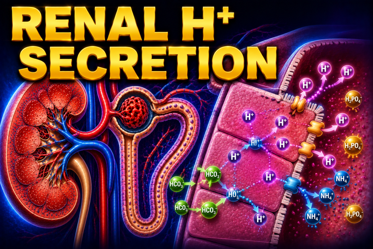
- The cells of the proximal tubules and distal tubules secrete hydrogen ions (H⁺).
- Hydrogen ion secretion also occurs in the collecting ducts.
- In the proximal tubule, H⁺ is mainly secreted by the Na⁺–H⁺ exchanger (NHE3).
- This is an example of secondary active transport.
- The Na⁺, K⁺-ATPase pump on the basolateral membrane pumps Na⁺ from the cell into the interstitial fluid.
- This keeps the Na⁺ concentration inside the cell low.
- The low intracellular Na⁺ creates a driving force for Na⁺ to enter the cell from the tubular lumen through the Na⁺–H⁺ exchanger.
- As Na⁺ enters the cell, H⁺ is secreted into the tubular lumen.
Figure: Figure 39–1
- The secreted H⁺ combines with filtered bicarbonate (HCO₃⁻) to form carbonic acid (H₂CO₃).
- Carbonic anhydrase on the apical membrane rapidly converts H₂CO₃ into carbon dioxide (CO₂) and water (H₂O).
- CO₂ and H₂O rapidly enter the proximal tubular cell because the apical membrane is permeable to them.
- About 80% of the filtered bicarbonate (HCO₃⁻) is reabsorbed in the proximal tubule.
- Inside the tubular cell, carbonic anhydrase again combines CO₂ and H₂O to form H₂CO₃.
- H₂CO₃ then dissociates into H⁺ and HCO₃⁻.
- The H⁺ is secreted back into the tubular lumen through the Na⁺–H⁺ exchanger.
- The newly formed HCO₃⁻ diffuses into the interstitial fluid.
- For every H⁺ secreted into the tubular lumen:
- One Na⁺ enters the interstitial fluid.
- One HCO₃⁻ enters the interstitial fluid.
- Carbonic anhydrase is essential for these reactions.
- Carbonic anhydrase inhibitors reduce H⁺ secretion by the proximal tubule.
- These drugs also reduce the reactions that depend on carbonic anhydrase.
- Some evidence suggests that other transporters also secrete H⁺ in the proximal tubule.
- However, their role is small and remains controversial.
- The Na⁺–H⁺ exchanger is the major mechanism of H⁺ secretion in the proximal tubule.
Distal Tubules and Collecting Ducts
- In the distal tubules and collecting ducts, H⁺ secretion is largely independent of Na⁺ in the tubular lumen.
- Most H⁺ is secreted by an ATP-driven proton (H⁺) pump.
- Aldosterone increases H⁺ secretion by stimulating this proton pump.
- Intercalated (I) cells are responsible for acid secretion in this part of the nephron.
- These cells contain abundant carbonic anhydrase.
- They also contain numerous tubulovesicular structures.
- The H⁺-ATPase pumps are present in these vesicles as well as in the apical membrane.
- During acidosis, more H⁺ pumps are inserted into the apical membrane from the tubulovesicles.
- This increases H⁺ secretion into the tubular lumen.
- Some H⁺ is also secreted by the H⁺–K⁺ ATPase pump.
- Intercalated (I) cells also contain Anion Exchanger 1 (AE1) in the basolateral membrane.
- AE1 functions as a chloride (Cl⁻)–bicarbonate (HCO₃⁻) exchanger.
- This transporter moves HCO₃⁻ into the interstitial fluid while Cl⁻ enters the cell.
KEY CONCEPT
- The proximal tubule secretes H⁺ mainly through the Na⁺–H⁺ exchanger (NHE3), which depends on the Na⁺ gradient created by the Na⁺, K⁺-ATPase. Carbonic anhydrase converts H₂CO₃ into CO₂ and H₂O outside the cell and reforms H₂CO₃ inside the cell, allowing continuous H⁺ secretion and HCO₃⁻ reabsorption. In the distal tubules and collecting ducts, H⁺ is secreted mainly by ATP-driven proton pumps that are stimulated by aldosterone and become more numerous during acidosis.
Conceptual Examples
- Example 1: In the proximal tubule, H⁺ is secreted into the tubular lumen through the Na⁺–H⁺ exchanger, while Na⁺ and HCO₃⁻ are returned to the blood, helping conserve bicarbonate.
- Example 2: During metabolic acidosis, more H⁺ pumps are inserted into the apical membrane of intercalated cells, allowing the kidneys to secrete more H⁺ and restore normal body pH.
- Example 3: A patient taking a carbonic anhydrase inhibitor (e.g., acetazolamide) secretes less H⁺ and reabsorbs less HCO₃⁻, resulting in increased bicarbonate loss in the urine.
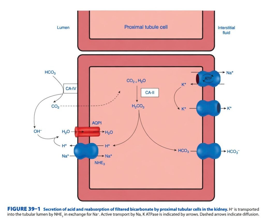
Figure 39-1: Secretion of Acid and Reabsorption of Filtered Bicarbonate by the Proximal Tubule
Easiest and Most Conceptual Explanation
This figure explains one of the kidney’s most important jobs:
The proximal tubule reabsorbs almost all filtered bicarbonate (HCO₃⁻) while secreting hydrogen ions (H⁺) into the tubular lumen.
⭐ One-Line Concept
Step 1: Understand the Layout
The figure shows one proximal tubule cell.
Left Side
Tubular lumen
- Contains the filtered fluid (future urine).
Middle
Proximal tubule cell
This is where all the reactions occur.
Right Side
Interstitial fluid
This leads to the blood (peritubular capillaries).
Step 2: Important Proteins and Their Locations
1. NHE3 (Na⁺/H⁺ Exchanger)
Location: Apical (luminal) membrane
Function
- Moves Na⁺ into the cell
- Moves H⁺ into the tubular lumen
Think of it as a swap:
Na⁺ comes in → H⁺ goes out
2. CA-IV (Carbonic Anhydrase IV)
Location: Outside the cell, on the luminal surface.
Function
Converts
Carbonic acid (H₂CO₃)
↓
CO₂ + H₂O
This reaction occurs in the tubular lumen.
3. AQP1 (Aquaporin-1)
Location: Apical membrane
Function
Allows water (H₂O) to move easily across the membrane.
4. CA-II (Carbonic Anhydrase II)
Location: Inside the proximal tubule cell.
Function
Converts
CO₂ + H₂O
↓
H₂CO₃
↓
H⁺ + HCO₃⁻
5. Na⁺/K⁺ ATPase
Location: Basolateral membrane
Function
Uses ATP to pump:
- 3 Na⁺ out of the cell
- 2 K⁺ into the cell
This maintains a low intracellular Na⁺ concentration, allowing NHE3 to keep working.
6. Basolateral HCO₃⁻ Transporter
Location: Basolateral membrane
Function
Moves newly formed bicarbonate (HCO₃⁻) into the interstitial fluid and then into the blood.
7. K⁺ Channel
Location: Basolateral membrane
Function
Allows K⁺ to leave the cell after it enters through the Na⁺/K⁺ ATPase.
Step 3: Follow the Entire Process
Step 1: Filtered Bicarbonate Reaches the Tubular Lumen
The glomerulus filters bicarbonate into the tubular fluid.
So, the lumen initially contains:
- HCO₃⁻
Step 2: NHE3 Secretes H⁺
The Na⁺/H⁺ exchanger (NHE3):
- Takes Na⁺ into the cell.
- Secretes H⁺ into the lumen.
So, hydrogen ions are continuously added to the tubular fluid.
Step 3: H⁺ Meets Filtered HCO₃⁻
In the lumen:
H⁺ + HCO₃⁻
↓
H₂CO₃ (carbonic acid)
Step 4: CA-IV Converts Carbonic Acid
Luminal carbonic anhydrase IV rapidly converts:
H₂CO₃
↓
CO₂ + H₂O
Step 5: CO₂ Diffuses into the Cell
The figure uses a dashed arrow from the lumen into the cell.
Meaning of the Dashed Arrow
It represents simple diffusion.
CO₂ is a small gas and easily diffuses across the cell membrane.
Step 6: CA-II Reforms Carbonic Acid
Inside the cell:
CO₂ + H₂O
↓
H₂CO₃
Step 7: Carbonic Acid Splits
H₂CO₃ immediately dissociates into:
- H⁺
- HCO₃⁻
Step 8: The H⁺ Is Recycled
The newly formed H⁺ is not lost.
Instead, NHE3 secretes it back into the lumen.
Therefore,
the same H⁺ is used repeatedly.
Step 9: The New HCO₃⁻ Enters the Blood
The newly formed bicarbonate leaves the cell through the basolateral HCO₃⁻ transporter.
It enters:
Interstitial fluid → Blood
Thus, bicarbonate is effectively reabsorbed.
Why Doesn’t HCO₃⁻ Simply Enter the Cell Directly?
Because:
The apical membrane is relatively impermeable to bicarbonate.
Therefore, bicarbonate must first be converted to CO₂, which crosses the membrane easily.
Understanding Every Arrow
Solid Black Arrows
These indicate:
- Direction of ion transport
- Direction of chemical reactions
Dashed Black Arrow
Shows:
Diffusion of CO₂
No ATP is required.
ATP Label
Appears beside the Na⁺/K⁺ ATPase.
Meaning
This transporter requires energy from ATP.
It performs primary active transport.
Understanding Each Chemical Reaction
In the Lumen
H⁺ + HCO₃⁻
↓
H₂CO₃
↓
CO₂ + H₂O (via CA-IV)
Inside the Cell
CO₂ + H₂O
↓
H₂CO₃ (via CA-II)
↓
H⁺ + HCO₃⁻
Easy Story
Imagine the bicarbonate molecule wants to return to the blood but cannot pass through the cell membrane.
So it changes its “clothes.”
Bicarbonate (HCO₃⁻)
↓
Becomes
CO₂
↓
CO₂ easily enters the cell.
↓
Inside the cell, it changes back into
HCO₃⁻
↓
The new bicarbonate leaves the cell and returns to the blood.
This is like using a disguise to cross a security gate.
Important Clinical Point
If carbonic anhydrase is inhibited (for example, by acetazolamide):
- Less CO₂ is formed in the lumen.
- Less bicarbonate is reabsorbed.
- More bicarbonate remains in the urine.
- Urine becomes more alkaline.
- Metabolic acidosis may develop.
High-Yield Summary Table
| Structure | Location | Main Function |
|---|---|---|
| NHE3 | Apical membrane | Exchanges Na⁺ into the cell for H⁺ into the lumen |
| CA-IV | Luminal surface | Converts H₂CO₃ into CO₂ + H₂O |
| CO₂ | Diffuses across the apical membrane | Enters the cell by simple diffusion |
| CA-II | Cytoplasm | Converts CO₂ + H₂O into H₂CO₃, then H⁺ + HCO₃⁻ |
| Na⁺/K⁺ ATPase | Basolateral membrane | Pumps Na⁺ out and K⁺ into the cell using ATP |
| HCO₃⁻ transporter | Basolateral membrane | Moves newly formed bicarbonate into the blood |
| AQP1 | Apical membrane | Allows water movement |
Sequence to Remember
Filtered HCO₃⁻
↓
H⁺ secreted by NHE3
↓
H₂CO₃ forms
↓
CA-IV converts H₂CO₃ → CO₂ + H₂O
↓
CO₂ diffuses into the cell
↓
CA-II converts CO₂ + H₂O → H₂CO₃
↓
H₂CO₃ dissociates → H⁺ + HCO₃⁻
↓
H⁺ is recycled into the lumen
↓
HCO₃⁻ enters the blood
↓
Net effect: Filtered bicarbonate is reabsorbed
Key Concept (Figure 39-1)
Figure 39-1 illustrates how the proximal tubule reabsorbs filtered bicarbonate while secreting hydrogen ions. Hydrogen ions are secreted into the tubular lumen by the Na⁺/H⁺ exchanger (NHE3), where they combine with filtered bicarbonate to form carbonic acid. Carbonic anhydrase IV (CA-IV) converts carbonic acid into CO₂ and H₂O, and CO₂ diffuses into the proximal tubule cell. Inside the cell, carbonic anhydrase II (CA-II) converts CO₂ and H₂O back into carbonic acid, which dissociates into H⁺ and HCO₃⁻. The H⁺ is recycled back into the lumen by NHE3, while the newly formed bicarbonate is transported across the basolateral membrane into the blood. The Na⁺/K⁺ ATPase maintains the sodium gradient that drives this process. Thus, the kidney effectively reabsorbs filtered bicarbonate without transporting luminal bicarbonate directly across the apical membrane.
FATE OF H⁺ IN THE URINE
- The amount of H⁺ secreted into the urine depends on what happens to H⁺ after it enters the tubular fluid.
- The kidney can secrete H⁺ only up to a maximum concentration.
- The maximum H⁺ gradient corresponds to a urine pH of about 4.5.
- At urine pH 4.5, the H⁺ concentration in urine is about 1000 times higher than in plasma.
- Therefore, pH 4.5 is the lowest (limiting) urine pH that the kidneys can normally produce.
- This limiting pH is usually reached in the collecting ducts.
- If there were no urinary buffers, free H⁺ would quickly accumulate in the urine.
- The rapid increase in free H⁺ would stop further H⁺ secretion.
- Urinary buffers bind free H⁺, allowing the kidneys to continue secreting more acid.
- Three important reactions remove free H⁺ from the tubular fluid.
Figure: Figure 39–2
- Reaction 1: H⁺ combines with HCO₃⁻ to form CO₂ and H₂O.
- Reaction 2: H⁺ combines with HPO₄²⁻ to form H₂PO₄⁻ (titratable acid).
- Reaction 3: H⁺ combines with NH₃ to form NH₄⁺ (ammonium).
KEY CONCEPT
- The kidneys can lower urine pH only to about 4.5. Without urinary buffers, H⁺ would rapidly accumulate and stop further acid secretion. Buffers remove free H⁺ by converting it into CO₂ + H₂O, H₂PO₄⁻, or NH₄⁺, allowing continuous H⁺ secretion and acid excretion.
Conceptual Examples
- Example 1: H⁺ combines with bicarbonate (HCO₃⁻) to form CO₂ and H₂O, helping remove free H⁺ from the tubular fluid.
- Example 2: H⁺ binds with phosphate (HPO₄²⁻) to form H₂PO₄⁻ (titratable acid), allowing additional acid to be excreted in urine.
- Example 3: H⁺ combines with ammonia (NH₃) to form NH₄⁺ (ammonium), which is trapped in the urine and excreted, helping eliminate excess acid from the body.
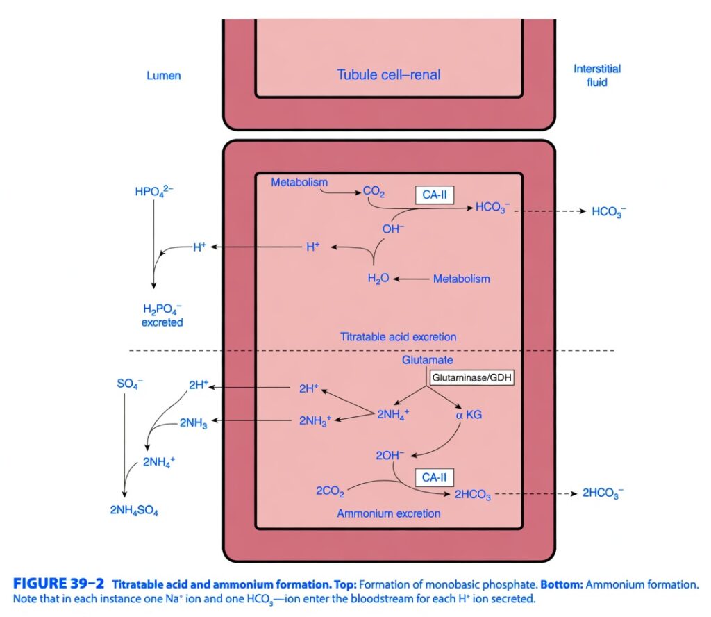
Figure 39-2: Titratable Acid and Ammonium Formation
Fate of H⁺ in the Urine – Easiest and Most Conceptual Explanation
⭐ One-Line Concept
The kidney keeps secreting H⁺ by immediately “hiding” it inside urinary buffers (phosphate and ammonia), preventing free H⁺ from accumulating and allowing more acid to be excreted.
First Understand the Main Idea
The kidney secretes H⁺ (acid) into the tubular fluid.
However, there is one major problem.
If too much free H⁺ accumulates in urine:
- Urine becomes extremely acidic.
- Urine reaches pH 4.5.
- At this point, the kidney cannot secrete any more H⁺ because the H⁺ concentration gradient is too high.
So the kidney needs buffers that immediately bind free H⁺.
These buffers act like acid sponges.
They remove free H⁺ from urine, allowing the kidney to continue secreting more H⁺.Why is pH 4.5 Important?
Normal plasma pH ≈ 7.4
Maximum urine acidity ≈ pH 4.5
This means:
- Urine has about 1000 times more H⁺ than plasma.
This is the lowest urine pH humans can produce.
Without urinary buffers, this limit would be reached very quickly.
Three Ways the Kidney Removes Free H⁺
1. Bicarbonate Buffer
H⁺ + HCO₃⁻
↓
H₂CO₃
↓
CO₂ + H₂O
This mechanism mainly reabsorbs filtered bicarbonate.
2. Phosphate Buffer (Titratable Acid)
HPO₄²⁻ + H⁺
↓
H₂PO₄⁻
This acid is excreted in urine.
3. Ammonia Buffer
NH₃ + H⁺
↓
NH₄⁺
Ammonium is trapped in urine and excreted.
Understanding the Figure
The figure has two separate sections.
TOP HALF
Titratable Acid Formation (Phosphate Buffer)
This explains how phosphate removes H⁺.
Step 1
Inside the tubular cell:
Cell metabolism produces
CO₂
Step 2
Water is also present.
CO₂ + H₂O
↓
Carbonic acid
Step 3
CA-II (Carbonic Anhydrase II)
This intracellular enzyme converts:
CO₂ + H₂O
↓
H₂CO₃
↓
H⁺ + HCO₃⁻
Step 4
The Newly Formed H⁺
H⁺ is secreted into the tubular lumen.
Step 5
What Happens in the Tubular Fluid?
Filtered phosphate is present as
HPO₄²⁻
The secreted H⁺ combines with it.
HPO₄²⁻ + H⁺
↓
H₂PO₄⁻
This is called:
Monobasic phosphate
or
Titratable acid
Step 6
H₂PO₄⁻ Cannot Diffuse Back
Therefore,
It remains in urine and is excreted.
Step 7
What Happens to the Newly Formed HCO₃⁻?
The bicarbonate produced inside the cell moves into the blood.
So,
For every H⁺ excreted as phosphate,
one new HCO₃⁻ is added to the blood.
Bottom Half
Ammonium Formation
This is the most important mechanism during chronic acidosis.
Step 1
Inside the proximal tubule cell:
The amino acid
Glutamate
is metabolized.
The enzymes involved are:
- Glutaminase
- Glutamate dehydrogenase (GDH)
Step 2
Glutamate Produces
Two important products:
A
2 NH₄⁺
↓
Can become NH₃ inside the cell.
B
α-Ketoglutarate (α-KG)
This enters metabolism.
Step 3
Metabolism of α-KG
Produces:
CO₂
↓
CA-II converts it into
2 HCO₃⁻
These bicarbonate ions enter the blood.
Again,
Two new bicarbonate ions are added to plasma.
Step 4
NH₃ Diffuses into the Tubular Fluid
Ammonia (NH₃) easily crosses the membrane.
Step 5
Secreted H⁺ Meets NH₃
NH₃ + H⁺
↓
NH₄⁺
Step 6
NH₄⁺ Is Trapped
NH₄⁺ cannot easily cross the membrane.
Therefore,
It stays inside urine.
Step 7
Urine Contains Sulfate
Sulfate (SO₄²⁻) combines with NH₄⁺.
2 NH₄⁺ + SO₄²⁻
↓
(NH₄)₂SO₄
This is excreted in urine.
Why Is NH₄⁺ Called “Trapped”?
NH₃ is uncharged.
It diffuses easily.
After combining with H⁺:
NH₃
↓
NH₄⁺
Now it is charged.
Charged molecules cannot easily diffuse back.
So the acid is permanently trapped inside urine.
This is called
Diffusion trapping (ion trapping).
Why Does This Help?
Imagine pouring acid into a bucket.
If the bucket becomes full of free acid,
you cannot add more.
Now imagine putting a sponge inside the bucket.
The sponge absorbs the acid,
creating more space.
Now you can pour in more acid.
Urinary buffers work exactly like that sponge.
They bind free H⁺,
keeping urine pH from reaching 4.5 too quickly.
Meaning of Every Label in the Figure
HPO₄²⁻
Filtered phosphate buffer.
Accepts H⁺.
H₂PO₄⁻
Monobasic phosphate.
Excreted in urine.
This is the titratable acid.
Glutamate
Main amino acid used to produce ammonia.
Glutaminase / GDH
Enzymes that convert glutamate into:
- NH₄⁺
- α-Ketoglutarate
α-KG (α-Ketoglutarate)
Metabolized to generate bicarbonate.
NH₃
Ammonia.
Diffuses easily into urine.
NH₄⁺
Ammonium.
Trapped inside urine.
Excreted.
SO₄²⁻
Sulfate.
Combines with NH₄⁺ to form ammonium sulfate.
CA-II
Carbonic anhydrase II.
Forms H⁺ and HCO₃⁻ inside the tubular cell.
CO₂
Produced by metabolism.
Used to generate bicarbonate.
HCO₃⁻
Moves into blood.
Replaces bicarbonate lost during acid buffering.
Understanding the Arrows
Solid Arrows
Show:
- Chemical reactions
- Transport of substances
- Direction of movement
Dashed Horizontal Arrow
Shows bicarbonate leaving the tubular cell and entering the blood.
Why Does Blood Gain Bicarbonate?
Whenever H⁺ leaves the body in urine,
the body effectively gains bicarbonate.
Therefore:
Every H⁺ excreted = One new HCO₃⁻ added to blood.
Comparison of the Two Buffers
| Feature | Phosphate Buffer | Ammonia Buffer |
|---|---|---|
| Buffer | HPO₄²⁻ | NH₃ |
| Product | H₂PO₄⁻ | NH₄⁺ |
| Excreted in urine | Yes | Yes |
| New bicarbonate formed | Yes | Yes (more effective) |
| Importance | Normal acid excretion | Very important during chronic acidosis |
Simple Flow Diagram
1. Titratable Acid Pathway
CO₂ + H₂O
↓
CA-II
↓
H⁺ + HCO₃⁻
↓
H⁺ secreted into urine
↓
HPO₄²⁻ + H⁺
↓
H₂PO₄⁻
↓
Excreted in urine
↓
HCO₃⁻ enters blood
2. Ammonium Pathway
Glutamate
↓
Glutaminase + GDH
↓
NH₃ + α-Ketoglutarate
↓
NH₃ enters urine
↓
NH₃ + H⁺
↓
NH₄⁺
↓
Trapped in urine
↓
Excreted
↓
α-Ketoglutarate metabolism
↓
2 HCO₃⁻
↓
Blood
Easy Story to Remember
Think of the kidney as a garbage truck carrying acid (H⁺).
If loose acid fills the truck, there is no room for more.
So the kidney packs the acid into sealed containers before throwing it away:
- Phosphate box → H₂PO₄⁻ (Titratable acid)
- Ammonia box → NH₄⁺ (Ammonium)
Packing the acid prevents free H⁺ from accumulating, allowing the kidney to keep loading and excreting more acid.
Key Concept (Figure 39-2)
Figure 39-2 shows the two major urinary buffering systems that allow continuous acid secretion by the kidney. Secreted H⁺ combines with filtered phosphate (HPO₄²⁻) to form H₂PO₄⁻ (titratable acid), which is excreted in urine. H⁺ also combines with ammonia (NH₃), produced from glutamate metabolism, to form NH₄⁺ (ammonium), which becomes trapped in the tubular fluid and is excreted. In both pathways, carbonic anhydrase II (CA-II) generates intracellular H⁺ and HCO₃⁻. The H⁺ is secreted into the lumen, while the newly formed HCO₃⁻ enters the bloodstream, replenishing plasma bicarbonate. These urinary buffers remove free H⁺ from the tubular fluid, prevent urine pH from rapidly reaching its minimum limit of 4.5, and allow the kidneys to continue excreting acid effectively.
REACTION WITH BUFFERS
- The three important buffers involved in kidney acid handling and H⁺ secretion are:
- Bicarbonate (HCO₃⁻)
- Dibasic phosphate (HPO₄²⁻)
- Ammonia (NH₃)
- With a normal diet, about 40% of nonvolatile acid (≈30 mEq/day) is excreted as titratable acid through the phosphate buffer system.
- About 60% of nonvolatile acid (≈50 mEq/day) is excreted as ammonium (NH₄⁺).
- The pK′ values of the buffer systems are:
- Bicarbonate buffer = 6.1
- Phosphate buffer = 6.8
- Ammonia buffer = 9.0
- The normal plasma concentration of bicarbonate (HCO₃⁻) is about 24 mEq/L.
- The normal plasma concentration of phosphate is about 1.5 mEq/L.
- Because bicarbonate is present in a much higher concentration, most secreted H⁺ in the proximal tubule reacts with HCO₃⁻.
- H⁺ combines with HCO₃⁻ to form carbonic acid (H₂CO₃).
- Carbonic anhydrase converts H₂CO₃ into CO₂ and H₂O.
- CO₂ and H₂O enter the proximal tubular cell.
- The CO₂ entering the cell becomes available to form H₂CO₃ again.
- Because most H⁺ is removed from the tubular fluid, the tubular fluid pH changes very little.
- This is the mechanism by which bicarbonate (HCO₃⁻) is reabsorbed.
- For every one HCO₃⁻ removed from the tubular fluid, one newly formed HCO₃⁻ enters the blood from the tubular cell.
- The bicarbonate entering the blood is not the same bicarbonate molecule that was filtered.
- About 4500 mEq of bicarbonate (HCO₃⁻) are filtered and reabsorbed each day.
Figure: Figure 39–2
- Secreted H⁺ also combines with dibasic phosphate (HPO₄²⁻) to form monobasic phosphate (H₂PO₄⁻).
- This reaction occurs mainly in the distal tubules and collecting ducts.
- It occurs there because phosphate becomes highly concentrated as water is reabsorbed.
- A small amount of H⁺ also combines with other buffer anions.
- The ammonia buffer system allows H⁺ to combine with NH₃ to form NH₄⁺.
- This reaction occurs in the proximal tubule and in the distal tubules.
- The pK′ of the ammonia buffer system is 9.0.
- The ammonia buffer contributes very little to titratable acidity because it is titrated only from the urine pH to pH 7.4.
- Every H⁺ that combines with urinary buffers contributes to urinary titratable acidity.
- Titratable acidity is measured by determining how much alkali is needed to raise the urine pH back to 7.4.
- Titratable acidity measures only part of the acid secreted because it does not include H₂CO₃ that has been converted into CO₂ and H₂O.
- Reabsorption of bicarbonate (HCO₃⁻) is essential for maintaining normal acid–base balance.
- Loss of one HCO₃⁻ ion in the urine has the same effect as adding one H⁺ ion to the blood.
- The kidneys can also produce new bicarbonate (HCO₃⁻).
- New bicarbonate is formed when H⁺ is excreted as NH₄⁺ or as titratable acid.
- This newly formed bicarbonate enters the blood.
- These bicarbonate ions are newly produced and are not the same bicarbonate ions that were originally filtered.
KEY CONCEPT
- The kidneys use three main buffers—bicarbonate, phosphate, and ammonia—to remove H⁺ from the tubular fluid. Most H⁺ first reacts with bicarbonate in the proximal tubule, allowing bicarbonate to be reabsorbed. In the distal nephron, phosphate and ammonia help excrete additional H⁺. When H⁺ is excreted as NH₄⁺ or titratable acid, the kidneys also produce new bicarbonate, helping maintain normal acid–base balance.
Conceptual Examples
- Example 1: In the proximal tubule, H⁺ combines with bicarbonate (HCO₃⁻), allowing filtered bicarbonate to be reabsorbed into the blood.
- Example 2: In the distal tubule, H⁺ combines with phosphate (HPO₄²⁻) to form H₂PO₄⁻, which is excreted in the urine as titratable acid.
- Example 3: H⁺ combines with ammonia (NH₃) to form NH₄⁺, which is excreted in the urine while new bicarbonate is added to the blood, helping correct acidosis.
AMMONIA SECRETION
- Renal tubular cells produce ammonium (NH₄⁺) and bicarbonate (HCO₃⁻).
- Inside the cells, NH₄⁺ is in equilibrium with ammonia (NH₃) and H⁺.
- The pK′ of this reaction is 9.0.
- At pH 7.0, the ratio of NH₃ to NH₄⁺ is 1:100.
- This means that for every 1 NH₃ molecule, there are about 100 NH₄⁺ ions.
- NH₃ is lipid-soluble and easily diffuses across cell membranes.
- NH₃ diffuses from the tubular cells into the interstitial fluid and tubular urine.
- In the urine, NH₃ combines with H⁺ to form NH₄⁺.
- The NH₄⁺ remains trapped in the urine and is excreted.
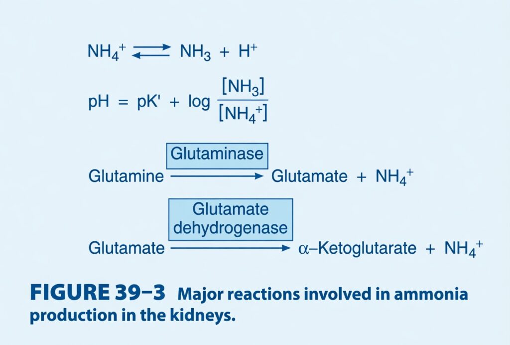
Figure: Figure 39–3
- The main source of NH₄⁺ is the conversion of glutamine into glutamate.
- This reaction is catalyzed by the enzyme glutaminase, which is abundant in renal tubular cells.
- Glutamate is then converted into α-ketoglutarate by the enzyme glutamate dehydrogenase.
- This reaction produces additional NH₄⁺.
- Further metabolism of α-ketoglutarate uses 2 H⁺ ions.
- This process produces 2 new HCO₃⁻ ions.
- In chronic acidosis, NH₄⁺ excretion increases even when urine pH remains the same.
- This happens because more NH₃ enters the tubular urine.
- More NH₃ removes more H⁺ from the tubular fluid.
- As a result, H⁺ secretion by the renal tubules increases.
- This also increases H⁺ excretion in the urine.
- The amount of phosphate buffer filtered by the glomerulus cannot be increased.
- Therefore, acid excretion through the phosphate buffer system is limited.
- Production of NH₄⁺ by the renal tubules is the only way the kidneys can excrete the normal and increased amounts of nonvolatile acid produced in the body.
- In the inner medullary collecting duct, NH₃ enters the urine mainly by nonionic diffusion.
- After entering the urine, NH₃ combines with H⁺ to form NH₄⁺.
- This maintains the concentration gradient that allows more NH₃ to diffuse into the urine.
- In the proximal tubule, nonionic diffusion of NH₄⁺ is less important.
- Here, NH₄⁺ can be secreted directly into the tubular lumen.
- NH₄⁺ often replaces H⁺ on the Na⁺–H⁺ exchanger during secretion.
- Salicylates and many other weak acids and weak bases are also secreted by nonionic diffusion.
- The rate at which these drugs enter the tubular fluid depends on the urine pH.
- Therefore, the amount of these drugs excreted changes according to the urine pH.
KEY CONCEPT
- The kidneys produce NH₄⁺ from glutamine metabolism. NH₃ diffuses into the tubular fluid, where it combines with H⁺ to form NH₄⁺, which becomes trapped in the urine and is excreted. This process removes acid from the body and produces new bicarbonate (HCO₃⁻), making ammonia the most important system for excreting nonvolatile acids, especially during chronic acidosis.
Conceptual Examples
- Example 1: During chronic metabolic acidosis, the kidneys produce more NH₃, which binds more H⁺ to form NH₄⁺, increasing acid excretion.
- Example 2: NH₃ diffuses into the tubular urine, combines with H⁺, and forms NH₄⁺, which becomes trapped in the urine and is eliminated from the body.
- Example 3: The metabolism of glutamine produces NH₄⁺ for acid excretion and new HCO₃⁻, which enters the blood and helps restore normal acid–base balance.
PH CHANGES ALONG THE NEPHRON
- The pH of the tubular fluid changes as it moves along the nephron.
- In the proximal tubule, the tubular fluid pH decreases moderately.
- Most of the secreted H⁺ has very little effect on the tubular fluid pH.
- This is because H⁺ combines with HCO₃⁻ to form H₂CO₃.
- H₂CO₃ is rapidly converted into CO₂ and H₂O.
- Therefore, free H⁺ is removed from the tubular fluid, so the pH changes only slightly.
- In contrast, the distal tubule has a lower capacity to secrete H⁺.
- However, H⁺ secretion in the distal tubule has a much greater effect on urine pH.
KEY CONCEPT
- The proximal tubule secretes a large amount of H⁺, but tubular pH decreases only slightly because H⁺ is buffered by bicarbonate. The distal tubule secretes less H⁺, but because there is less buffering, it produces a much greater decrease in urine pH.
Conceptual Examples
- Example 1: In the proximal tubule, secreted H⁺ combines with HCO₃⁻, so the tubular fluid pH falls only a little.
- Example 2: In the distal tubule, even a small amount of H⁺ secretion causes the urine pH to decrease markedly because there is less bicarbonate available for buffering.
FACTORS AFFECTING ACID SECRETION
- Renal acid secretion is affected by changes in:
- Intracellular PCO₂
- Potassium (K⁺) concentration
- Carbonic anhydrase level
- Adrenocortical hormone concentration
- When intracellular PCO₂ is high (respiratory acidosis), more H₂CO₃ is formed inside the tubular cells.
- More H₂CO₃ is available to buffer hydroxyl (OH⁻) ions.
- As a result, H⁺ secretion increases.
- When PCO₂ decreases, less H₂CO₃ is formed.
- Therefore, H⁺ secretion decreases.
- Potassium (K⁺) depletion increases H⁺ secretion.
- This is because loss of K⁺ causes intracellular acidosis, even if the plasma pH is increased.
- Excess potassium (K⁺) inside the cells decreases H⁺ secretion.
- When carbonic anhydrase is inhibited, less H₂CO₃ is formed.
- As a result, H⁺ secretion decreases.
- Aldosterone and other adrenocortical steroids increase Na⁺ reabsorption in the renal tubules.
- These hormones also increase the secretion of H⁺ and K⁺.
KEY CONCEPT
- Renal H⁺ secretion increases with high PCO₂, potassium depletion, and aldosterone. It decreases with low PCO₂, excess intracellular potassium, and carbonic anhydrase inhibition. These factors help regulate acid–base balance by controlling the amount of acid secreted by the kidneys.
Conceptual Examples
- Example 1: In respiratory acidosis, high PCO₂ produces more H₂CO₃, so the kidneys secrete more H⁺.
- Example 2: A patient with low K⁺ (hypokalaemia) develops increased renal H⁺ secretion because potassium loss causes intracellular acidosis.
- Example 3: A patient taking a carbonic anhydrase inhibitor forms less H₂CO₃, so the kidneys secrete less H⁺.
- Example 4: Aldosterone increases Na⁺ reabsorption and simultaneously increases H⁺ and K⁺ secretion in the renal tubules.
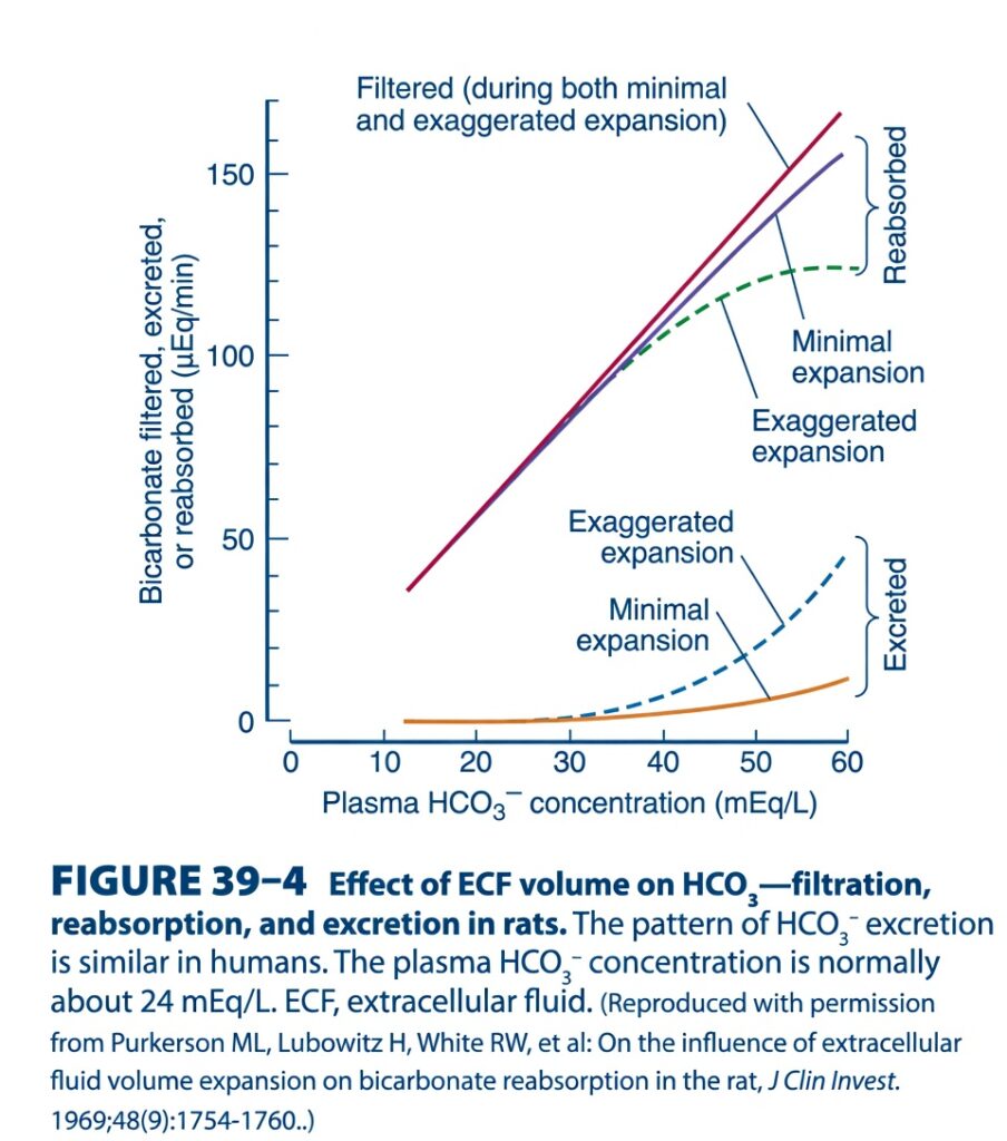
Figure 39-4: Effect of Extracellular Fluid (ECF) Volume on HCO₃⁻ Filtration, Reabsorption, and Excretion
Easiest and Most Conceptual Explanation
⭐ One-Line Concept
The kidneys always filter bicarbonate according to its blood concentration, but how much they reabsorb depends on ECF volume. When ECF expands, the kidneys reabsorb less bicarbonate and excrete more in urine.
Step 1: Understand the Graph Axes
X-axis (Horizontal)
Plasma HCO₃⁻ concentration (mEq/L)
- Shows the bicarbonate level in the blood.
- Moving right means more bicarbonate in plasma.
Y-axis (Vertical)
Bicarbonate filtered, reabsorbed, or excreted (µEq/min)
- Higher on the graph = more bicarbonate being handled by the kidneys.
Step 2: Meaning of Each Curve
1. Red Line – Filtered Bicarbonate
What does it show?
This is the amount of bicarbonate filtered by the glomerulus.
Key point
- As plasma bicarbonate increases, filtered bicarbonate increases in a straight line.
- This line is the same during:
- Minimal ECF expansion
- Exaggerated ECF expansion
Why?
Because filtration mainly depends on:
- Plasma bicarbonate concentration
- GFR
ECF expansion does not significantly change the filtered load in this figure.
Easy concept
More bicarbonate in blood → More bicarbonate enters the kidney filter.
2. Purple Line – Reabsorbed (Minimal Expansion)
What does it show?
How much bicarbonate the kidney reabsorbs when ECF volume is normal or only slightly expanded.
Key points
- Reabsorption rises as more bicarbonate is filtered.
- The kidney reabsorbs almost all filtered bicarbonate.
Only a tiny amount appears in urine.
Easy concept
When body fluid volume is nearly normal, the kidney tries to save bicarbonate.
3. Green Dashed Line – Reabsorbed (Exaggerated Expansion)
What does it show?
Bicarbonate reabsorption during marked ECF expansion.
Why is it lower?
When ECF volume is greatly increased:
- The body does not need to conserve sodium and bicarbonate.
- The proximal tubule decreases bicarbonate reabsorption.
Therefore:
Less bicarbonate returns to the blood.
Easy concept
Too much body fluid → Kidney becomes less interested in saving bicarbonate.
4. Orange Line – Excreted (Minimal Expansion)
What does it show?
Amount of bicarbonate lost in urine during minimal ECF expansion.
Key points
- Very little bicarbonate is excreted.
- Excretion increases only slightly at very high plasma bicarbonate levels.
Easy concept
Normal volume → Almost no bicarbonate is wasted.
5. Blue Dashed Line – Excreted (Exaggerated Expansion)
What does it show?
Bicarbonate excretion during marked ECF expansion.
Key points
- Excretion rises much earlier.
- Much more bicarbonate is lost in urine.
Why?
Because less bicarbonate is reabsorbed.
Easy concept
Expanded ECF → More bicarbonate is thrown away in urine.
Understanding the Brackets
Upper Bracket – “Reabsorbed”
This compares:
- Purple line (minimal expansion)
- Green dashed line (exaggerated expansion)
Meaning
The vertical gap shows:
How much bicarbonate is reabsorbed.
Notice:
The green dashed line is lower.
This means:
Less bicarbonate is reabsorbed during exaggerated ECF expansion.
Lower Bracket – “Excreted”
This compares:
- Orange line
- Blue dashed line
Meaning
The blue dashed line is much higher.
This means:
Much more bicarbonate is excreted when ECF volume is greatly expanded.
Understanding the Solid and Dashed Lines
Solid Lines
Represent:
- Normal filtration
- Minimal ECF expansion
Dashed Green Line
Represents:
Reabsorption during exaggerated ECF expansion
↓
Lower than normal.
Dashed Blue Line
Represents:
Excretion during exaggerated ECF expansion
↓
Higher than normal.hy Doesn’t the Filtered Line Change?
Because:
The glomerulus continues filtering bicarbonate according to:
- Plasma bicarbonate concentration
- GFR
The main change with ECF expansion is tubular reabsorption, not filtration.
What Happens at Normal Plasma HCO₃⁻?
Normal plasma bicarbonate ≈ 24 mEq/L
At this level:
- Almost all filtered bicarbonate is reabsorbed.
- Very little bicarbonate is excreted.
This helps maintain normal blood pH.
What Happens When Plasma HCO₃⁻ Increases?
As plasma bicarbonate rises:
Step 1
More bicarbonate is filtered.
↓
Step 2
The kidney tries to reabsorb it.
↓
Step 3
Eventually, the tubules cannot reabsorb all of it.
↓
Step 4
Extra bicarbonate appears in urine.
What Changes During ECF Expansion?
Normally:
Filtered HCO₃⁻
↓
Most is reabsorbed
↓
Very little is excreted
During exaggerated ECF expansion:
Filtered HCO₃⁻
↓
Less is reabsorbed
↓
Much more is excreted
Why Does ECF Expansion Reduce HCO₃⁻ Reabsorption?
Expanded ECF means:
- More sodium in the body
- More water in the body
The kidney responds by reducing proximal Na⁺ reabsorption.
Because bicarbonate reabsorption is closely linked to sodium reabsorption:
↓ Na⁺ reabsorption
↓
↓ HCO₃⁻ reabsorption
↓
↑ HCO₃⁻ excretion
Simple Story
Imagine bicarbonate is a valuable item being collected by workers.
Normal ECF
The workers collect almost every item.
Almost nothing is left behind.
Expanded ECF
The warehouse is already full.
The workers stop collecting every item.
Many are left behind and thrown away.
That is exactly what happens to bicarbonate during ECF expansion.
Quick Comparison
| Minimal ECF Expansion | Exaggerated ECF Expansion |
|---|---|
| Filtered bicarbonate is normal | Filtered bicarbonate is essentially unchanged |
| Most bicarbonate is reabsorbed | Less bicarbonate is reabsorbed |
| Very little bicarbonate is excreted | Much more bicarbonate is excreted |
| Conserves bicarbonate | Eliminates excess bicarbonate |
Flow Diagram
Normal / Minimal Expansion
Normal plasma HCO₃⁻
↓
Filtered normally
↓
Almost completely reabsorbed
↓
Very little bicarbonate in urine
Exaggerated ECF Expansion
ECF volume increases
↓
Proximal Na⁺ reabsorption decreases
↓
HCO₃⁻ reabsorption decreases
↓
More bicarbonate remains in the tubule
↓
More bicarbonate is excreted in urine
Key Concept (Figure 39-4)
Figure 39-4 shows that the filtered load of bicarbonate increases directly with plasma bicarbonate concentration and is essentially the same during both minimal and exaggerated ECF expansion. The major difference is in tubular reabsorption. During minimal ECF expansion, the kidneys reabsorb nearly all filtered bicarbonate, so only a very small amount is excreted. During exaggerated ECF expansion, proximal tubular sodium and bicarbonate reabsorption decrease, causing the reabsorbed bicarbonate curve to fall and the excreted bicarbonate curve to rise. Thus, ECF expansion promotes bicarbonate loss in urine without significantly changing the amount filtered, helping regulate extracellular fluid volume and acid–base balance.
CLINICAL BOX 39–1
Implications of Urinary pH Changes
- The urine pH in humans normally ranges from 4.5 to 8.0.
- The urine pH depends on the combined effects of:
- H⁺ secretion
- NH₄⁺ production
- HCO₃⁻ excretion
- Urine with a pH different from body fluids plays an important role in maintaining electrolyte balance and acid–base balance.
- When acid is added to the body, it is buffered in the plasma and cells.
- The overall buffering reaction is: HA + NaHCO₃ → NaA + H₂CO₃
- H₂CO₃ is then converted into CO₂ and H₂O.
- The CO₂ is exhaled through the lungs.
- NaA enters the glomerular filtrate.
- When Na⁺ in the urine is replaced by H⁺, Na⁺ is conserved in the body.
- For every H⁺ excreted with phosphate or as NH₄⁺, one new HCO₃⁻ is added to the blood.
- This replenishes the body’s bicarbonate buffer.
- When base is added to the body fluids, OH⁻ is buffered.
- This increases the plasma HCO₃⁻ concentration.
- When plasma HCO₃⁻ rises above 28 mEq/L, the urine becomes alkaline.
- The excess HCO₃⁻ is then excreted in the urine.
- The maximum rate of H⁺ secretion by the renal tubules increases as arterial PCO₂ increases.
- Therefore, HCO₃⁻ reabsorption is also affected by arterial PCO₂.
- This relationship is explained in more detail in the main text.
KEY CONCEPT
- Urine pH changes help maintain acid–base balance. During acidosis, the kidneys conserve Na⁺, excrete H⁺ with phosphate and NH₄⁺, and add new HCO₃⁻ to the blood. During alkalosis, excess HCO₃⁻ is excreted, making the urine alkaline. H⁺ secretion and HCO₃⁻ reabsorption are also influenced by arterial PCO₂.
Conceptual Examples
- Example 1: During metabolic acidosis, the kidneys excrete H⁺ as NH₄⁺ and phosphate, while new HCO₃⁻ enters the blood, helping restore normal pH.
- Example 2: During metabolic alkalosis, plasma HCO₃⁻ rises above 28 mEq/L, so the kidneys excrete excess HCO₃⁻, making the urine alkaline.
- Example 3: During respiratory acidosis (high PCO₂), the kidneys increase H⁺ secretion and HCO₃⁻ reabsorption, helping compensate for the increased acid load.
THERAPEUTIC HIGHLIGHTS
Sulfonamides inhibit carbonic anhydrase and sulfonamide
derivatives have been used clinically as diuretics because of
their inhibitory effects on carbonic anhydrase in the kidney
(see Chapter 37).
DEFENSE OF H⁺ CONCENTRATION
- The main goal of acid–base balance is to maintain the normal H⁺ concentration in the extracellular fluid (ECF).
- The main problem is not buffer base, fixed cation, or similar terms.
- The real focus is maintaining a normal H⁺ concentration in the ECF.
- The mechanisms that regulate the composition of the ECF are especially important for H⁺.
- This is because the cells of the body are very sensitive to changes in H⁺ concentration.
- The intracellular H⁺ concentration is different from the extracellular H⁺ concentration.
- Intracellular H⁺ concentration can be measured using:
- Microelectrodes
- pH-sensitive fluorescent dyes
- Phosphorus magnetic resonance
- Intracellular H⁺ concentration is regulated by several intracellular processes.
- However, it is also affected by changes in the H⁺ concentration of the ECF.
- The pH scale is a useful way to express H⁺ concentration in the body.
- This is because the H⁺ concentration is extremely low compared with other positively charged ions (cations).
- The normal Na⁺ concentration in arterial plasma is about 140 mEq/L.
- The normal H⁺ concentration is about 0.00004 mEq/L.
Table: Table 39–1
- pH is the negative logarithm of the H⁺ concentration.
- For an H⁺ concentration of 0.00004 mEq/L, the pH is 7.4.
- A decrease in pH by 1 unit means a 10-fold increase in H⁺ concentration.
- For example, when pH decreases from 7.0 to 6.0, the H⁺ concentration increases 10 times.
- The pH of blood refers to the pH of true plasma.
- True plasma is plasma that has reached equilibrium with red blood cells.
- Red blood cells contain hemoglobin.
- Hemoglobin is one of the most important buffers in the blood.
KEY CONCEPT
- The primary purpose of acid–base regulation is to keep the H⁺ concentration of the extracellular fluid within the normal range. Even small changes in H⁺ can affect cell function. Because H⁺ concentration is very low, it is expressed as pH. A decrease of one pH unit represents a 10-fold increase in H⁺ concentration. Hemoglobin in red blood cells is an important blood buffer that helps maintain normal pH.
Conceptual Examples
- Example 1: When blood pH falls from 7.4 to 7.3, the H⁺ concentration increases, making the body more acidic.
- Example 2: If blood pH decreases from 7.0 to 6.0, the H⁺ concentration increases 10 times, showing that even a small pH change represents a large change in acidity.
- Example 3: Hemoglobin inside red blood cells binds H⁺ and acts as an important buffer, helping keep the blood pH close to 7.4.
H⁺ BALANCE
- The normal pH of arterial plasma is 7.40.
- The pH of venous plasma is slightly lower than arterial plasma.
- Acidosis is present when the arterial pH is below 7.40.
- Alkalosis is present when the arterial pH is above 7.40.
- Small changes of up to 0.05 pH unit usually do not cause harmful effects.
- The H⁺ concentration compatible with life ranges from 0.00002 mEq/L to 0.0001 mEq/L.
- This corresponds to a pH range of 7.70 to 7.00.
- In the liver, amino acids are used for gluconeogenesis.
- This process produces NH₄⁺ and HCO₃⁻ from the amino and carboxyl groups of amino acids.
Figure: Figure 39–5
- Most NH₄⁺ is converted into urea.
- The H⁺ produced during this process is buffered inside the cells by HCO₃⁻.
- Therefore, very little NH₄⁺ and HCO₃⁻ enter the bloodstream.
- Metabolism of sulfur-containing amino acids produces sulfuric acid (H₂SO₄).
- Metabolism of phosphorylated amino acids, such as phosphoserine, produces phosphoric acid (H₃PO₄).
- These strong acids enter the bloodstream.
- They produce a major H⁺ load that must be buffered in the extracellular fluid (ECF).
- The normal H⁺ load from amino acid metabolism is about 50 mEq/day.
- Tissue metabolism also produces CO₂.
- Most CO₂ combines with water to form H₂CO₃.
- This produces a total H⁺ load of more than 12,500 mEq/day.
- Most CO₂ is removed by the lungs.
- Therefore, only a small amount of H⁺ remains to be excreted by the kidneys.
- Extra acid loads commonly occur during strenuous exercise because of lactic acid production.
- Diabetic ketosis produces acetoacetic acid and β-hydroxybutyric acid.
- Acidifying salts, such as NH₄Cl and CaCl₂, also increase the body’s acid load.
- These salts have the same overall effect as adding HCl to the body.
- Diseased kidneys can also cause acidosis because they cannot excrete normal amounts of acid.
- Fruits are the main dietary source of alkali.
- Fruits contain sodium (Na⁺) and potassium (K⁺) salts of weak organic acids.
- These organic acid anions are metabolized to CO₂.
- This leaves NaHCO₃ and KHCO₃ in the body.
- These substances increase the body’s alkali.
- NaHCO₃ and other alkalinizing salts may also be taken in large amounts.
- A more common cause of alkalosis is loss of gastric acid during vomiting.
- Vomiting removes gastric juice rich in HCl.
- Loss of HCl has the same effect as adding alkali to the body.
KEY CONCEPT
- The body normally maintains arterial blood pH around 7.40. Acids are continuously produced from amino acid metabolism, CO₂ production, exercise, diabetes, and acidifying salts. Most CO₂ is removed by the lungs, while the kidneys excrete the remaining H⁺. Alkali mainly comes from fruits and alkalinizing salts. Vomiting causes alkalosis because loss of HCl is equivalent to adding alkali to the body.
Conceptual Examples
- Example 1: During strenuous exercise, lactic acid is produced, increasing the body’s H⁺ load.
- Example 2: In diabetic ketoacidosis, acetoacetic acid and β-hydroxybutyric acid increase the body’s acidity.
- Example 3: A patient with chronic kidney disease cannot excrete enough H⁺, leading to acidosis.
- Example 4: A patient with persistent vomiting loses HCl from the stomach, so the body becomes alkalotic because losing acid is equivalent to gaining alkali.
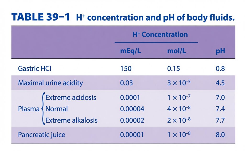
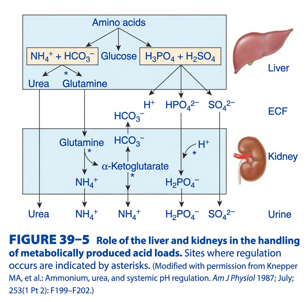
Figure 39-5: Role of the Liver and Kidneys in Handling Metabolically Produced Acid Loads
Easiest and Most Conceptual Explanation
⭐ One-Line Concept
The liver produces acids and determines whether nitrogen becomes urea or glutamine, while the kidneys remove acid from the body and return new bicarbonate (HCO₃⁻) to the blood to maintain normal pH.
First Understand the Layout
The figure is divided into three levels.
Top Box
Liver
The liver processes amino acids.
Middle Space
ECF (Extracellular Fluid)
This is the fluid between the liver and kidneys where substances travel in the blood.
Bottom Box
Kidney
The kidney removes acid from the body and regulates blood bicarbonate.
Step 1: What Happens in the Liver?
Everything starts with:
Amino Acids
The liver metabolizes amino acids.
This produces three important products.
Pathway 1
NH₄⁺ + HCO₃⁻
Ammonium and bicarbonate are produced.
From here, the liver has two choices.
Option A
Convert NH₄⁺ into:
Urea
Urea is released into the blood.
↓
Kidney
↓
Urine
Option B
Convert NH₄⁺ into:
Glutamine
Glutamine is released into the blood.
↓
Kidney
↓
Used for ammonium production.
Pathway 2
Glucose
Some amino acids are converted into glucose.
This pathway is shown only for completeness and is not directly involved in acid excretion.
Pathway 3
H₃PO₄ + H₂SO₄
Metabolism of sulfur- and phosphorus-containing amino acids produces:
- Phosphoric acid (H₃PO₄)
- Sulfuric acid (H₂SO₄)
These are fixed (non-volatile) acids.
They cannot be removed by the lungs.
Therefore,
They must be excreted by the kidneys.
Step 2: What Enters the ECF?
The liver releases several substances into the extracellular fluid.
These include:
- Urea
- Glutamine
- H⁺
- HCO₃⁻
- HPO₄²⁻
- SO₄²⁻
These substances travel through the blood to the kidneys.
Step 3: What Happens in the Kidney?
The kidney receives these substances and processes them in different ways.
Pathway 1
Glutamine
Glutamine enters the kidney.
Inside the kidney,
Glutamine is metabolized.
This produces:
- NH₄⁺
- α-Ketoglutarate
NH₄⁺
Ammonium is secreted into urine.
This removes acid from the body.
α-Ketoglutarate
α-Ketoglutarate is metabolized.
This generates:
HCO₃⁻
The bicarbonate enters the blood.
Therefore,
The kidney adds new bicarbonate to the body.
Pathway 2
Phosphate Buffer
Phosphate arrives as:
HPO₄²⁻
Inside the tubular lumen,
Secreted H⁺ combines with phosphate.
HPO₄²⁻ + H⁺
↓
H₂PO₄⁻
This is called:
Titratable acid
It is excreted in urine.
Pathway 3
Sulfate
Sulfate reaches the kidney as:
SO₄²⁻
It is excreted in urine.
It represents sulfuric acid that has already been neutralized.
Pathway 4
Urea
Urea enters the kidney.
↓
Filtered
↓
Excreted in urine.
Understanding Every Arrow
Arrow: Amino Acids → NH₄⁺ + HCO₃⁻
The liver produces ammonium and bicarbonate during amino acid metabolism.
Arrow: NH₄⁺ + HCO₃⁻ → Urea
The liver converts ammonium into urea.
This is the normal pathway.
Arrow: NH₄⁺ + HCO₃⁻ → Glutamine
The liver can also package ammonium into glutamine.
Glutamine safely transports ammonia to the kidneys.
Arrow: H₃PO₄ + H₂SO₄ → H⁺
These strong acids release hydrogen ions.
Arrow: H₃PO₄ + H₂SO₄ → HPO₄²⁻
Phosphate buffer reaches the kidney.
Arrow: H₃PO₄ + H₂SO₄ → SO₄²⁻
Sulfate reaches the kidney.
Arrow: Glutamine → NH₄⁺
The kidney produces ammonium.
This is excreted.
Arrow: Glutamine → α-Ketoglutarate
This intermediate enters metabolism.
Arrow: α-Ketoglutarate → HCO₃⁻
New bicarbonate is produced.
This bicarbonate enters the blood.
Arrow: HPO₄²⁻ + H⁺ → H₂PO₄⁻
Phosphate buffers H⁺.
The acid is safely excreted.
Meaning of the Asterisks (*)
The figure states:
“Sites where regulation occurs are indicated by asterisks.”
The asterisks mark important regulatory points.
Asterisk 1
Liver
Choice between:
- Urea production
- Glutamine production
During acidosis:
↓ Urea formation
↑ Glutamine formation
This provides more glutamine for the kidneys.
Asterisk 2
Kidney
Glutamine metabolism
During acidosis:
The kidney increases glutamine metabolism.
↓
Produces more NH₄⁺.
↓
Excretes more acid.
Asterisk 3
α-Ketoglutarate Metabolism
Regulates production of new bicarbonate.
More metabolism
↓
More HCO₃⁻ enters blood.
Asterisk 4
Phosphate Buffer
Regulates how much H⁺ is buffered by phosphate.
Why Does Acidosis Increase Glutamine?
Imagine the body has too much acid.
Instead of making urea,
the liver sends more glutamine to the kidneys.
The kidneys then convert glutamine into:
- NH₄⁺ (acid removal)
- HCO₃⁻ (base replacement)
Thus,
Both acid removal and bicarbonate production increase.
Easy Story
Imagine:
The Liver = Factory
The factory processes amino acids.
It can pack nitrogen into:
- Urea
- Glutamine
The Blood = Delivery Truck
It transports:
- Glutamine
- Urea
- Phosphate
- Sulfate
to the kidneys.
The Kidney = Waste Disposal Plant
The kidney:
- Throws acid into urine.
- Creates new bicarbonate.
- Sends bicarbonate back into the blood.
This keeps blood pH normal.
Simple Flow Diagram
Liver
Amino acids
↓
NH₄⁺ + HCO₃⁻
↓
Two pathways
- Urea → Kidney → Urine
- Glutamine → Kidney
↓
Glutamine
↓
Kidney
↓
NH₄⁺ → Urine
↓
α-Ketoglutarate
↓
HCO₃⁻
↓
Bloodixed Acid Pathway
Amino acids
↓
H₃PO₄ + H₂SO₄
↓
H⁺ + HPO₄²⁻ + SO₄²⁻
↓
Kidney
↓
HPO₄²⁻ + H⁺
↓
H₂PO₄⁻
↓
Urine
↓
SO₄²⁻
↓
Urine
High-Yield Summary Table
| Substance | Liver | Kidney | Final Result |
|---|---|---|---|
| Urea | Produced | Excreted | Removes nitrogen |
| Glutamine | Produced | Converted to NH₄⁺ | Removes acid |
| NH₄⁺ | Produced indirectly | Excreted | Acid elimination |
| α-Ketoglutarate | — | Metabolized | Produces new HCO₃⁻ |
| HCO₃⁻ | Produced | Returned to blood | Replaces lost buffer |
| HPO₄²⁻ | Delivered to kidney | Buffers H⁺ | Excreted as H₂PO₄⁻ |
| SO₄²⁻ | Produced from sulfuric acid | Excreted | Removes fixed acid |
Key Concept (Figure 39-5)
Figure 39-5 illustrates the coordinated role of the liver and kidneys in maintaining acid–base balance. The liver metabolizes amino acids, producing ammonium (NH₄⁺), bicarbonate (HCO₃⁻), glucose, phosphoric acid (H₃PO₄), and sulfuric acid (H₂SO₄). Nitrogen is either converted into urea or packaged as glutamine. Glutamine is transported to the kidneys, where it is metabolized to produce NH₄⁺, which is excreted in urine, and α-ketoglutarate, which generates new bicarbonate (HCO₃⁻) that returns to the bloodstream. The kidneys also excrete H₂PO₄⁻ (titratable acid) and SO₄²⁻, removing fixed acids from the body. During acidosis, the liver shifts toward glutamine production, and the kidneys increase ammonium excretion and bicarbonate generation, helping restore normal blood pH.
MADE BY EASIEST AND SELF LKEARNING CEO AND FOUNDER DR SHEEN.
