



STRUCTURE
- Scientists have long believed that the body contains hormones that increase sodium (Na⁺) excretion (natriuretic hormones).
- Two important natriuretic hormones are produced by the heart.
- The muscle cells of the atria contain secretory granules.
- The ventricles also contain secretory granules, but in much smaller amounts.
- The number of these granules increases when:
- Sodium chloride (NaCl) intake increases.
- Extracellular fluid (ECF) volume expands.
- Extracts from atrial tissue increase sodium excretion (natriuresis).
Atrial Natriuretic Peptide (ANP)
- The first natriuretic hormone discovered in the heart was Atrial Natriuretic Peptide (ANP).
- ANP is a polypeptide hormone.
- It contains a characteristic ring made of 17 amino acids.
- This ring is formed by a disulfide bond between two cysteine amino acids.
- The circulating form of ANP contains 28 amino acids.
- ANP is produced from a larger precursor protein containing 151 amino acids.
- This precursor includes a 24-amino-acid signal peptide.
- ANP is also found in other tissues, including the brain.
- In the brain, ANP exists in two smaller forms than the circulating hormone.
B-Type Natriuretic Peptide (BNP)
- A second natriuretic hormone is B-type Natriuretic Peptide (BNP), also called Brain Natriuretic Peptide.
- BNP was first isolated from the brain of pigs.
- In humans, BNP is present in both the brain and the heart.
- The human heart, especially the ventricles, contains more BNP than the brain.
- The circulating form of BNP contains 32 amino acids.
- BNP has the same 17-amino-acid ring as ANP.
- However, some amino acids within the ring are different from those in ANP.
C-Type Natriuretic Peptide (CNP)
- The third member of this hormone family is C-type Natriuretic Peptide (CNP).
- CNP was named because it was the third natriuretic peptide to be discovered.
- One form of CNP contains 22 amino acids.
- Another larger form contains 53 amino acids.
- CNP is found in:
- Brain
- Pituitary gland
- Kidneys
- Vascular endothelial cells
- Very little CNP is present in the heart or in the bloodstream.
- CNP mainly acts as a paracrine hormone, meaning it acts locally on nearby cells rather than through the circulation.
KEY CONCEPT
- The heart produces natriuretic hormones that help regulate body fluid volume and sodium balance. The main members are Atrial Natriuretic Peptide (ANP), B-type Natriuretic Peptide (BNP), and C-type Natriuretic Peptide (CNP). ANP is mainly produced by the atria, BNP is abundant in the ventricles, and CNP mainly acts locally in tissues as a paracrine mediator rather than as a circulating hormone.
Conceptual Examples
- Example 1: After drinking a large amount of salty fluid, the atria stretch because of increased blood volume. This stimulates the release of ANP, which promotes sodium and water excretion, helping return blood volume to normal.
- Example 2: In heart failure, the ventricles become stretched and release more BNP. Clinically, BNP levels are measured in the blood to help assess the severity of heart failure, while CNP mainly acts locally within tissues and contributes very little to the circulating hormone levels.
ACTIONS
- ANP (Atrial Natriuretic Peptide) and BNP (B-type Natriuretic Peptide) circulate in the blood and act mainly on the kidneys.
- Injected CNP (C-type Natriuretic Peptide) produces similar effects.
Effects on the Kidneys
- ANP, BNP, and CNP increase sodium (Na⁺) excretion in the urine (natriuresis).
- They dilate the afferent arterioles of the glomerulus.
- They relax the mesangial cells of the glomerulus.
- Both of these actions increase the Glomerular Filtration Rate (GFR).
- They also act directly on the renal tubules.
- In the renal tubules, they decrease sodium (Na⁺) reabsorption.
- As a result, more sodium and water are excreted in the urine.
Effects on Blood Vessels and Blood Pressure
- These peptides increase capillary permeability.
- More fluid moves from the blood into the tissues (extravasation).
- This decreases blood volume and lowers blood pressure.
- They relax vascular smooth muscle in both:
- Arterioles
- Venules
- CNP produces greater venous dilation than ANP and BNP.
Effects on Hormones
- ANP, BNP, and CNP inhibit renin secretion.
- They reduce the activity of the renin–angiotensin–aldosterone system (RAAS).
- They also oppose the blood pressure–raising (pressor) effects of:
- Catecholamines
- Angiotensin II
Effects in the Brain
- ANP is present in neurons of the brain.
- ANP-containing nerve pathways extend from the anteromedial hypothalamus to cardiovascular control centres in the lower brainstem.
- In the brain, ANP generally produces effects opposite to those of angiotensin II.
- ANP helps oppose the actions of:
- Vasopressin (ADH)
- Angiotensin II
- These neural pathways promote sodium excretion (natriuresis).
KEY CONCEPT
- ANP, BNP, and CNP help reduce blood volume and blood pressure. They increase sodium and water excretion by increasing GFR and decreasing sodium reabsorption in the renal tubules. They also dilate blood vessels, inhibit renin secretion, oppose the effects of angiotensin II and catecholamines, and, in the brain, ANP counteracts the actions of vasopressin and angiotensin II to promote natriuresis.
Conceptual Examples
- Example 1: After receiving a large intravenous saline infusion, the atria stretch and release ANP. ANP dilates the afferent arteriole, increases GFR, reduces sodium reabsorption, and increases sodium and water excretion, helping restore normal blood volume.
- Example 2: A patient with high blood pressure and excess body fluid has increased ANP and BNP release. These hormones inhibit renin, oppose angiotensin II, relax blood vessels, and promote natriuresis, helping lower blood pressure and extracellular fluid volume.
NATRIURETIC PEPTIDE RECEPTORS
- There are three main types of natriuretic peptide receptors (NPRs):
- NPR-A
- NPR-B
- NPR-C
NPR-A Receptor
- NPR-A is a cell membrane receptor.
- It has a cytoplasmic domain that functions as guanylyl cyclase.
- When activated, it produces cyclic guanosine monophosphate (cGMP).
- ANP (Atrial Natriuretic Peptide) has the highest affinity for the NPR-A receptor.
NPR-B Receptor
- NPR-B is also a cell membrane receptor.
- Like NPR-A, it contains a cytoplasmic guanylyl cyclase domain.
- Activation of NPR-B also increases intracellular cGMP.
- CNP (C-type Natriuretic Peptide) has the highest affinity for the NPR-B receptor.
NPR-C Receptor
- NPR-C is the third type of natriuretic peptide receptor.
- It binds all three natriuretic peptides:
- ANP
- BNP
- CNP
- Unlike NPR-A and NPR-B, NPR-C has a very short (truncated) cytoplasmic domain.
- Some studies suggest that NPR-C acts through G-proteins.
- Through G-proteins, NPR-C may:
- Activate phospholipase C (PLC).
- Inhibit adenylyl cyclase.
- However, the exact role of NPR-C is still debated.
- Many researchers believe that NPR-C mainly functions as a clearance receptor.
- As a clearance receptor, NPR-C removes natriuretic peptides from the bloodstream.
- It can later release these peptides, helping maintain a stable blood concentration of the hormones.
KEY CONCEPT
- Natriuretic peptides act through three receptors. NPR-A mainly binds ANP, and NPR-B mainly binds CNP. Both receptors activate guanylyl cyclase and increase intracellular cGMP, producing the biological effects of the hormones. NPR-C binds ANP, BNP, and CNP but mainly functions as a clearance receptor that helps regulate circulating natriuretic peptide levels, although it may also have limited intracellular signaling through G-proteins.
Conceptual Examples
- Example 1: When ANP is released because of increased blood volume, it binds mainly to NPR-A, increasing cGMP inside target cells. This promotes natriuresis, vasodilation, and a reduction in blood pressure.
- Example 2: After ANP, BNP, and CNP have produced their effects, NPR-C binds these hormones and removes them from the bloodstream, helping maintain stable circulating hormone levels and preventing excessive hormonal activity.
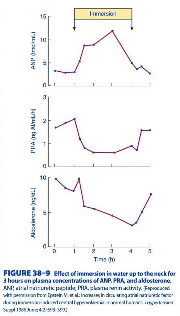
Figure 38-9 – Effect of Neck-Deep Water Immersion on ANP, Plasma Renin Activity (PRA), and Aldosterone
Easy Conceptual Explanation (Step-by-Step)
This figure demonstrates how immersing a person in water up to the neck for 3 hours affects three important hormones that regulate blood volume, sodium, and water balance.
The graph teaches one simple physiological principle:
Water immersion shifts blood from the legs into the chest (central circulation), making the body think it has too much blood.
The body responds by:
- Increasing ANP (to remove salt and water).
- Suppressing Renin (PRA).
- Suppressing Aldosterone.
First Understand What Happens During Water Immersion (Completely Submerging something in liquid)
Imagine standing in water up to your neck.
The surrounding water presses on your legs and abdomen.
↓
Blood is squeezed from the veins of the legs toward the chest.
↓
More blood reaches:
- Right atrium
- Left atrium
- Heart
↓
The atria become stretched.
The body interprets this as:
“Blood volume is too high!”
Even though the total blood volume has not changed.
This is called:
Central Hypervolemia
Timeline
The yellow bar at the top shows:
Immersion lasted from approximately 1 hour to 4 hours.
Everything happening inside this period is the body’s response to central hypervolemia.
Graph 1 – ANP (Top Graph)
Y-axis
ANP concentration (fmol/mL)
Higher graph
↓
More ANP released.
Before Immersion
ANP is around 3 fmol/mL.
Normal resting level.
During Immersion
The curve suddenly rises.
Why?
Because:
Water pressure
↓
Blood shifts into chest
↓
Atria stretch
↓
ANP secretion increases.
Notice:
The highest point occurs around 3 hours.
This is because:
Maximum atrial stretch
↓
Maximum ANP release.
After Immersion Ends
Water pressure disappears.
↓
Blood redistributes normally.
↓
Atrial stretch decreases.
↓
ANP falls back toward normal.
Easy Memory
More atrial stretch = More ANP
Why Does ANP Increase?
ANP stands for:
Atrial Natriuretic Peptide
Released from:
Atrial muscle cells
Stimulus:
Atrial stretching.
Purpose:
Get rid of excess volume.
ANP causes:
✅ Sodium excretion
↓
Water follows sodium
↓
Urine increases
↓
Blood volume decreases.
Graph 2 – Plasma Renin Activity (PRA)
Now look at the middle graph.
Before Immersion
PRA is relatively high.
Around:
2 ng AI/mL/h.
Normal resting value.
During Immersion
PRA falls dramatically.
Why?
Because the kidneys think:
“There is already plenty of blood.”
Therefore:
Renin secretion is inhibited.
Flow:
Central hypervolemia
↓
Renal perfusion appears adequate
↓
Less renin released
↓
PRA falls.
Notice
The lowest PRA occurs while immersion continues.
After Immersion
Blood shifts back to the legs.
↓
Central volume decreases.
↓
Kidneys restart renin secretion.
↓
PRA rises again.
Easy Memory
More blood volume → Less renin
Why Does Renin Fall?
Normally renin is released when:
- Blood pressure decreases
- Blood volume decreases
- Sodium delivery decreases
Here,
None of these occur.
Instead,
The kidney senses:
“I have plenty of blood.”
↓
No need to conserve sodium.
↓
Renin secretion stops.
Graph 3 – Aldosterone
Now look at the bottom graph.
Before Immersion
Aldosterone is relatively high.
During Immersion
It steadily falls.
Why?
Because:
Renin falls.
↓
Angiotensin II falls.
↓
Less stimulation of adrenal cortex.
↓
Less aldosterone secretion.
Remember the sequence.
Renin
↓
Angiotensin I
↓
Angiotensin II
↓
Aldosterone.
If renin decreases,
everything downstream decreases.
After Immersion
Renin begins increasing.
↓
Angiotensin II increases.
↓
Aldosterone also rises.
Why Does Aldosterone Decrease?
Normally aldosterone saves sodium.
During immersion,
the body wants to lose sodium.
Therefore,
it suppresses aldosterone.
Put All Three Graphs Together
Step 1
Water immersion
↓
Compression of legs
↓
Blood moves to chest.
Step 2
Atria stretch.
↓
ANP increases.
Step 3
Kidneys detect adequate circulation.
↓
Renin decreases.
Step 4
Less renin
↓
Less angiotensin II
↓
Less aldosterone.
Step 5
Overall result
The kidneys excrete:
- More sodium
- More water
↓
Blood volume returns toward normal.
Complete Flow Diagram
Neck-deep water immersion
            ↓
Blood shifts from legs to chest
            ↓
Central hypervolemia
            ↓
Atrial stretch
            ↓
↑ ANP
            ↓
↓ Renin
            ↓
↓ Angiotensin II
            ↓
↓ Aldosterone
            ↓
↓ Sodium reabsorption
            ↓
↑ Sodium excretion
            ↓
↑ Water excretion
            ↓
Blood volume returns toward normal
Easy Story
Imagine your heart is a water tank.
Normally,
it contains the correct amount of water.
Suddenly,
water immersion squeezes extra blood into the tank.
The heart says:
“I’m too full!”
So it presses the emergency drain button.
That drain button is:
ANP
At the same time,
it switches OFF the water-saving system:
- Renin OFF
- Angiotensin II OFF
- Aldosterone OFF
Result:
The kidneys flush out extra salt and water.
Comparison Table
| During Water Immersion | What Happens? | Why? |
|---|---|---|
| Blood shifts to chest | ↑ Central blood volume | External water pressure compresses peripheral veins |
| Atrial stretch | ↑ | More venous return |
| ANP | ↑↑ | Stretch of atria stimulates ANP release |
| Renin (PRA) | ↓ | Kidney senses adequate circulating volume |
| Angiotensin II | ↓ | Less renin available |
| Aldosterone | ↓ | Less angiotensin II stimulation |
| Sodium excretion | ↑ | ANP increases natriuresis and low aldosterone reduces sodium reabsorption |
| Water excretion | ↑ | Water follows sodium, producing diuresis |
High-Yield Exam Points
- Neck-deep water immersion causes central hypervolemia by shifting blood from the peripheral veins to the thoracic circulation.
- Atrial stretch stimulates ANP secretion.
- ANP promotes natriuresis and diuresis.
- Central hypervolemia suppresses renin release, reducing plasma renin activity (PRA).
- Reduced renin decreases angiotensin II and aldosterone secretion.
- After immersion ends, ANP falls while renin and aldosterone return toward baseline.
One-Line Summary
Neck-deep water immersion creates central hypervolemia, causing atrial stretch that increases ANP while simultaneously suppressing renin and aldosterone, leading to increased sodium and water excretion to restore normal blood volume.
Key Concept
Figure 38-9 illustrates the hormonal response to central hypervolemia produced by neck-deep water immersion. Hydrostatic pressure shifts blood from the lower limbs into the thoracic circulation, increasing venous return and atrial stretch. This stimulates atrial natriuretic peptide (ANP) secretion, which promotes natriuresis and diuresis. At the same time, the perceived increase in effective circulating volume suppresses plasma renin activity (PRA), leading to reduced angiotensin II formation and decreased aldosterone secretion. The combined effect is increased renal excretion of sodium and water, reducing circulating volume back toward normal. When immersion ends, central blood volume decreases, ANP levels fall, and renin and aldosterone gradually return to baseline.
A, K ATPase-INHIBITING FACTOR
- Another natriuretic factor is present in the blood.
- This factor increases sodium (Na⁺) excretion (natriuresis).
- It produces this effect by inhibiting the sodium–potassium ATPase (Na⁺, K⁺-ATPase) pump.
- Unlike ANP and BNP, this factor raises blood pressure instead of lowering it.
- Current evidence suggests that this factor may be the digitalis-like steroid called ouabain.
- It is believed to be produced by the adrenal glands.
- Its exact physiological role is still not fully understood.
DEFENSE OF SPECIFIC IONIC COMPOSITION
- The body has special regulatory mechanisms to maintain the normal concentration of specific ions in the extracellular fluid (ECF).
- These mechanisms also regulate glucose and other important non-ionized substances required for metabolism.
Regulation of Calcium (Ca²⁺)
- The ionized calcium (Ca²⁺) concentration in the ECF is tightly regulated.
- Calcium levels are maintained by feedback mechanisms involving:
- Parathyroid glands
- Calcitonin-secreting cells
- These glands adjust the secretion of Parathyroid Hormone (PTH) and calcitonin to keep calcium levels normal.
Regulation of Magnesium (Mg²⁺)
- The concentration of magnesium (Mg²⁺) is also closely regulated.
- However, the exact mechanisms controlling magnesium homeostasis are not yet completely understood.
Regulation of Sodium (Na⁺) and Potassium (K⁺)
- The regulation of sodium (Na⁺) and potassium (K⁺) is closely linked to:
- Extracellular fluid (ECF) volume
- Extracellular fluid tonicity (osmolarity)
- These regulatory mechanisms have already been discussed earlier in the chapter.
Effect of Hydrogen Ions (H⁺)
- The concentrations of sodium (Na⁺) and potassium (K⁺) are also influenced by the hydrogen ion (H⁺) concentration.
- Changes in H⁺ concentration alter the body’s acid–base balance (pH).
- The pH is one of the major factors determining the composition of anions in the extracellular fluid (ECF).
- The regulation of acid–base balance is discussed in Chapter 39.
KEY CONCEPT
- Besides ANP and BNP, another circulating natriuretic factor inhibits the Na⁺, K⁺-ATPase pump, increases sodium excretion, and may raise blood pressure. The body also maintains a constant extracellular ionic composition through specific regulatory mechanisms. Calcium is controlled by PTH and calcitonin, magnesium is tightly regulated by mechanisms that are not yet fully understood, sodium and potassium are regulated mainly through ECF volume and osmolarity, and hydrogen ion concentration (pH) influences the balance of many extracellular ions.
Conceptual Examples
- Example 1: A circulating ouabain-like factor inhibits the Na⁺, K⁺-ATPase pump, causing increased sodium excretion, but unlike ANP, it can also increase blood pressure.
- Example 2: When blood calcium decreases, the parathyroid glands release Parathyroid Hormone (PTH), while calcitonin secretion decreases, helping restore the normal calcium concentration in the extracellular fluid.
MADE BY EASIEST SELF LEARNING CEO AND FOUNDER DR SHEEN.
