


- Buffering is essential for maintaining a normal H⁺ concentration (H⁺ homeostasis).
- Buffering is defined in Chapter 1.
- It is discussed in Chapter 36 in relation to gas transport.
- This discussion emphasizes the roles of:
- Proteins
- Hemoglobin
- The carbonic anhydrase system in the blood
- Carbonic anhydrase is also present in high concentrations in gastric acid-secreting cells.
- Carbonic anhydrase is also present in renal tubular cells.
- Carbonic anhydrase is a protein with a molecular weight of 30,000.
- Each carbonic anhydrase molecule contains one zinc atom.
- Carbonic anhydrase is inhibited by:
- Cyanide
- Azide
- Sulfide
- In the body, buffering is not limited to the blood.
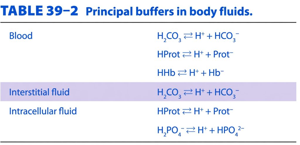
- The main buffers in the blood, interstitial fluid, and intracellular fluid are listed in Table 39–2.
Table: Table 39–2
- The main buffers in cerebrospinal fluid and urine are the bicarbonate and phosphate buffer systems.
- In metabolic acidosis, only about 15–20% of the acid load is buffered by the H₂CO₃–HCO₃⁻ system in the extracellular fluid (ECF).
- Most of the remaining acid is buffered inside the cells.
- In metabolic alkalosis, about 30–35% of the OH⁻ load is buffered inside the cells.
- In respiratory acidosis and respiratory alkalosis, almost all buffering occurs inside the cells.
- In animal cells, the main regulators of intracellular pH are HCO₃⁻ transporters.
- These transporters include:
- The Cl⁻–HCO₃⁻ exchanger (AE1)
- Three Na⁺–HCO₃⁻ cotransporters
- One K⁺–HCO₃⁻ cotransporter
KEY CONCEPT
- Buffering is the body’s first defense against changes in H⁺ concentration. Although blood buffers are important, most buffering during metabolic and respiratory acid–base disorders occurs inside the cells. Carbonic anhydrase plays a key role in buffering and is abundant in blood, gastric cells, and renal tubular cells. Intracellular pH is mainly regulated by HCO₃⁻ transporters.
Conceptual Examples
- Example 1: During metabolic acidosis, only 15–20% of the excess acid is buffered by the ECF bicarbonate system, while most buffering occurs inside the cells.
- Example 2: During respiratory acidosis, almost all buffering occurs intracellularly, helping reduce changes in blood H⁺ concentration.
- Example 3: Carbonic anhydrase rapidly converts CO₂ and H₂O into H₂CO₃ and back again, helping maintain normal acid–base balance in the blood, stomach, and kidneys.
SUMMARY
- When a strong acid is added to the blood, the main buffer reactions shift to the left.
- As a result, the blood levels of the three main buffer anions decrease:
- Hemoglobin (Hb⁻)
- Protein (Prot⁻)
- Bicarbonate (HCO₃⁻)
- The anions of the added acid are filtered into the renal tubules.
- These anions are accompanied mainly by sodium (Na⁺) to maintain electrical neutrality.
- The renal tubules replace Na⁺ with H⁺.
- At the same time, equal amounts of Na⁺ and HCO₃⁻ are reabsorbed into the blood.
- This process conserves Na⁺.
- It also removes acid from the body.
- It restores the normal level of buffer anions in the blood.
- When CO₂ is added to the blood, similar reactions occur.
- However, CO₂ forms carbonic acid (H₂CO₃).
- As a result, the plasma HCO₃⁻ concentration increases instead of decreasing.
KEY CONCEPT
- The kidneys help maintain acid–base balance by replacing Na⁺ with H⁺ in the renal tubules, reabsorbing Na⁺ and HCO₃⁻, and excreting acid. When a strong acid is added, buffer anions decrease and are later restored by the kidneys. When CO₂ increases, carbonic acid is formed, causing plasma HCO₃⁻ to increase.
Conceptual Examples
- Example 1: When a strong acid enters the blood, HCO₃⁻ is used as a buffer, so its level falls. The kidneys then reabsorb new HCO₃⁻ and excrete H⁺, restoring normal acid–base balance.
- Example 2: During respiratory acidosis, CO₂ increases, forming H₂CO₃. As a result, the plasma HCO₃⁻ concentration increases, helping buffer the extra acid.
RENAL COMPENSATION TO RESPIRATORY ACIDOSIS & ALKALOSIS
- An increase in arterial PCO₂ caused by decreased ventilation produces respiratory acidosis.
- A decrease in arterial PCO₂ produces respiratory alkalosis.
- The initial changes occur before any compensatory mechanism starts.
- These early changes are called uncompensated respiratory acidosis or uncompensated respiratory alkalosis.
- In both conditions, the kidneys respond to compensate.
- The kidneys help move the blood pH back toward normal.
- Renal HCO₃⁻ reabsorption depends on the amount of HCO₃⁻ filtered by the kidneys.
- The filtered amount of HCO₃⁻ depends on:
- Glomerular filtration rate (GFR)
- Plasma HCO₃⁻ concentration
- HCO₃⁻ reabsorption also depends on the rate of H⁺ secretion by the renal tubular cells.
- HCO₃⁻ is reabsorbed in exchange for H⁺.
- The rate of H⁺ secretion is directly proportional to the arterial PCO₂.
- Higher PCO₂ provides more CO₂ inside the tubular cells.
- More CO₂ forms more H₂CO₃ inside the cells.
- More H₂CO₃ produces more H⁺.
- As a result, more H⁺ is secreted into the tubular fluid.
- When PCO₂ is high, the inside of most cells becomes more acidic.
- During respiratory acidosis, renal H⁺ secretion increases.
- This removes more H⁺ from the body.
- Although plasma HCO₃⁻ is already increased, the kidneys reabsorb even more HCO₃⁻.
- This further increases the plasma HCO₃⁻ concentration.
- This is the renal compensation for respiratory acidosis.
- As plasma HCO₃⁻ increases, chloride (Cl⁻) excretion also increases.
- Therefore, the plasma Cl⁻ concentration decreases.
- During respiratory alkalosis, arterial PCO₂ is low.
- Low PCO₂ decreases renal H⁺ secretion.
- As a result, HCO₃⁻ reabsorption decreases.
- More HCO₃⁻ is excreted in the urine.
- This further lowers the already reduced plasma HCO₃⁻ concentration.
- These changes help return the blood pH toward normal.
KEY CONCEPT
- The kidneys compensate for respiratory acid–base disorders by changing H⁺ secretion and HCO₃⁻ reabsorption. In respiratory acidosis (high PCO₂), the kidneys secrete more H⁺ and reabsorb more HCO₃⁻, increasing plasma bicarbonate. In respiratory alkalosis (low PCO₂), the kidneys secrete less H⁺ and excrete more HCO₃⁻, lowering plasma bicarbonate and helping restore normal pH.
Conceptual Examples
- Example 1: A patient with chronic hypoventilation develops respiratory acidosis. The kidneys increase H⁺ secretion and reabsorb more HCO₃⁻, helping raise the blood pH toward normal.
- Example 2: A patient with hyperventilation develops respiratory alkalosis. The kidneys decrease H⁺ secretion and excrete more HCO₃⁻, helping lower the blood pH back toward normal.
METABOLIC ACIDOSIS
- Metabolic acidosis occurs when acids stronger than hemoglobin (Hb) and other buffer acids are added to the blood.
- Metabolic alkalosis occurs when the H⁺ concentration decreases because alkali is added or acid is lost from the body.
- For example, when sulfuric acid (H₂SO₄) is added to the blood, the released H⁺ is buffered.
- As a result, the levels of the following plasma buffer anions decrease:
- Hemoglobin (Hb⁻)
- Protein (Prot⁻)
- Bicarbonate (HCO₃⁻)
- The H₂CO₃ formed is converted into H₂O and CO₂.
- The CO₂ is rapidly excreted by the lungs.
- This is the state of uncompensated metabolic acidosis.
- In reality, the increase in plasma H⁺ stimulates respiration.
- As respiration increases, the arterial PCO₂ decreases instead of increasing or remaining constant.
- This decrease in PCO₂ is the respiratory compensation for metabolic acidosis.
- Respiratory compensation raises the blood pH toward normal.
- The kidneys then begin renal compensation.
- The kidneys excrete the excess H⁺ from the body.
- The kidneys also restore the body’s buffer systems to normal.
KEY CONCEPT
- Metabolic acidosis occurs when excess acid is added or bicarbonate is lost. The added H⁺ is first buffered, lowering Hb⁻, Prot⁻, and HCO₃⁻ levels. The lungs then compensate by increasing ventilation and lowering PCO₂. Finally, the kidneys excrete excess H⁺ and restore the body’s bicarbonate and buffer systems.
Conceptual Examples
- Example 1: A patient with lactic acidosis develops metabolic acidosis. The lungs increase breathing to remove CO₂, and later the kidneys excrete more H⁺ to restore normal pH.
- Example 2: When H₂SO₄ enters the blood, the released H⁺ is buffered by HCO₃⁻, causing the plasma bicarbonate level to decrease. Later, the lungs remove CO₂ and the kidneys regenerate HCO₃⁻, helping return acid–base balance to normal.
RENAL COMPENSATION
- In metabolic acidosis, the anions that replace HCO₃⁻ in the blood are filtered by the kidneys.
- These anions are filtered together with cations, mainly sodium (Na⁺), to maintain electrical neutrality.
- The renal tubular cells secrete H⁺ into the tubular fluid in exchange for Na⁺.
- For every H⁺ secreted:
- One Na⁺ is reabsorbed into the blood.
- One HCO₃⁻ is added back to the blood.
- Without urinary buffers, the urine would quickly reach its lowest possible pH (4.5).
- In that case, only a small amount of H⁺ could be secreted.
- Urinary buffers bind (tie up) secreted H⁺, allowing much more H⁺ to be excreted.
- Secreted H⁺ reacts with HCO₃⁻ to form CO₂ and H₂O.
- This process allows bicarbonate (HCO₃⁻) to be reabsorbed.
- Secreted H⁺ also reacts with HPO₄²⁻ to form H₂PO₄⁻.
- Secreted H⁺ also reacts with NH₃ to form NH₄⁺.
- These buffering reactions allow large amounts of H⁺ to be secreted safely.
- As more H⁺ is excreted, larger amounts of HCO₃⁻ are returned to or added back to the body’s depleted bicarbonate stores.
- These processes also allow more Na⁺ and other cations to be reabsorbed.
- Only when the acid load becomes extremely large are cations lost in the urine together with the acid anions.
- This can cause increased urine output (diuresis).
- It can also reduce the body’s cation stores.
- During chronic acidosis, the liver increases glutamine synthesis.
- This process uses some NH₄⁺ that would normally be converted into urea.
Figure: Figure 39–5
- The newly formed glutamine is transported to the kidneys.
- In the kidneys, glutamine provides an additional source of NH₄⁺.
- Over several days, NH₃ secretion increases.
- This is called adaptation of NH₃ secretion.
- This adaptation improves renal compensation during chronic acidosis.
- The metabolism of glutamine in the kidneys also produces α-ketoglutarate.
- α-Ketoglutarate is further metabolized to produce HCO₃⁻.
- The newly formed HCO₃⁻ enters the bloodstream.
- This bicarbonate helps buffer the excess acid.
Figure: Figure 39–5
- When a strong acid such as H₂SO₄ is added to the blood, the overall reaction is:
2NaHCO3+H2SO4→Na2SO4+2H2CO3
- For every 1 mole of H⁺ added, 1 mole of NaHCO₃ is consumed.
- The kidneys effectively reverse this reaction by regenerating bicarbonate:
Na2SO4+2H2CO3→2NaHCO3+2H++SO42−
- The regenerated HCO₃⁻ is returned to the blood.
- The H⁺ and SO₄²⁻ are excreted in the urine.
- H₂SO₄ itself is not excreted directly.
- Instead, H⁺ leaves the body as:
- Titratable acid
- NH₄⁺ (ammonium)
- During metabolic acidosis, the lungs increase ventilation, causing PCO₂ to decrease.
- The lower PCO₂ reduces renal H⁺ secretion.
- Therefore, respiratory compensation slightly inhibits the kidney’s acid-secreting response.
- However, the lower PCO₂ also decreases the filtered load of HCO₃⁻.
- Because of this, the overall inhibitory effect on renal compensation is small.
KEY CONCEPT
- In metabolic acidosis, the kidneys restore acid–base balance by secreting H⁺, reabsorbing Na⁺, and regenerating HCO₃⁻. Urinary buffers (bicarbonate, phosphate, and ammonia) allow large amounts of H⁺ to be excreted without making the urine excessively acidic. During chronic acidosis, increased glutamine metabolism produces more NH₃ and new HCO₃⁻, making renal compensation much more effective. Although respiratory compensation lowers PCO₂ and slightly reduces H⁺ secretion, its overall effect on kidney compensation is minimal.
Conceptual Examples
- Example 1: A patient with chronic metabolic acidosis increases glutamine production. The kidneys convert glutamine into NH₃ and new HCO₃⁻, allowing more H⁺ to be excreted as NH₄⁺ while replenishing bicarbonate stores.
- Example 2: Phosphate (HPO₄²⁻) and ammonia (NH₃) act as urinary buffers. They bind secreted H⁺ to form H₂PO₄⁻ and NH₄⁺, enabling the kidneys to excrete large amounts of acid without the urine pH falling below its minimum limit.
- Example 3: When H₂SO₄ is added to the body, bicarbonate is consumed to buffer the acid. The kidneys then regenerate bicarbonate and excrete H⁺ and sulfate, restoring normal acid–base balance.
METABOLIC ALKALOSIS
- In metabolic alkalosis, the plasma HCO₃⁻ concentration increases.
- The blood pH also increases.
- The respiratory compensation is a decrease in ventilation (hypoventilation).
- This decrease in ventilation occurs because the H⁺ concentration falls.
- Slower breathing causes arterial PCO₂ to increase.
- The increased PCO₂ helps lower the blood pH toward normal.
- At the same time, the plasma HCO₃⁻ concentration increases even further.
- The amount of respiratory compensation is limited.
- The carotid and aortic chemoreceptors monitor arterial PO₂.
- If arterial PO₂ falls significantly, these chemoreceptors stimulate the respiratory center.
- As a result, breathing increases, preventing excessive hypoventilation.
- Therefore, respiratory compensation in metabolic alkalosis cannot continue indefinitely.
- In metabolic alkalosis, the kidneys receive a larger filtered load of HCO₃⁻.
- More renal H⁺ secretion is used to reabsorb this increased amount of filtered HCO₃⁻.
- If the plasma HCO₃⁻ concentration rises above 26–28 mEq/L, not all HCO₃⁻ can be reabsorbed.
- Excess HCO₃⁻ is then excreted in the urine.
- This urinary loss of HCO₃⁻ helps reduce alkalosis.
- The increased PCO₂ slightly promotes renal H⁺ secretion.
- This tends to oppose (inhibit) the kidney’s compensatory response.
- However, this inhibitory effect is relatively small.
KEY CONCEPT
- Metabolic alkalosis is characterized by increased plasma HCO₃⁻ and elevated blood pH. The lungs compensate by reducing ventilation, increasing PCO₂, and bringing the pH closer to normal. This compensation is limited because low arterial PO₂ stimulates the carotid and aortic chemoreceptors to increase breathing. The kidneys further compensate by excreting excess HCO₃⁻ once the plasma HCO₃⁻ level exceeds 26–28 mEq/L. Although the rise in PCO₂ slightly increases renal H⁺ secretion, its effect on renal compensation is minimal.
Conceptual Examples
- Example 1: A patient with persistent vomiting loses large amounts of HCl, causing metabolic alkalosis. The lungs slow breathing, increasing PCO₂, while the kidneys excrete excess HCO₃⁻ to help restore normal pH.
- Example 2: A patient taking large amounts of sodium bicarbonate (NaHCO₃) develops metabolic alkalosis. If the plasma HCO₃⁻ rises above 26–28 mEq/L, the kidneys begin excreting bicarbonate in the urine, helping reduce the alkalosis.
- Example 3: During metabolic alkalosis, hypoventilation cannot continue indefinitely because a fall in arterial PO₂ activates the carotid and aortic chemoreceptors, which stimulate breathing and limit respiratory compensation.
MADE BY EASIEST AND SELF LEARNING DR SHEEN.
