


- Aldosterone secretion is closely linked to ECF electrolytes, ECF volume, blood volume, arterial pressure, and kidney function.
- Control of aldosterone from the zona glomerulosa is largely independent of cortisol and androgen control in the other cortical zones.
- ↑ K⁺ in ECF → greatly ↑ aldosterone secretion.
- ↑ Angiotensin II → greatly ↑ aldosterone secretion.
- ↑ Na⁺ in ECF → slightly ↓ aldosterone secretion.
- ↑ ANP → ↓ aldosterone secretion.
- ACTH is necessary for normal aldosterone secretion, but usually has little control over its rate.
- The two strongest regulators are K⁺ and angiotensin II.
- Even a small rise in K⁺ can cause a several-fold increase in aldosterone.
- ↓ renal blood flow or ↓ Na⁺ loss → ↑ angiotensin II → ↑ aldosterone.
- Aldosterone then acts on the kidneys to:
- Excrete excess K⁺
- Increase blood volume and arterial pressure
- Help return the renin-angiotensin system toward normal.
- These feedback mechanisms are essential for maintaining life.
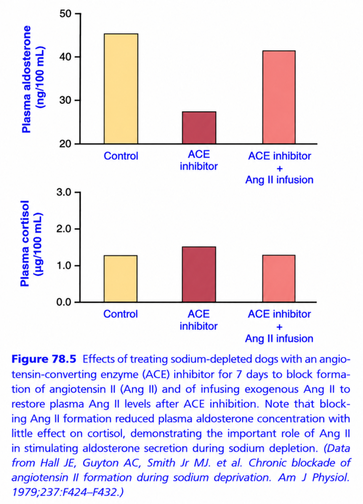
- After a low-sodium diet, aldosterone rises; blocking angiotensin II formation markedly lowers aldosterone without significantly changing cortisol (Fig. 78.5).
- This shows the major role of angiotensin II when Na⁺ intake and ECF volume are low.
- ANP, Na⁺ itself, and ACTH usually have smaller effects.
- A rare 10–20% fall in ECF Na⁺ may increase aldosterone by about 50%.
- ↑ plasma volume → atrial stretch → ↑ ANP → ↓ aldosterone → ↑ sodium excretion.
- ACTH has a mainly permissive role: a small amount is usually enough for normal aldosterone secretion, but complete absence can greatly reduce it.
KEY CONCEPT
K⁺ ↑ and Angiotensin II ↑ → Aldosterone ↑ → renal K⁺ excretion + Na⁺/water retention → blood volume and pressure ↑.
CONCEPTUAL EXAMPLES
- High K⁺ → ↑ Aldosterone → K⁺ excreted.
- Low Na⁺/low renal blood flow → ↑ Ang II → ↑ Aldosterone.
- High blood volume → ↑ ANP → ↓ Aldosterone.
- No ACTH → Aldosterone secretion falls significantly.
Short Essay: Role of Angiotensin II in Aldosterone Secretion
During sodium depletion, the renin–angiotensin system becomes activated. Angiotensin II (Ang II) plays an important role in stimulating aldosterone secretion from the adrenal cortex. When an ACE inhibitor is given, formation of Ang II decreases, causing plasma aldosterone to fall markedly. However, when Ang II is infused directly during ACE inhibition, aldosterone levels rise again toward normal. Plasma cortisol changes very little in comparison. Therefore, this experiment demonstrates that Angiotensin II is a major stimulus for aldosterone secretion, particularly during sodium depletion, whereas it has little effect on cortisol secretion.
p🚨 EXAM TRAP
❌ ACE inhibitor directly destroys aldosterone.
✅ ACE inhibitor reduces Ang II formation, which reduces aldosterone stimulation.
❌ Giving Ang II cannot work during ACE inhibition.
✅ It can work because you’re giving Ang II directly, bypassing ACE.
🏆 10-SECOND GRAPH MEMORY
ACE inhibitor → ↓ Ang II → ↓ Aldosterone
ACE inhibitor + Ang II → Aldosterone comes back ↑
Cortisol ≈ unchanged
🔑 GOLDEN CONCLUSION:
During sodium depletion, Angiotensin II is a major stimulus for aldosterone secretion, while having relatively little effect on cortisol secretion.
FUNCTIONS OF GLUCOCORTICOIDS
- Mineralocorticoids can keep an acutely adrenalectomized animal alive, but the animal is still far from normal.
- Without glucocorticoids, protein, carbohydrate, and fat metabolism become severely disturbed.
- The animal also cannot tolerate physical or mental stress normally.
- Even minor illnesses, such as respiratory infections, may become fatal.
- Therefore, glucocorticoids are essential for long-term survival, just as mineralocorticoids are essential for acute survival.
- Cortisol (hydrocortisone) provides at least 95% of the glucocorticoid activity of the adrenal cortex.
- Corticosterone provides a small but significant amount of the remaining glucocorticoid activity.
KEY CONCEPT
Mineralocorticoids → acute survival; Glucocorticoids → normal metabolism + ability to survive stress and illness.
CONCEPTUAL EXAMPLES
- No glucocorticoids → disturbed protein, carbohydrate, and fat metabolism.
- No glucocorticoids → poor stress resistance → minor illness can become fatal.
- Cortisol = main glucocorticoid (~95%).
EFFECTS OF CORTISOL ON CARBOHYDRATE METABOLISM
- Cortisol strongly increases gluconeogenesis → formation of glucose from amino acids and other substances.
- It can increase hepatic gluconeogenesis 6–10 times.
- Cortisol does this mainly by acting on the liver and opposing insulin.
- Cortisol:
- ↑ Liver enzymes needed to convert amino acids → glucose.
- ↑ Amino acid release from muscle → more amino acids reach the liver for glucose production.
- Opposes insulin, which normally inhibits hepatic glucose production.
- Therefore, the liver produces more glucose.
- Increased gluconeogenesis also increases liver glycogen storage, helping epinephrine and glucagon release glucose when needed, such as between meals.
- Cortisol also decreases glucose utilization by most cells.
- It decreases GLUT4 movement to the cell membrane, especially in skeletal muscle → insulin resistance.
- It also reduces insulin-signaling pathways involved in glucose uptake and utilization.
- Therefore: ↑ glucose production + ↓ glucose utilization → ↑ blood glucose.
- High blood glucose stimulates insulin secretion, but glucocorticoids make tissues less sensitive to insulin.
- High cortisol also increases fatty-acid availability, which can further impair insulin action.
- If blood glucose rises to ≥50% above normal, the condition may be called adrenal diabetes.
- Insulin lowers glucose only moderately in adrenal diabetes because the tissues are insulin-resistant.
EFFECTS OF CORTISOL ON PROTEIN METABOLISM
- Cortisol reduces protein stores in most cells, except the liver.
- It does this by ↓ protein synthesis + ↑ protein breakdown.
- Cortisol also decreases RNA formation and protein synthesis, especially in muscle and lymphoid tissue.
- Excess cortisol can cause severe muscle weakness.
- It can also greatly reduce immune function of lymphoid tissue.
- In contrast, cortisol increases protein synthesis in the liver.
- Liver-produced plasma proteins also increase.
- This occurs partly because cortisol increases amino acid transport into liver cells and increases liver enzymes needed for protein synthesis.
KEY CONCEPT
Cortisol → ↑ gluconeogenesis + ↓ cellular glucose use → ↑ blood glucose; meanwhile → ↓ protein stores in most tissues but ↑ liver/plasma proteins.
CONCEPTUAL EXAMPLES
- Cortisol ↑ → muscle amino acids ↑ → liver gluconeogenesis ↑ → blood glucose ↑.
- Cortisol ↑ → GLUT4 movement ↓ → muscle glucose uptake ↓ → insulin resistance.
- Excess cortisol → protein breakdown ↑ → muscle weakness.
- Liver is the exception → liver proteins ↑.
Cortisol Increases Blood Amino Acids, Decreases Transport of Amino Acids Into Extrahepatic Cells, and Enhances Transport Into Hepatic Cells
- Cortisol decreases amino acid entry into muscle and other extrahepatic cells.
- Therefore, intracellular amino acids fall → protein synthesis decreases.
- At the same time, protein breakdown continues → amino acids leave these tissues → blood amino acids increase.
- Thus, cortisol mobilizes amino acids from peripheral tissues, reducing their protein stores.
- Cortisol also increases amino acid entry into liver cells.
- The liver then uses these amino acids to:
- ↑ Deamination
- ↑ Liver protein synthesis
- ↑ Plasma protein formation
- ↑ Gluconeogenesis → glucose formation
- Therefore, cortisol essentially moves amino acids from peripheral tissues to the liver, while increasing the liver’s ability to use them.
KEY CONCEPT
Cortisol → muscle/peripheral amino acids ↓ inside cells → blood amino acids ↑ → liver uptake ↑ → proteins + glucose formation ↑.
CONCEPTUAL EXAMPLES
- Muscle → amino acids released → blood amino acids ↑.
- Blood → liver amino acids ↑ → gluconeogenesis ↑ → glucose ↑.
- Peripheral tissues → protein stores ↓.
- Liver → protein synthesis and plasma protein formation ↑.
EFFECTS OF CORTISOL ON FAT METABOLISM
- Cortisol mobilizes fatty acids from adipose tissue → free fatty acids in blood increase.
- These fatty acids are then used more for energy.
- Cortisol also directly increases fatty-acid oxidation in cells.
- Part of fat mobilization occurs because cortisol decreases glucose entry into fat cells.
- Less glucose → less α-glycerophosphate → triglyceride storage decreases → fatty acids are released.
- Thus, cortisol shifts the body from glucose use → fatty-acid use, especially during starvation or stress.
- This shift develops over several hours and is less rapid and powerful than the shift caused by decreased insulin.
- Increased fatty-acid use helps conserve glucose and glycogen for long-term energy needs.
- Excess cortisol can cause obesity, especially fat accumulation in the chest and head.
- This produces a buffalo-like torso and rounded “moon face.”
- The exact cause is unclear, but increased food intake and faster fat formation in some tissues may contribute.
KEY CONCEPT
Cortisol → fat mobilization + fatty-acid oxidation ↑ → fat becomes an energy source → glucose/glycogen are conserved.
CONCEPTUAL EXAMPLES
- Cortisol ↑ → adipose fatty acids ↑ → blood free fatty acids ↑.
- Starvation/stress → fatty-acid use ↑ → glucose conserved.
- Excess cortisol → abnormal fat deposition → moon face + buffalo-like torso.
CORTISOL IS IMPORTANT IN RESISTING STRESS AND INFLAMMATION
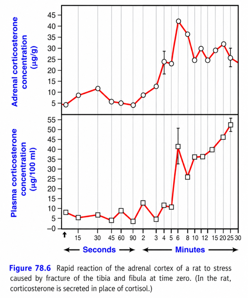
- Almost any physical or neurogenic stress → ↑ ACTH immediately → ↑ cortisol within minutes.
- After fracture of two leg bones, corticosteroid secretion in rats increased 6-fold within 4–20 minutes (Fig. 78.6).
- Stressors that increase cortisol include:
- Trauma
- Infection
- Extreme heat or cold
- Norepinephrine/sympathomimetic drugs
- Surgery
- Necrotizing substances
- Physical restraint
- Debilitating diseases
- Cortisol helps during stress by rapidly mobilizing amino acids and fats from storage.
- These substances provide energy and materials to make glucose and other essential compounds.
- Amino acids can help damaged tissues make new proteins and essential substances needed for cell survival and repair.
- Cortisol preferentially mobilizes labile proteins before basic functional proteins such as muscle contractile proteins and neuronal proteins.
- Its strong anti-inflammatory effects are also important in resisting stress.
- Tissue damage from trauma, infection, or other causes → inflammation.
- Excessive inflammation can itself cause major tissue damage, as in rheumatoid arthritis.
- Large amounts of cortisol can prevent inflammation or rapidly reduce inflammation that has already started.
- The main stages of inflammation are:
- Damaged cells release histamine, bradykinin, proteolytic enzymes, prostaglandins, and leukotrienes.
- Blood flow increases → erythema.
- Capillary permeability increases → plasma leaks into tissues → nonpitting edema.
- Leukocytes enter the damaged area.
- After days to weeks → fibrous tissue grows and helps healing.
- High cortisol has two major anti-inflammatory actions:
- Prevents inflammation from developing.
- Resolves existing inflammation and promotes healing.
- Cortisol stabilizes lysosomal membranes → fewer proteolytic enzymes are released → inflammation decreases.
- Cortisol decreases capillary permeability → less plasma escapes into tissues.
- Cortisol decreases leukocyte migration and phagocytosis.
- Cortisol reduces prostaglandins and leukotrienes, decreasing vasodilation, capillary permeability, and leukocyte movement.
- Cortisol suppresses lymphocyte reproduction, especially T lymphocytes, reducing immune reactions.
- Cortisol reduces fever mainly by decreasing interleukin-1 release from white blood cells → less stimulation of the hypothalamic temperature-control system.
- Therefore, cortisol produces a broad reduction in inflammation.
- Even after inflammation is established, cortisol can reduce it within hours to a few days.
- Healing is also supported by:
- Amino acids → tissue repair
- ↑ Gluconeogenesis → extra glucose
- ↑ Fatty acids → cellular energy
- Possible inactivation/removal of inflammatory products
- Cortisol is especially useful against severe inflammation in rheumatoid arthritis, rheumatic fever, and acute glomerulonephritis.
- Glucocorticoid treatment can make inflammation begin to subside within about 24 hours, although it does not correct the underlying disease.
KEY CONCEPT
Stress → ACTH ↑ → Cortisol ↑ → fuels available for energy/repair + inflammation suppressed.
CONCEPTUAL EXAMPLES
- Fracture → ACTH ↑ → cortisol ↑ rapidly → stress resistance.
- Tissue injury → cortisol ↑ → lysosome stabilization + ↓ inflammatory mediators → inflammation ↓.
- Cortisol ↑ → amino acids + fatty acids mobilized → energy and repair ↑.
- Rheumatoid arthritis → glucocorticoids → inflammation ↓.
STRESS → ADRENAL CORTICOSTERONE
🎯 BIG IDEA
A sudden injury/stress makes the adrenal cortex respond very quickly.
Stress → adrenal cortex activated → corticosterone ↑ → hormone enters blood → plasma corticosterone ↑
In humans, the corresponding major glucocorticoid is cortisol; in this rat experiment, it is corticosterone.
📊 1. TOP GRAPH = ADRENAL CORTICOSTERONE
Y-axis: corticosterone inside the adrenal cortex
X-axis: time after the fracture:
Seconds → Minutes
What happens?
At time 0, the tibia/fibula is fractured → 🚨 stress begins
The adrenal cortex quickly starts responding.
- Early seconds: relatively low
- By 3–6 min: huge rise
- Around 6 min: peak ≈ 42 μg/g
- Then it fluctuates but remains elevated.
👉 Meaning: the adrenal gland rapidly increases hormone production/secretion in response to stress.
📊 2. BOTTOM GRAPH = PLASMA CORTICOSTERONE
Y-axis: corticosterone in the blood
Now watch the delay:
Before stress:
Blood level ≈ 5–10 μg/100 mL
After fracture:
Initially, only a small change.
Then around 6 minutes:
🚀 Huge increase → ~40 μg/100 mL
Later:
~35 → 40 → 46 → 52 μg/100 mL
👉 Hormone made by the adrenal gland has entered the bloodstream.
⭐ WHY DOES THE BLOOD RESPONSE LAG?
Think of a factory + delivery truck:
🏭 Adrenal cortex = factory
🧪 Makes corticosterone
🚚 Blood = delivery system
So:
Stress 🚨
↓
Adrenal cortex responds first
↓
Corticosterone production/release ↑
↓
Hormone enters blood
↓
Plasma corticosterone ↑
🔴 WHAT DOES THE RED LINE MEAN?
The red line simply connects the measured hormone levels over time.
It lets you see the trend:
low → rising → sharp peak → fluctuating high levels
⚪ Circles on top
= individual measurements of adrenal corticosterone
⬜ Squares below
= individual measurements of plasma corticosterone
Error bars
= variation/uncertainty around each measurement.
🧠 THE MOST IMPORTANT PART
🚨 One sudden stress can produce a VERY RAPID adrenal response.
Fracture at time 0
⬇️
Adrenal cortex activated
⬇️
Corticosterone ↑
⬇️
Blood corticosterone ↑↑
⬇️
Body prepares to deal with stress.
🔥 10-SECOND MEMORY
“Stress hits → adrenal responds within minutes → corticosterone floods the blood.”
Exam concept:
Acute stress → rapid activation of the adrenal cortex → marked increase in glucocorticoid secretion.
Other Effects of Cortisol
- Cortisol blocks inflammation during allergic reactions: the antigen–antibody reaction itself is not stopped, but cortisol reduces the inflammatory response that causes many serious effects.
- Therefore, cortisol can prevent shock or death from anaphylaxis.
- Cortisol decreases eosinophils and lymphocytes in blood, beginning within minutes and becoming marked within hours.
- Therefore, lymphocytopenia and eosinopenia can indicate excessive cortisol production.
- Large doses of cortisol cause lymphoid tissue atrophy → fewer T cells and antibodies → decreased immunity.
- This can allow severe infections to develop, but the same immune-suppressing effect makes glucocorticoids useful for preventing rejection of transplanted tissues.
- Cortisol also stimulates erythropoietin → increases RBC production; excess cortisol may cause polycythemia, whereas absence of cortisol may cause anemia.
- Cellular Mechanism of Cortisol Action: cortisol is lipid-soluble, so it diffuses through the cell membrane and binds an intracellular cytoplasmic receptor.
- The cortisol–receptor complex enters the nucleus and acts on glucocorticoid response elements (GREs) in DNA, changing gene transcription.
- This changes mRNA and protein synthesis, producing cortisol’s physiological effects.
- Therefore, most cortisol effects are slow: about 45–60 minutes to begin and several hours to days for full development.
- At high concentrations, glucocorticoids may also produce rapid nongenomic effects on cell-membrane ion transport.
- Activated glucocorticoid receptors can also move to mitochondria, where they may regulate genes involved in oxidative phosphorylation and energy metabolism.
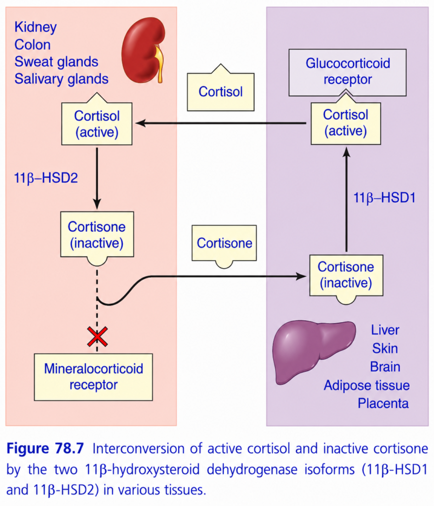
- Modulation of Glucocorticoid Effects By 11β-Hydroxysteroid Dehydrogenase: local 11β-HSD enzymes control how strongly tissues respond to cortisol.
- 11β-HSD2: converts cortisol → inactive cortisone, protecting mineralocorticoid receptors from cortisol.
- 11β-HSD2 is present in the renal tubules, colon, sweat glands, salivary glands, and placenta (Fig. 78.7).
- If 11β-HSD2 is deficient, licorice is excessive, or cortisol is extremely high, cortisol can activate mineralocorticoid receptors → sodium retention + hypertension + hypokalemia.
- 11β-HSD1: converts inactive cortisone → active cortisol, thereby amplifying glucocorticoid effects.
- 11β-HSD1 is expressed in the liver, brain, adipose tissue, skeletal muscle, lung, and skin.
- Thus, 11β-HSD2 reduces cortisol action, whereas 11β-HSD1 increases cortisol action.
- These enzymes act like tissue “gate-keepers” of glucocorticoid effects.
- Increased 11β-HSD1 in adipose tissue may contribute to insulin resistance and diabetes, while increased brain 11β-HSD1 has been associated with cognitive decline in aging.
KEY CONCEPT
Cortisol = ↓ inflammation + ↓ immunity + ↑ RBC production; its cellular effects are mainly gene-mediated, while 11β-HSD1 activates cortisol and 11β-HSD2 inactivates it.
CONCEPTUAL EXAMPLES
- High cortisol → ↓ lymphocytes/eosinophils → ↓ immunity.
- 11β-HSD2 deficiency → cortisol activates mineralocorticoid receptors → Na⁺ retention + hypertension + hypokalemia.
- 11β-HSD1 activity → cortisone → cortisol → stronger glucocorticoid action.
🧠 Cortisol ↔ Cortisone
The whole picture in ONE idea:
11β-HSD1 = activates cortisol 🔥
11β-HSD2 = deactivates cortisol 🛑
🟣 RIGHT SIDE — ACTIVATION
In tissues like:
Liver, skin, brain, adipose tissue, placenta
Cortisone (inactive)
⬇️ 11β-HSD1
Cortisol (active)
⬇️
Glucocorticoid receptor
⬇️
Cortisol effects
🧠 Think: HSD1 = “1 → ON”
🟠 LEFT SIDE — PROTECTION
In:
Kidney, colon, sweat glands, salivary glands
Cortisol (active)
⬇️ 11β-HSD2
Cortisone (inactive)
Now cortisol cannot strongly activate the mineralocorticoid receptor.
Why?
Because these tissues need to respond mainly to aldosterone, not cortisol.
🛡️ HSD2 protects the mineralocorticoid receptor from cortisol.
🎯 WHY IS THIS IMPORTANT?
Cortisol can bind the mineralocorticoid receptor, just like aldosterone.
So the kidney uses:
11β-HSD2 = protective enzyme
It converts:
Cortisol → Cortisone
Therefore:
Cortisol ❌ → Mineralocorticoid receptor
and:
Aldosterone ✅ → Mineralocorticoid receptor
🧩 STORY
Imagine cortisol is an active key 🔑.
HSD1:
Finds an inactive key (cortisone) and makes it active.
Cortisone → Cortisol 🔥
HSD2:
Takes the active key and disables it.
Cortisol → Cortisone 🛑
The kidney does this because it wants the aldosterone key to control the mineralocorticoid receptor.
🚨 HIGH-YIELD MEMORY
| Enzyme | Conversion | Main idea |
|---|---|---|
| 11β-HSD1 | Cortisone → Cortisol | ACTIVATES |
| 11β-HSD2 | Cortisol → Cortisone | INACTIVATES / PROTECTS |
⭐ MASTER LINE:
HSD1 turns cortisol ON; HSD2 turns cortisol OFF—especially in the kidney to protect aldosterone action.
