


- Mineralocorticoids are lifesaving because they maintain Na⁺, Cl⁻, K⁺, extracellular fluid volume, and blood volume.
- Without mineralocorticoids:
- Na⁺ + Cl⁻ ↓ → large loss from the body.
- K⁺ ↑ in extracellular fluid → hyperkalemia.
- ↓ NaCl → ↓ extracellular fluid volume → ↓ blood volume.
- ↓ blood volume → ↓ cardiac output → shock-like state → death.
- Complete adrenal cortical hormone loss may cause death within 3–14 days without salt therapy or mineralocorticoids.
- Aldosterone prevents this sequence, so mineralocorticoids are called the acute “lifesaving” portion of adrenocortical hormones.
- Aldosterone is the major mineralocorticoid, producing about 90% of mineralocorticoid activity.
- Cortisol also has mineralocorticoid activity, but:
- Aldosterone is about 3000× more potent than cortisol.
- Cortisol concentration is about 2000× higher than aldosterone.
- Cortisol can bind mineralocorticoid receptors, but renal 11β-HSD2 normally prevents this effect by converting:
- Cortisol → Cortisone
- Cortisone has little ability to activate mineralocorticoid receptors.
- 11β-HSD2 also helps prevent cortisol from activating these receptors through effects on the cell’s redox state.
- If 11β-HSD2 is genetically deficient, cortisol produces strong mineralocorticoid effects → apparent mineralocorticoid excess (AME).
- AME resembles aldosterone excess, but plasma aldosterone is very low.
- Large amounts of licorice can also cause AME because glycyrrhetinic acid blocks 11β-HSD2.
KEY CONCEPT
Aldosterone protects circulation by preventing NaCl loss and excessive K⁺ accumulation → maintains extracellular fluid and blood volume → prevents shock.
CONCEPTUAL EXAMPLES
- No mineralocorticoid → NaCl loss → ↓ blood volume → ↓ cardiac output → shock → death.
- 11β-HSD2 normal → cortisol → cortisone → mineralocorticoid receptor protected.
- 11β-HSD2 deficient → cortisol acts like aldosterone → AME despite very low aldosterone.
- Licorice → blocks 11β-HSD2 → cortisol acts on mineralocorticoid receptors → AME.
RENAL AND CIRCULATORY EFFECTS OF ALDOSTERONE
- Aldosterone → ↑ Na⁺ reabsorption + ↑ K⁺ secretion mainly in principal cells of collecting tubules, and also in distal tubules and collecting ducts.
- Therefore, aldosterone saves Na⁺ in the body while increasing K⁺ loss in urine.
- Excess aldosterone: Na⁺ loss in urine can fall to only a few mEq/day, while K⁺ loss can temporarily increase several-fold → ↑ body Na⁺ and ↓ body K⁺.
- No aldosterone: Na⁺ loss may reach 10–20 g/day, while K⁺ is strongly retained.
- Although aldosterone strongly retains Na⁺, plasma Na⁺ rises only slightly because retained Na⁺ pulls water with it → ECF volume increases more than Na⁺ concentration.
- Slight ↑ ECF Na⁺ also → ↑ thirst + ↑ ADH → more water intake and renal water reabsorption.
- Excess aldosterone → ↑ ECF volume → ↑ arterial pressure.
- If ECF volume remains elevated for >1–2 days, increased arterial pressure causes:
- Pressure natriuresis → ↑ Na⁺ excretion.
- Pressure diuresis → ↑ water excretion.
- ↓ Angiotensin II + ↑ natriuretic hormones such as ANP → further Na⁺ and water loss.
- Thus, after ECF volume rises about 5–15%, arterial pressure rises about 15–25 mm Hg, and renal Na⁺/water excretion return toward normal despite continued excess aldosterone (Fig. 78.3).
- This return of Na⁺ and water excretion to normal is called aldosterone escape.
- After aldosterone escape, Na⁺ and water intake = output, so further retention stops; however, hypertension persists while aldosterone remains high.
- Severe aldosterone deficiency → ↓ Na⁺ reabsorption → ↑ Na⁺ loss → ↓ ECF volume → ↓ blood pressure → ↓ cardiac output.
- ↓ ECF volume and blood pressure → ↑ ADH + ↑ thirst, which reduce water loss but also contribute to hyponatremia.
- If aldosterone secretion becomes zero, massive Na⁺ loss causes severe dehydration + low blood volume → circulatory shock → death within a few days without treatment.
KEY CONCEPT
Aldosterone saves Na⁺ and loses K⁺ → Na⁺ retention pulls water → ↑ ECF volume → ↑ blood pressure.
Excess → aldosterone escape; deficiency → Na⁺ wasting → ↓ volume/BP → shock.
CONCEPTUAL EXAMPLES
- ↑ Aldosterone → ↑ Na⁺ reabsorption → ↑ water retention → ↑ ECF volume → ↑ BP.
- Excess aldosterone → BP rises → kidneys excrete more Na⁺/water → aldosterone escape.
- ↓ Aldosterone → Na⁺ wasting → ↓ ECF volume → ↓ BP → shock.
- Aldosterone = “Na⁺ saver + K⁺ excreter.”
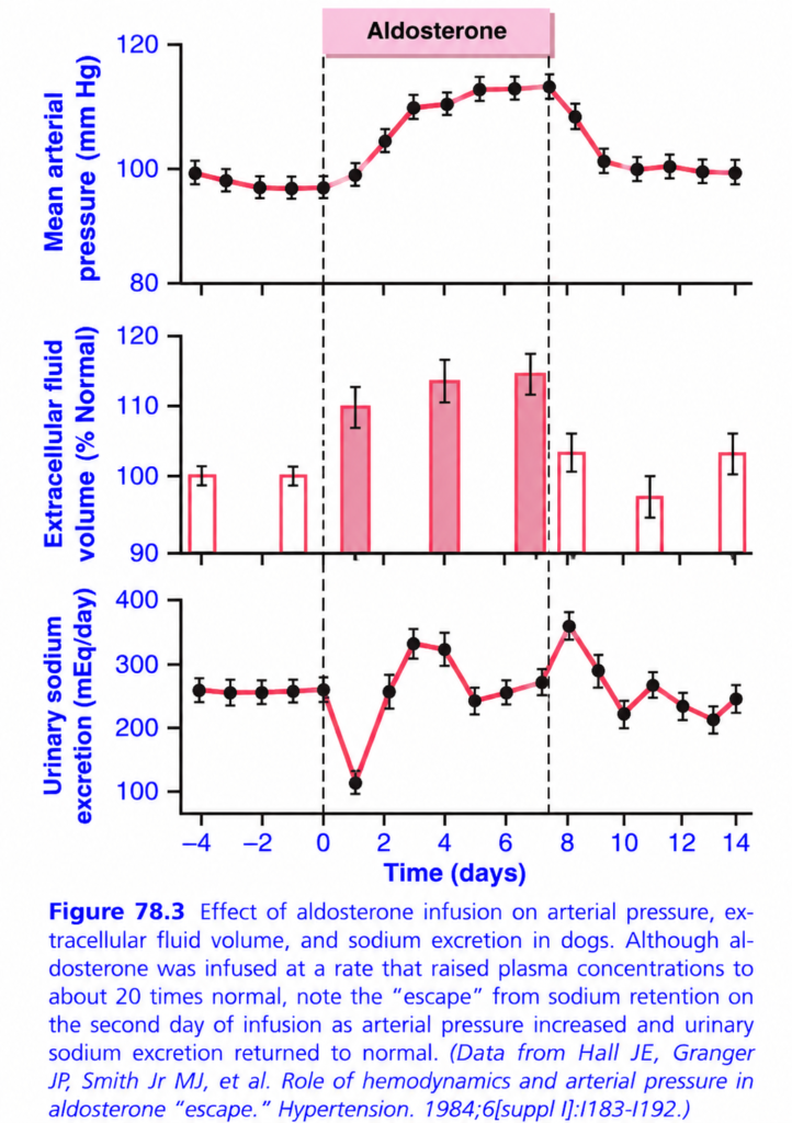
ALDOSTERONE “ESCAPE” GRAPH
🎯 BIG IDEA
Aldosterone initially saves Na⁺ → water follows → ECF volume ↑ → BP ↑ → kidneys then start dumping ( getting rid of something )Na⁺ again.
👉 This recovery of Na⁺ excretion despite continued aldosterone = ALDOSTERONE ESCAPE.
🟦 First: What do the 3 graphs show?
1️⃣ TOP = 🩸 Mean arterial pressure
During aldosterone infusion:
~98 → ~112 mmHg
➡️ BP rises.
After aldosterone stops:
➡️ BP gradually returns toward normal.
2️⃣ MIDDLE = 💧 Extracellular fluid (ECF) volume
Before infusion:
100% = normal
After aldosterone:
100% → ~110–115%
➡️ Aldosterone causes Na⁺ retention
➡️ Water follows Na⁺
➡️ ECF volume expands
After stopping:
➡️ Volume gradually falls toward normal.
3️⃣ BOTTOM = 🧂 Urinary sodium excretion
This is the most important graph.
Before aldosterone:
➡️ ~250 mEq/day
Day 0–1:
Aldosterone starts.
Na⁺ excretion ↓↓↓
➡️ Kidneys retain sodium.
Then — THE AMAZING PART ⭐
By about Day 2:
Na⁺ excretion rises back toward normal
❗ Even though aldosterone is STILL being infused!
This is the:
🚨 ALDOSTERONE ESCAPE
🧠 WHY DOES ESCAPE HAPPEN?
Think:
Aldosterone
⬇️
Kidney retains Na⁺ 🧂
⬇️
Water follows Na⁺ 💧
⬇️
ECF volume ↑
⬇️
Blood pressure ↑ 🩸
⬇️
Kidneys receive higher pressure
⬇️
More Na⁺ + water excreted 🚽
⬇️
Na⁺ excretion returns toward normal
So the body says:
“Aldosterone is trying to save sodium, but the increased blood pressure makes the kidneys throw sodium out anyway!”
🔥 What do the dashed lines mean?
First dashed line = Day 0
➡️ Aldosterone infusion STARTS
Second dashed line = ~Day 7
➡️ Aldosterone infusion STOPS
The pink region between them = aldosterone is being continuously infused.
⭐ THE MOST IMPORTANT CONTRAST
Early aldosterone:
Na⁺ retention ↑ → ECF ↑ → BP ↑
After ~2 days:
BP ↑ → renal Na⁺ excretion ↑ → “escape” from Na⁺ retention
Therefore:
Aldosterone continues, but sodium retention does NOT continue indefinitely.
🧠 10-SECOND MASTER MEMORY
ALDOSTERONE → Na⁺ SAVE → WATER SAVE → VOLUME ↑ → BP ↑ → Na⁺ LOSS ↑
“Aldosterone holds sodium, but high BP makes the kidney escape.” 🧂➡️💧➡️🩸➡️🚽
⭐ Exam killer point:
Aldosterone escape = return of urinary Na⁺ excretion toward normal despite continued aldosterone administration, mainly because volume expansion and increased arterial pressure promote renal Na⁺ excretion.
Excess Aldosterone Causes Hypokalemia and Muscle Weakness; Aldosterone Deficiency Causes Hyperkalemia and Cardiac Toxicity
- Excess aldosterone → ↑ K⁺ excretion in urine + ↑ K⁺ movement into cells → ↓ plasma K⁺ = hypokalemia.
- Plasma K⁺ may fall from about 4.5 mEq/L → as low as 2 mEq/L.
- When plasma K⁺ falls below about ½ of normal, severe muscle weakness may develop.
- Low K⁺ changes the electrical excitability of nerve and muscle membranes, so normal action potentials cannot be transmitted properly → muscle weakness.
- Aldosterone deficiency → ↓ K⁺ excretion → ↑ plasma K⁺ = hyperkalemia.
- When K⁺ rises 60–100% above normal, serious cardiac toxicity develops:
- ↓ strength of heart contraction
- Arrhythmias
- Further K⁺ rise → heart failure
- Excess aldosterone also increases H⁺ secretion by intercalated cells of the cortical collecting tubules.
- ↑ H⁺ secretion → ↑ renal HCO₃⁻ reabsorption → ↓ H⁺ concentration in extracellular fluid → metabolic alkalosis.
KEY CONCEPT
Too much aldosterone → K⁺ ↓ + H⁺ ↓ → muscle weakness + metabolic alkalosis.
Too little aldosterone → K⁺ ↑ → cardiac toxicity → possible heart failure.
CONCEPTUAL EXAMPLES
- Aldosterone ↑ → K⁺ lost in urine → hypokalemia → muscle weakness.
- Aldosterone ↓ → K⁺ retained → hyperkalemia → arrhythmia/heart failure.
- Aldosterone ↑ → H⁺ secretion ↑ → HCO₃⁻ retention ↑ → metabolic alkalosis.
ALDOSTERONE STIMULATES SODIUM AND POTASSIUM TRANSPORT IN SWEAT GLANDS, SALIVARY GLANDS, AND INTESTINAL EPITHELIAL CELLS
- Aldosterone acts on sweat and salivary glands much like the kidney: it ↑ NaCl reabsorption and ↑ K⁺ secretion in their ducts.
- Sweat glands: aldosterone conserves body salt, especially during hot environments when sweating is high.
- Salivary glands: aldosterone helps conserve salt when large amounts of saliva are lost.
- Intestine, especially colon: aldosterone ↑ Na⁺ absorption, preventing excessive sodium loss in stools.
- Without aldosterone → ↓ intestinal Na⁺ absorption → ↓ absorption of Cl⁻, other anions, and water → diarrhea → further salt loss.
CELLULAR MECHANISM OF ALDOSTERONE ACTION
- Aldosterone is lipid-soluble, so it easily enters tubular epithelial cells.
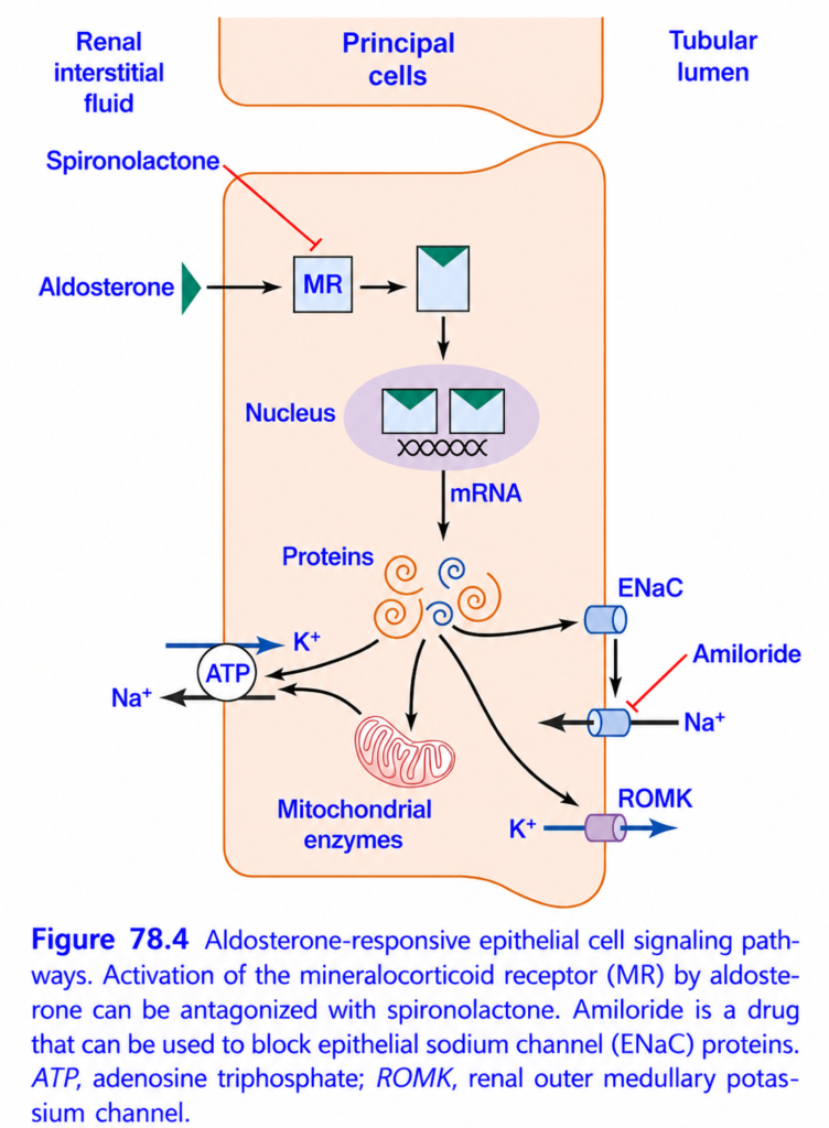
- Inside the cell, aldosterone binds to a specific mineralocorticoid receptor (MR).
- 11β-HSD2 normally protects MR from cortisol by converting cortisol → cortisone, which does not readily bind MR (Fig. 78.4).
- The aldosterone–receptor complex → nucleus → DNA activation → mRNA formation for proteins involved in Na⁺ and K⁺ transport.
- mRNA → ribosomes → production of:
- Na⁺-K⁺ ATPase
- Epithelial Na⁺ channels
- K⁺ channels
- Other transport-related enzymes and proteins.
- Na⁺-K⁺ ATPase increases Na⁺/K⁺ exchange at the basolateral membrane.
- Na⁺ channels allow Na⁺ to move from tubular fluid into the cell.
- K⁺ channels allow K⁺ to move from the cell into the tubular lumen.
- Therefore, aldosterone ultimately increases Na⁺ reabsorption and K⁺ secretion.
- Aldosterone does not act immediately because new proteins must first be produced:
- ~30 min: new RNA appears.
- ~45 min: Na⁺/K⁺ transport begins increasing.
- Several hours: effects reach maximum.
KEY CONCEPT
Aldosterone → MR → DNA → mRNA → transport proteins → ↑ Na⁺ reabsorption + ↑ K⁺ secretion.
CONCEPTUAL EXAMPLES
- Hot weather → sweating → aldosterone → ↑ NaCl reabsorption → salt conserved.
- Aldosterone ↑ in colon → ↑ Na⁺ + water absorption → less salt loss in stool.
- Aldosterone enters cell → MR → DNA → new Na⁺/K⁺ transport proteins → transport increases after hours.
Possible Nongenomic Actions of Aldosterone and Other Steroid Hormones
- Aldosterone and other steroids may produce two types of effects:
- Genomic effects: develop slowly, with a latency of about 45–60 minutes, because they require gene transcription and new protein synthesis.
- Nongenomic effects: develop rapidly, within seconds to minutes.
- Nongenomic effects are thought to occur when steroids bind to cell-membrane receptors linked to second-messenger systems, similar to peptide hormones.
- Aldosterone can increase cAMP in vascular smooth muscle and renal collecting-tubule epithelial cells in less than 2 minutes → too fast to require new gene transcription and protein synthesis.
- In some cells, aldosterone rapidly activates the phosphatidylinositol second-messenger system.
- The exact receptors responsible for these rapid effects are not yet determined, and their physiological importance is not well understood.
- Overall, nongenomic effects appear much less important than genomic effects in regulating Na⁺, K⁺, and acid-base balance.
KEY CONCEPT
Genomic = slow → genes → new proteins.
Nongenomic = fast → membrane receptors → second messengers.
CONCEPTUAL EXAMPLES
- Aldosterone → gene transcription → new proteins → effect after ~45–60 min = genomic.
- Aldosterone → membrane receptor → cAMP → effect within <2 min = nongenomic.
