


Definition
- Pneumonia is an inflammatory disease of the lungs.
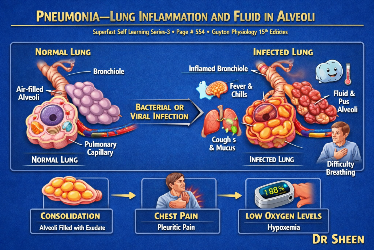
- In pneumonia, some or all of the alveoli become filled with fluid and blood cells instead of air. (Fig. 43.5)
- This filling of the alveoli interferes with normal gas exchange.
Bacterial Pneumonia
- A common type is bacterial pneumonia, which is most often caused by pneumococci.
Development of the Disease
- The infection begins in the alveoli.
- The pulmonary (alveolar) membrane becomes inflamed.
- Because of inflammation, the membrane becomes highly porous (more permeable).
- As a result:
- Fluid leaks from the blood into the alveoli.
- Red blood cells leak into the alveoli.
- White blood cells also enter the alveoli to fight the infection.
- Consequently, the infected alveoli gradually fill with fluid and inflammatory cells.
- The infection spreads from one alveolus to another through the movement of bacteria or viruses.
- As the disease progresses:
- Large areas of the lung may become involved.
- Sometimes an entire lobe or even a whole lung is affected.
- These affected regions become consolidated, meaning they are:
- Filled with fluid.
- Filled with inflammatory cells and cellular debris.
- Unable to participate effectively in gas exchange.
Effect on Gas Exchange
- In pneumonia, the gas exchange function of the lungs gradually decreases as the disease progresses.
Early Stage
- In the early stage, only one lung or one part of a lung may be affected.
- In the diseased area:
- Alveolar ventilation decreases because the alveoli are filled with fluid.
- Blood flow continues normally through the pulmonary capillaries.
Major Pulmonary Abnormalities
1. Reduced Respiratory Membrane Surface Area
- Fluid-filled alveoli cannot participate effectively in gas exchange.
- Therefore, the total surface area available for diffusion decreases.
- This reduces the exchange of oxygen and carbon dioxide.
2. Decreased Ventilation–Perfusion (V̇A/Q̇) Ratio
- The affected alveoli receive little or no ventilation.
- However, blood continues to flow through their capillaries.
- This causes a decreased ventilation–perfusion (V̇A/Q̇) ratio.
- Because blood passes through poorly ventilated alveoli, it:
- Receives less oxygen.
- Removes less carbon dioxide.
- As a result:
- Hypoxemia develops (low oxygen level in the blood).
- Hypercapnia develops (high carbon dioxide level in the blood).
Oxygen Saturation in Pneumonia
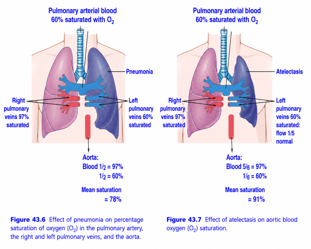
- Fig. 43.6 illustrates the effect of a low V̇A/Q̇ ratio.
- Blood flowing through the normal (aerated) lung becomes approximately 97% saturated with oxygen.
- Blood flowing through the affected (unaerated) lung becomes only about 60% saturated with oxygen.
- When these two blood streams mix in the left side of the heart, the average arterial oxygen saturation falls to about 78%.
- This oxygen saturation is far below the normal level, resulting in significant hypoxemia.
KEY CONCEPT
- Pneumonia is inflammation of the lungs in which alveoli become filled with fluid and blood cells (Fig. 43.5).
- Bacterial pneumonia is most commonly caused by pneumococci.
- Inflammation increases alveolar membrane permeability, allowing fluid, red blood cells, and white blood cells to enter the alveoli.
- The infection spreads from alveolus to alveolus, and large lung areas may become consolidated with fluid and cellular debris.
- Pneumonia reduces gas exchange by decreasing the respiratory membrane surface area and lowering the ventilation–perfusion (V̇A/Q̇) ratio.
- These changes produce hypoxemia (low blood O₂) and hypercapnia (high blood CO₂).
- Blood from normal alveoli is about 97% oxygen saturated, whereas blood from fluid-filled alveoli is only about 60% saturated; after mixing, arterial oxygen saturation falls to about 78% (Fig. 43.6).
ATELECTASIS—COLLAPSE OF THE ALVEOLI
Definition
- Atelectasis means collapse of the alveoli.
- It may involve:
- A small (localized) area of the lung, or
- An entire lung.
Common Causes of Atelectasis
The two most common causes are:
- Complete obstruction of an airway.
- Lack of surfactant in the fluid lining the alveoli.
Airway Obstruction Causes Lung Collapse
Causes of Airway Obstruction
Airway obstruction usually occurs because of:
- Blockage of many small bronchi by mucus, or
- Blockage of a major bronchus by a large mucus plug or a solid object such as a tumor.
What Happens After Airway Obstruction?
- Air becomes trapped in the alveoli beyond the obstruction.
- This trapped air is gradually absorbed into the blood flowing through the pulmonary capillaries.
- Absorption usually occurs within minutes to hours.
If the Lung Is Pliable (Flexible)
- If the lung tissue is soft and flexible, the alveoli simply collapse after the trapped air is absorbed.
- This produces atelectasis.
If the Lung Is Rigid
- If the lung is stiff because of fibrotic tissue, it cannot collapse easily.
- As the trapped air is absorbed:
- Very negative pressure develops inside the alveoli.
- This negative pressure pulls fluid from the pulmonary capillaries into the alveoli.
- Consequently, the alveoli become completely filled with edema fluid.
- When an entire lung collapses, this condition is called massive collapse of the lung.
Effects of Massive Atelectasis on Pulmonary Function
(Fig. 43.7 given above )
1. Collapse of Alveoli
- The collapsed lung tissue closes the alveoli, preventing ventilation.
2. Increased Pulmonary Vascular Resistance
- Lung collapse compresses and folds the pulmonary blood vessels.
- This increases resistance to blood flow through the affected lung.
- In addition:
- Hypoxia in the collapsed alveoli causes pulmonary vasoconstriction.
- Therefore, pulmonary vascular resistance increases even further.
3. Blood Flow Is Redirected
- Because vascular resistance increases, much less blood flows through the collapsed lung.
- Most of the blood is redirected to the healthy, ventilated lung.
- This helps maintain better oxygenation.
- In the example shown in Fig. 43.7:
- Five-sixths of the blood flows through the ventilated lung.
- Only one-sixth flows through the collapsed (unventilated) lung.
4. Ventilation–Perfusion Relationship
- Although one lung receives no ventilation, most blood is diverted to the functioning lung.
- Therefore:
- The overall ventilation–perfusion (V̇A/Q̇) ratio is only moderately reduced.
- Arterial oxygen saturation decreases only slightly (mild O₂ desaturation) despite complete collapse of one lung.
KEY CONCEPT
- Atelectasis is the collapse of alveoli and may involve part of a lung or an entire lung.
- The two major causes are complete airway obstruction and lack of surfactant.
- Airway obstruction allows trapped air to be absorbed into the blood, leading to alveolar collapse within minutes to hours.
- If the lung is rigid and cannot collapse, negative alveolar pressure draws fluid into the alveoli, producing pulmonary edema.
- Massive atelectasis increases pulmonary vascular resistance because lung collapse compresses blood vessels and hypoxia causes pulmonary vasoconstriction (Fig. 43.7). (given above )
- Most blood is redirected to the normal lung (about five-sixths), while only one-sixth passes through the collapsed lung.
- As a result, the overall V̇A/Q̇ ratio is only moderately impaired, and arterial oxygen desaturation is usually mild despite complete collapse of one lung.
LACK OF SURFACTANT AS A CAUSE OF LUNG COLLAPSE
Normal Role of Surfactant
- Surfactant is secreted by special alveolar epithelial cells.
- It is released into the fluid lining the inner surface of the alveoli.
- Surfactant reduces the surface tension of alveolar fluid by 2–10 times.
- By lowering surface tension, surfactant helps keep the alveoli open during breathing.
- Therefore, surfactant plays a major role in preventing alveolar collapse.
Surfactant Deficiency
- In some conditions, the amount of surfactant produced by the alveoli is greatly reduced.
- One important example is hyaline membrane disease, also called respiratory distress syndrome (RDS).
- This condition commonly occurs in premature newborn infants.
- Because too little surfactant is present:
- Surface tension of the alveolar fluid becomes several times higher than normal.
- The alveoli have a strong tendency to collapse (atelectasis).
- The alveoli may also fill with fluid.
Effects of Surfactant Deficiency
- Large portions of the lungs may collapse (atelectasis).
- As more alveoli collapse:
- Ventilation decreases markedly.
- Gas exchange becomes severely impaired.
- The infant develops severe respiratory distress.
- Without treatment:
- Many premature infants die from suffocation because large areas of the lungs become atelectatic.
Treatment
- Survival of premature infants has improved greatly with:
- Surfactant replacement therapy.
- Assisted mechanical ventilation.
- Administration of corticosteroids before birth.
- Other supportive treatments.
- Despite these advances, hyaline membrane disease remains an important cause of illness (morbidity) and death (mortality) in premature infants.
- The more premature the infant, the more severe the respiratory distress.
KEY CONCEPT
- Surfactant is produced by alveolar epithelial cells and lines the inner surface of the alveoli.
- It reduces alveolar surface tension by 2–10 fold, helping to prevent alveolar collapse.
- Surfactant deficiency occurs in hyaline membrane disease (respiratory distress syndrome), especially in premature newborns.
- Without adequate surfactant, surface tension increases, causing alveolar collapse (atelectasis) and fluid accumulation.
- Severe surfactant deficiency leads to respiratory distress, impaired gas exchange, and may cause death if untreated.
- Surfactant therapy, assisted ventilation, and antenatal corticosteroids have significantly improved survival, although the disease remains a major cause of morbidity and mortality in preterm infants.
Made by self learning Dr sheen
