


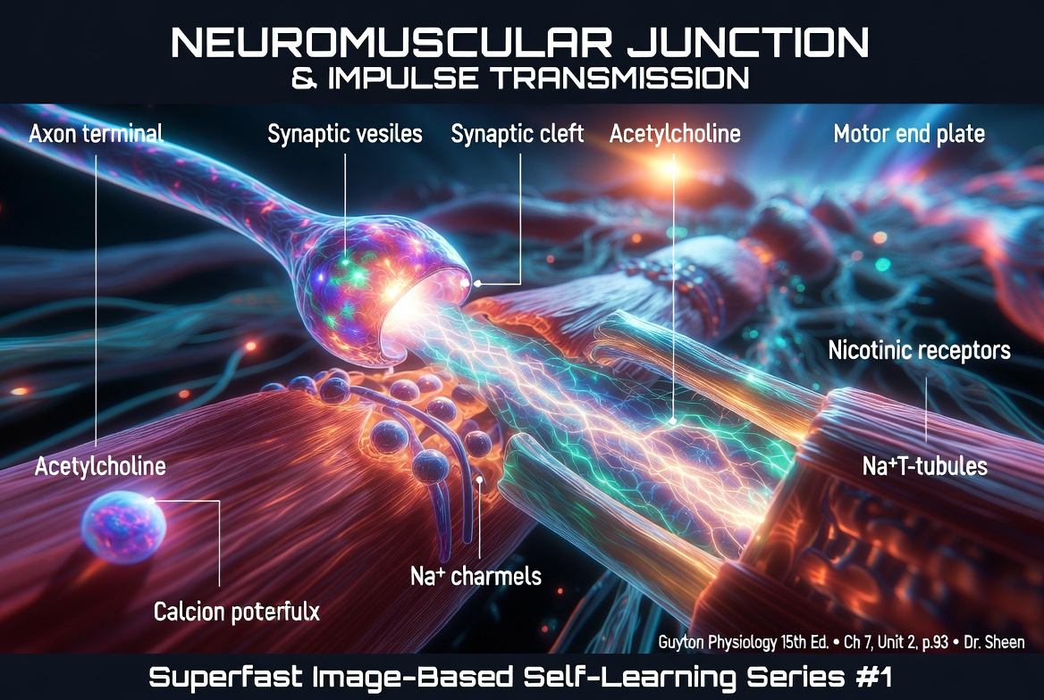
Neuromuscular Junction
- Skeletal muscle fibers are supplied (innervated) by large myelinated nerve fibers.
- These nerve fibers originate from large motor neurons (motoneurons) located in the anterior horns of the spinal cord.
- After entering the muscle belly, each nerve fiber branches into many smaller endings.
- These branches stimulate multiple skeletal muscle fibers.
- A single nerve fiber may supply from about 3 to several hundred muscle fibers.
- Each nerve ending forms a specialized connection with a muscle fiber called the neuromuscular junction.
- The neuromuscular junction is usually located near the midpoint of the muscle fiber.
- When a nerve impulse reaches the neuromuscular junction, it initiates an action potential in the muscle fiber.
- The muscle action potential travels in both directions:
- Toward one end of the muscle fiber, and
- Toward the opposite end of the muscle fiber.
- This allows the entire muscle fiber to become activated and contract.
- Almost every skeletal muscle fiber has only one neuromuscular junction.
- Only about 2% of muscle fibers have more than one neuromuscular junction.
KEY CONCEPT
- Skeletal muscle fibers are innervated by large myelinated motor nerve fibers arising from anterior horn motor neurons of the spinal cord.
- Each motor nerve branches to supply 3 to several hundred muscle fibers.
- Each nerve ending forms a neuromuscular junction near the middle of the muscle fiber.
- A nerve impulse at the neuromuscular junction generates a muscle action potential that spreads in both directions along the muscle fiber.
- Nearly all skeletal muscle fibers have a single neuromuscular junction.
THE NEUROMUSCULAR JUNCTION—THE MOTOR END PLATE
Structure of the Neuromuscular Junction
- The neuromuscular junction is the specialized site where a motor nerve communicates with a skeletal muscle fiber.
- Fig. 7.1A–B shows the neuromuscular junction between a large myelinated nerve fiber and a skeletal muscle fiber.
- As the nerve reaches the muscle:
- It divides into a complex network of branching nerve terminals.
- These nerve terminals invaginate (sink into) the surface of the muscle fiber.
- However, they remain outside the muscle fiber plasma membrane.
- This entire specialized structure is called the motor end plate.
- The motor end plate is covered by one or more Schwann cells.
- These Schwann cells insulate the neuromuscular junction from the surrounding tissue fluid.
Synaptic Gutter and Synaptic Cleft
- Fig. 7.1C shows the junction between a single axon terminal and the muscle fiber membrane.
- The invagination of the muscle membrane beneath the nerve terminal is called the:
- Synaptic gutter, or
- Synaptic trough.
- The space between the nerve terminal and the muscle membrane is called the:
- Synaptic space, or
- Synaptic cleft.
- The synaptic cleft is about 20–30 nanometers wide.
Subneural Clefts
- At the bottom of the synaptic gutter are many small folds of the muscle membrane.
- These folds are called subneural clefts.
- Subneural clefts greatly increase the surface area of the muscle membrane.
- This larger surface area allows acetylcholine to act more effectively on the muscle fiber.
Mitochondria and Acetylcholine Synthesis
- The axon terminal contains many mitochondria.
- These mitochondria produce ATP (adenosine triphosphate).
- ATP provides the energy required to synthesize acetylcholine (ACh).
- Acetylcholine is the neurotransmitter that stimulates the muscle fiber membrane and initiates muscle excitation.
Synaptic Vesicles
- Acetylcholine is synthesized in the cytoplasm of the axon terminal.
- It is then rapidly packaged into many small synaptic vesicles.
- A single motor end plate normally contains about 300,000 synaptic vesicles.
Acetylcholinesterase
- The synaptic cleft contains large amounts of the enzyme acetylcholinesterase.
- Acetylcholinesterase rapidly destroys acetylcholine within a few milliseconds after it is released from the synaptic vesicles.
- This terminates the action of acetylcholine, allowing the muscle fiber to relax and preparing the neuromuscular junction for the next nerve impulse.
KEY CONCEPT
- The neuromuscular junction is the specialized connection between a motor nerve ending and a skeletal muscle fiber (Fig. 7.1A–B).
- The branching nerve terminals invaginate into the muscle surface but remain outside the muscle fiber membrane, forming the motor end plate.
- Schwann cells surround the motor end plate and provide insulation.
- The synaptic gutter (trough) contains the nerve terminal, while the synaptic cleft is the 20–30 nm space between the nerve terminal and the muscle membrane (Fig. 7.1C).
- Subneural clefts increase the muscle membrane surface area, enhancing the action of acetylcholine.
- Mitochondria produce ATP needed for acetylcholine synthesis.
- Acetylcholine is synthesized in the axon terminal and stored in about 300,000 synaptic vesicles at each motor end plate.
- Acetylcholinesterase in the synaptic cleft rapidly breaks down acetylcholine within a few milliseconds after its release.
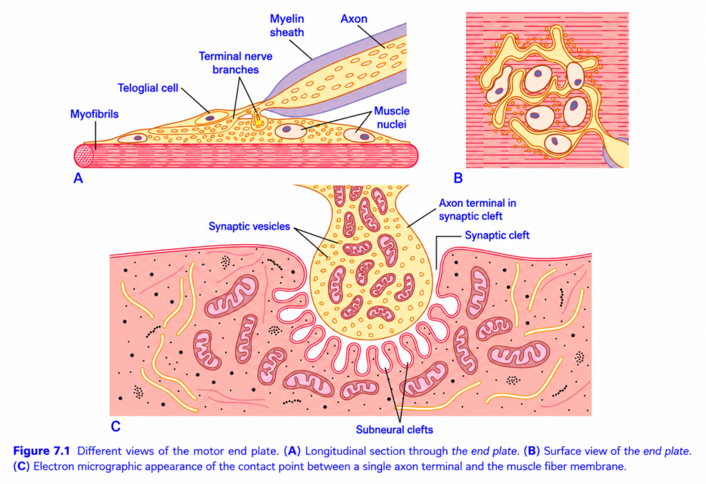
Motor End Plate (Neuromuscular Junction) – Figure 7.1
Easiest & Self learning Summary
This figure explains the Neuromuscular Junction (NMJ), also called the Motor End Plate, which is the communication site between a motor neuron and a skeletal muscle fiber.
Main Concept
Brain → Spinal cord → Motor neuron → Neuromuscular Junction (Motor End Plate) → Muscle Fiber → Muscle Contraction
The neuromuscular junction is where the nerve tells the muscle to contract.
Overall Structure of the Figure
The figure has three views of the same structure.
- A = Longitudinal (Side) View
- B = Surface (Top) View
- C = Electron Microscope (Highly Magnified) View
All three illustrate the same motor end plate from different perspectives.
A. Longitudinal (Side) View of the Motor End Plate
This is a side view showing how the nerve reaches the muscle.
1. Myelinated Axon
The nerve impulse travels through a motor axon.
The upper part of the axon is covered by a myelin sheath.
Function
- Insulates the nerve.
- Increases the speed of impulse conduction.
Concept
Think of myelin as the plastic insulation around an electrical wire.
2. Loss of Myelin Near the Muscle
As the nerve approaches the muscle,
the myelin sheath disappears.
Why?
The nerve terminal must directly communicate with the muscle membrane.
3. Terminal Nerve Branches
Near the muscle, one axon divides into many terminal branches.
Each branch contacts the muscle.
Function
- Distributes the nerve impulse.
- Forms the neuromuscular junction.
4. Teloglial Cell (Terminal Schwann Cell)
The nerve ending is covered by a teloglial (terminal Schwann) cell.
Function
- Protects the nerve terminal.
- Provides structural and metabolic support.
- Helps maintain and repair the neuromuscular junction.
5. Muscle Nuclei
These are the nuclei of the skeletal muscle fiber.
Function
- Control protein synthesis.
- Maintain the muscle fiber.
6. Myofibrils
These are the contractile elements inside the muscle.
They contain:
- Actin
- Myosin
Function
Produce muscle contraction after receiving the nerve signal.
Flow in Part A
Motor axon
      ↓
Myelin ends
      ↓
Axon branches
      ↓
Terminal nerve endings
      ↓
Muscle fiber
B. Surface (Top) View
This is what the motor end plate looks like from above.
Notice the branching pattern of the nerve terminal.
Concept
One nerve ending spreads over a small area of the muscle surface, allowing efficient stimulation of the muscle fiber.
Think of it like:
Tree trunk → Branches → Twigs
The axon branches so that one nerve terminal can communicate effectively with the muscle membrane.
C. Electron Microscope View (Highest Magnification)
This panel shows the microscopic details of the neuromuscular junction.
1. Axon Terminal
The bulb-shaped swelling is the axon terminal.
This is the last part of the motor neuron.
Function
Stores and releases acetylcholine (ACh).
2. Synaptic Vesicles
Small round vesicles inside the axon terminal.
Contents
Each vesicle contains acetylcholine (ACh).
Function
When a nerve impulse arrives,
the vesicles release ACh into the synaptic cleft.
3. Mitochondria
Many mitochondria are present inside the axon terminal.
Why so many?
Releasing neurotransmitters requires energy.
Mitochondria produce the ATP needed for:
- Vesicle movement
- Neurotransmitter release
- Recycling of vesicles
4. Synaptic Cleft
The narrow gap between the nerve ending and muscle membrane.
Important Point
The nerve does not directly touch the muscle.
There is a tiny space called the synaptic cleft.
Function
Acetylcholine diffuses across this gap.
5. Muscle Membrane (Motor End Plate)
The muscle membrane lies opposite the nerve terminal.
It contains many acetylcholine receptors.
Function
Receives the chemical signal from the nerve.
6. Subneural Clefts (Junctional Folds)
The muscle membrane is deeply folded beneath the nerve terminal.
These folds are called subneural clefts or junctional folds.
Why are they important?
They:
- Greatly increase surface area.
- Contain numerous acetylcholine receptors.
- Make nerve-to-muscle transmission faster and more efficient.
Easy Concept
Imagine a flat sheet versus a folded sheet.
The folded sheet has much more surface area.
Similarly, these folds provide more space for receptors.
How Muscle Contraction Occurs
Step 1
A nerve impulse reaches the axon terminal.
↓
Step 2
Synaptic vesicles release acetylcholine.
↓
Step 3
Acetylcholine diffuses across the synaptic cleft.
↓
Step 4
Acetylcholine binds to receptors on the motor end plate.
↓
Step 5
The muscle membrane depolarizes.
↓
Step 6
An action potential spreads through the muscle fiber.
↓
Step 7
Calcium is released inside the muscle.
↓
Step 8
Actin and myosin interact.
↓
Step 9
Muscle contracts.
Easy Flow Diagram
Brain
   ↓
Motor neuron
   ↓
Axon terminal
   ↓
Acetylcholine released
   ↓
Synaptic cleft
   ↓
ACh receptors on motor end plate
   ↓
Muscle action potential
   ↓
Calcium release
   ↓
Muscle contraction
Simple Real-Life Analogy
Imagine a person pressing a doorbell.
- Finger = Nerve impulse
- Doorbell button = Axon terminal
- Electrical signal = Acetylcholine
- Doorbell wire = Muscle membrane
- Bell ringing = Muscle contraction
Without pressing the button, the bell does not ring.
Similarly, without acetylcholine release, the muscle cannot contract.
Important Points from Figure 7.1
- The motor end plate (neuromuscular junction) is the site where a motor neuron communicates with a skeletal muscle fiber.
- The axon is myelinated, but the myelin sheath ends before the nerve terminal.
- The axon divides into terminal nerve branches that contact the muscle.
- Terminal Schwann (teloglial) cells cover and support the nerve endings.
- The axon terminal contains numerous synaptic vesicles filled with acetylcholine and many mitochondria to supply ATP.
- The synaptic cleft separates the nerve ending from the muscle membrane; they never directly touch.
- The muscle membrane forms subneural (junctional) folds, increasing surface area and housing a high density of acetylcholine receptors.
- Binding of acetylcholine to these receptors initiates a muscle action potential, leading to calcium release and skeletal muscle contraction.
KEY CONCEPT (Figure 7.1)
The motor end plate (neuromuscular junction) is the specialized synapse between a somatic motor neuron and a skeletal muscle fiber. A nerve impulse travels down a myelinated axon, which loses its myelin near the muscle and branches into terminal endings. These terminals contain synaptic vesicles filled with acetylcholine and release it into the synaptic cleft. Acetylcholine binds to receptors concentrated within the subneural (junctional) folds of the muscle membrane, generating a muscle action potential that triggers skeletal muscle contraction. This specialized design ensures rapid, reliable, and efficient transmission of signals from nerve to muscle.
SECRETION OF ACETYLCHOLINE BY THE NERVE TERMINALS
Release of Acetylcholine
- When a nerve impulse (action potential) reaches the neuromuscular junction, it triggers the release of acetylcholine (ACh).
- About 125 acetylcholine-containing synaptic vesicles are released into the synaptic cleft with each nerve impulse.
Arrival of the Action Potential
- Fig. 7.2 shows an enlarged view of the neuromuscular junction.
- The nerve terminal (presynaptic membrane) is located above.
- The muscle membrane with its subneural clefts is located below.
Opening of Voltage-Gated Calcium Channels
- The inner surface of the nerve terminal membrane contains linear dense bars.
- On both sides of each dense bar are voltage-gated Ca²⁺ channels.
- When the action potential reaches the nerve terminal:
- The voltage-gated Ca²⁺ channels open.
- Ca²⁺ diffuses from the synaptic cleft into the nerve terminal.
Activation of Ca²⁺–Calmodulin–Dependent Protein Kinase
- The incoming Ca²⁺ binds to calmodulin.
- This activates Ca²⁺–calmodulin–dependent protein kinase.
- The activated kinase phosphorylates synapsin proteins.
Release of Synaptic Vesicles
- Synapsin proteins normally anchor acetylcholine vesicles to the cytoskeleton of the presynaptic terminal.
- Phosphorylation of synapsin releases the vesicles from the cytoskeleton.
- The freed acetylcholine vesicles move toward the active zone of the presynaptic membrane, which is adjacent to the dense bars.
Docking and Exocytosis
- The vesicles dock at specialized release sites on the presynaptic membrane.
- They then fuse with the nerve terminal membrane.
- Acetylcholine is released into the synaptic cleft by exocytosis.
- The released acetylcholine is then available to bind to receptors on the muscle membrane and initiate muscle excitation.
KEY CONCEPT
- Each nerve impulse releases about 125 acetylcholine-containing vesicles into the synaptic cleft.
- Action potentials open voltage-gated Ca²⁺ channels located beside the dense bars of the nerve terminal membrane (Fig. 7.2).
- Ca²⁺ enters the nerve terminal and activates Ca²⁺–calmodulin–dependent protein kinase.
- This kinase phosphorylates synapsin proteins, releasing acetylcholine vesicles from the cytoskeleton.
- The vesicles move to the active zone, dock with the presynaptic membrane, and release acetylcholine by exocytosis.
- Released acetylcholine diffuses across the synaptic cleft to stimulate the muscle fiber.
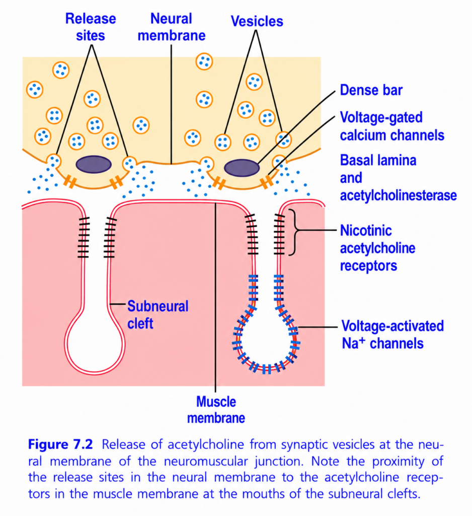
Release of Acetylcholine at the Neuromuscular Junction (Figure 7.2)
Easiest & Self learning Summary
This figure explains how a nerve communicates with a skeletal muscle by releasing acetylcholine (ACh) at the neuromuscular junction (NMJ).
It also shows why the structure of the NMJ is designed for rapid and efficient muscle contraction.
Main Concept
Nerve impulse → Calcium enters nerve terminal → Acetylcholine is released → Acetylcholine binds receptors → Sodium enters muscle → Muscle action potential → Muscle contracts
Overall View of the Figure
The figure has two main parts:
Upper Half = Nerve Terminal (Presynaptic Side)
Contains:
- Synaptic vesicles
- Release sites
- Voltage-gated calcium channels
- Dense bars
This is where acetylcholine is released.
Lower Half = Muscle Membrane (Postsynaptic Side)
Contains:
- Muscle membrane (motor end plate)
- Subneural clefts (junctional folds)
- Nicotinic acetylcholine receptors
- Voltage-gated sodium (Na⁺) channels
This is where the muscle receives the signal.
Understanding Each Label
1. Neural Membrane (Presynaptic Membrane)
This is the membrane of the axon terminal.
It is the last part of the motor neuron.
Function
- Stores acetylcholine.
- Releases acetylcholine into the synaptic cleft.
2. Synaptic Vesicles
These are the small circular sacs inside the nerve terminal.
They contain:
Acetylcholine (ACh)
Function
Store acetylcholine until a nerve impulse arrives.
Think of them as:
Small packets filled with neurotransmitter.
3. Release Sites (Active Zones)
These are specialized areas of the neural membrane where vesicles fuse.
Function
- Synaptic vesicles release acetylcholine here.
- ACh enters the synaptic cleft.
Important Concept
Acetylcholine is not released randomly.
It is released only at these active zones, directly opposite the muscle receptors.
This ensures fast and efficient transmission.
4. Dense Bar
The dense bar is a protein-rich structure at the release site.
Function
- Anchors synaptic vesicles.
- Organizes calcium channels.
- Helps vesicles fuse with the membrane.
Concept
It acts like a docking platform where vesicles are positioned before releasing acetylcholine.
5. Voltage-Gated Calcium (Ca²⁺) Channels
These channels are located beside each release site.
What happens?
When a nerve impulse arrives:
- The membrane depolarizes.
- Calcium channels open.
- Calcium enters the nerve terminal.
Why is calcium important?
Calcium triggers the fusion of synaptic vesicles with the neural membrane.
Without calcium:
❌ No acetylcholine release.
Flow So Far
Nerve impulse
      ↓
Voltage-gated Ca²⁺ channels open
      ↓
Calcium enters nerve terminal
      ↓
Synaptic vesicles fuse
      ↓
Acetylcholine released
6. Basal Lamina and Acetylcholinesterase
Between the nerve and muscle lies the basal lamina.
Embedded within it is the enzyme acetylcholinesterase (AChE).
Function of AChE
Breaks down acetylcholine into:
- Acetate
- Choline
Why?
If acetylcholine remained in the synaptic cleft, the muscle would continue contracting.
AChE quickly removes acetylcholine, allowing the muscle to relax and prepare for the next signal.
7. Muscle Membrane (Motor End Plate)
This is the specialized membrane of the skeletal muscle fiber facing the nerve terminal.
Function
Receives acetylcholine released from the nerve.
8. Subneural Clefts (Junctional Folds)
These are deep folds in the muscle membrane.
Why are they important?
They:
- Increase surface area.
- Contain a large number of acetylcholine receptors.
- Improve transmission efficiency.
Concept
More folds → More receptors → Stronger response.
9. Nicotinic Acetylcholine Receptors
These receptors are located at the mouths (tops) of the subneural clefts, directly opposite the release sites.
Function
Acetylcholine binds to these receptors.
This opens receptor channels, allowing mainly:
- Sodium (Na⁺) to enter the muscle.
- A small amount of potassium (K⁺) to leave.
This produces the end-plate potential, the first electrical change in the muscle membrane.
10. Voltage-Gated Sodium (Na⁺) Channels
These channels are concentrated deeper within the subneural clefts.
Function
When the end-plate potential is strong enough:
- Voltage-gated Na⁺ channels open.
- A muscle action potential is generated.
- The action potential spreads across the muscle membrane.
This initiates muscle contraction.
Complete Sequence of Events
Step 1
Brain sends a motor command.
↓
Step 2
Action potential reaches the axon terminal.
↓
Step 3
Voltage-gated Ca²⁺ channels open.
↓
Step 4
Calcium enters the nerve terminal.
↓
Step 5
Synaptic vesicles fuse with the neural membrane.
↓
Step 6
Acetylcholine is released through the release sites.
↓
Step 7
Acetylcholine diffuses across the synaptic cleft.
↓
Step 8
Acetylcholine binds to nicotinic receptors.
↓
Step 9
Na⁺ enters the muscle → End-plate potential develops.
↓
Step 10
Voltage-gated Na⁺ channels open.
↓
Step 11
Muscle action potential is generated.
↓
Step 12
The action potential triggers calcium release inside the muscle.
↓
Step 13
Muscle contracts.
↓
Step 14
Acetylcholinesterase breaks down acetylcholine.
↓
Step 15
The signal ends, and the muscle relaxes.
Easy Flow Diagram
Brain
      ↓
Motor neuron
      ↓
Action potential
      ↓
Ca²⁺ channels open
      ↓
Ca²⁺ enters nerve terminal
      ↓
Acetylcholine released
      ↓
ACh binds nicotinic receptors
      ↓
Na⁺ enters muscle
      ↓
Muscle action potential
      ↓
Calcium released inside muscle
      ↓
Muscle contraction
      ↓
Acetylcholinesterase destroys ACh
      ↓
Muscle relaxes
Why Are the Release Sites Opposite the Receptors?
The figure highlights that release sites in the nerve terminal are positioned directly above the nicotinic acetylcholine receptors at the mouths of the subneural clefts.
Importance
- Acetylcholine travels only a very short distance.
- Transmission is extremely rapid.
- Very little acetylcholine is wasted.
- Every nerve impulse efficiently activates the muscle.
Simple Real-Life Analogy
Imagine a mail delivery system:
- Synaptic vesicles = Envelopes containing messages (acetylcholine).
- Release sites = Mailboxes where letters are posted.
- Synaptic cleft = Short distance the letter travels.
- Nicotinic receptors = Mailboxes on the muscle waiting for the message.
- Voltage-gated Na⁺ channels = Electrical switch that starts the muscle response.
- Acetylcholinesterase = Recycling worker who removes the message after it has been read.
Important Points from Figure 7.2
- The neural (presynaptic) membrane contains synaptic vesicles, release sites (active zones), dense bars, and voltage-gated Ca²⁺ channels.
- Arrival of a nerve impulse opens voltage-gated Ca²⁺ channels, allowing calcium to enter the nerve terminal.
- Calcium triggers fusion of synaptic vesicles with the neural membrane, releasing acetylcholine into the synaptic cleft.
- The basal lamina contains acetylcholinesterase, which rapidly breaks down acetylcholine after it has acted.
- The muscle membrane forms subneural (junctional) clefts that increase surface area.
- Nicotinic acetylcholine receptors are concentrated at the mouths of the subneural clefts, directly opposite the release sites, ensuring rapid transmission.
- Voltage-gated Na⁺ channels are concentrated deeper in the folds. Activation of nicotinic receptors produces an end-plate potential, which opens these channels and generates a muscle action potential, leading to contraction.
KEY CONCEPT (Figure 7.2)
Figure 7.2 illustrates the highly specialized design of the neuromuscular junction for efficient nerve-to-muscle communication. A nerve impulse opens voltage-gated Ca²⁺ channels, allowing calcium to enter the nerve terminal and trigger the release of acetylcholine from synaptic vesicles at release sites. Acetylcholine diffuses across the synaptic cleft and binds to nicotinic acetylcholine receptors concentrated at the mouths of the subneural clefts, producing an end-plate potential. This activates nearby voltage-gated Na⁺ channels, generating a muscle action potential that initiates muscle contraction. The enzyme acetylcholinesterase, located in the basal lamina, rapidly degrades acetylcholine to terminate the signal and allow the muscle to relax.
ACETYLCHOLINE OPENS ION CHANNELS ON POSTSYNAPTIC MEMBRANES
Acetylcholine Receptors at the Neuromuscular Junction
- The muscle fiber membrane contains:
- Nicotinic acetylcholine (ACh) receptors, and
- Voltage-gated sodium (Na⁺) channels. (Fig. 7.2)
- Nicotinic acetylcholine receptors are concentrated near the openings of the subneural clefts, directly beneath the dense bars, where acetylcholine is released.
- Voltage-gated sodium channels are also located along the subneural clefts.
Types of Acetylcholine Receptors
- There are two major types of acetylcholine receptors:
1. Nicotinic Acetylcholine Receptors
- Found at:
- Neuromuscular junctions.
- Neurons.
- These receptors are ligand-gated ion channels.
- They open directly when acetylcholine binds.
2. Muscarinic Acetylcholine Receptors
- Found in:
- Brain.
- Heart.
- Smooth muscle.
- Endocrine glands.
- Exocrine glands.
- These receptors are G protein-coupled receptors (GPCRs).
- They act through second messenger systems rather than directly opening ion channels.
Structure of the Nicotinic Acetylcholine Receptor
- Each nicotinic acetylcholine receptor is a protein complex with a molecular weight of about 275,000.
Fetal Receptor
It contains five subunits:
- 2 alpha (α) subunits
- 1 beta (β) subunit
- 1 delta (δ) subunit
- 1 gamma (γ) subunit
Adult Receptor
- The gamma (γ) subunit is replaced by an epsilon (ε) subunit.
- Therefore, the adult receptor contains:
- 2 α
- 1 β
- 1 δ
- 1 ε
- These five protein subunits pass completely through the muscle membrane.
- They are arranged in a circular pattern, forming a central ion channel. (Fig. 7.3A)
Opening of the Acetylcholine-Gated Channel
- Normally, the channel remains closed. (Fig. 7.3A)
- Two acetylcholine molecules bind to the two α (alpha) subunits.
- This binding causes a conformational (shape) change in the receptor.
- As a result, the ion channel opens. (Fig. 7.3B)
Ions That Pass Through the Channel
- The acetylcholine-gated channel has a diameter of about 0.65 nanometer.
- It allows the following positive ions to pass:
- Sodium (Na⁺)
- Potassium (K⁺)
- Calcium (Ca²⁺)
- Negative ions, such as chloride (Cl⁻), cannot pass because negative charges at the channel entrance repel them.
Why Sodium Entry Is Greatest
- Although Na⁺ and K⁺ move simultaneously in opposite directions, far more Na⁺ enters the muscle fiber.
This occurs because:
- Na⁺ concentration is much higher outside the cell.
- K⁺ concentration is much higher inside the cell.
- The electrochemical driving force is:
- Na⁺: about 160 mV inward.
- K⁺: about 10 mV outward.
- Therefore, the inward movement of Na⁺ greatly exceeds the outward movement of K⁺.
- Patch-clamp studies show that one acetylcholine-gated channel can allow approximately 15,000–30,000 sodium ions to enter in just 1 millisecond.
Formation of the End Plate Potential
- Opening of the acetylcholine-gated channels allows large numbers of Na⁺ ions to enter the muscle fiber.
- The incoming Na⁺ carries positive charges into the muscle cell.
- This produces a local depolarization of the muscle membrane, called the end plate potential (EPP).
- The end plate potential is usually large enough to open nearby voltage-gated sodium channels.
- Opening of these channels allows even more Na⁺ to enter.
- This generates a muscle action potential.
- The action potential spreads along the entire muscle fiber membrane, leading to muscle contraction.
KEY CONCEPT
- Nicotinic acetylcholine receptors and voltage-gated Na⁺ channels are concentrated at the motor end plate (Fig. 7.2).
- Nicotinic receptors are ligand-gated ion channels, whereas muscarinic receptors are G protein-coupled receptors found in the brain, heart, smooth muscle, and glands.
- The adult nicotinic receptor consists of five subunits: 2α, 1β, 1δ, and 1ε; the fetal receptor contains a γ subunit instead of ε.
- Binding of two acetylcholine molecules to the two α subunits opens the receptor channel (Fig. 7.3A–B).
- The channel allows Na⁺, K⁺, and Ca²⁺ to pass but excludes Cl⁻.
- Na⁺ influx is much greater than K⁺ efflux because of the larger electrochemical driving force for Na⁺.
- Na⁺ entry creates the end plate potential (EPP), which opens adjacent voltage-gated Na⁺ channels, generates a muscle action potential, and initiates muscle contraction.
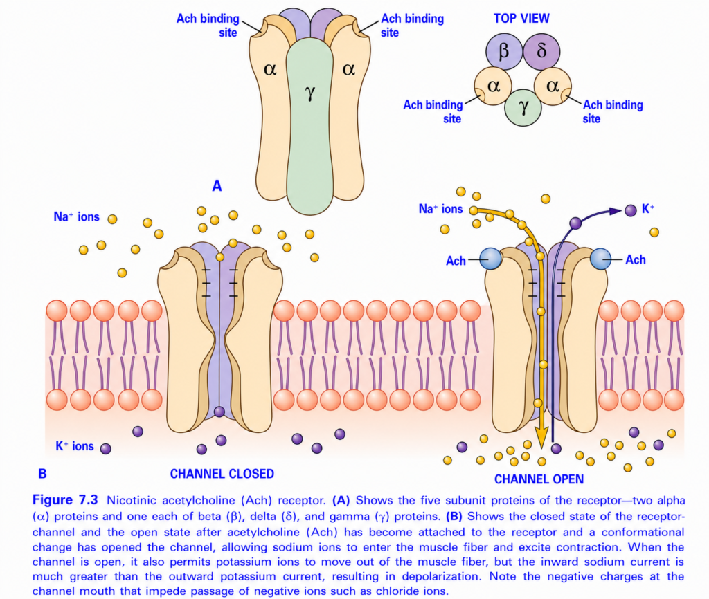
Nicotinic Acetylcholine (ACh) Receptor (Figure 7.3)
Easiest & Self learning Summary
This figure explains how acetylcholine (ACh) opens the nicotinic receptor channel, allowing sodium (Na⁺) to enter the muscle fiber, which starts muscle contraction.
Think of the receptor as a locked gate on the muscle membrane.
- Without ACh → Gate is closed.
- With ACh → Gate opens.
- Na⁺ enters → Muscle is excited → Muscle contracts.
Main Concept
Acetylcholine binds to nicotinic receptors → Receptor channel opens → Na⁺ enters muscle → Muscle depolarizes → Muscle action potential develops → Muscle contracts
Overall Structure of the Figure
The figure has two main parts:
A. Structure of the Nicotinic ACh Receptor
Shows:
- Five protein subunits
- Acetylcholine binding sites
- Top view of the receptor
B. Function of the Receptor
Shows:
- Closed channel
- Open channel
- Sodium entering
- Potassium leaving
PART A – Structure of the Nicotinic Receptor
1. The Nicotinic Receptor is a Protein Channel
The receptor is located in the motor end plate of the muscle membrane.
It is both:
- A receptor (binds acetylcholine)
- An ion channel (allows ions to pass)
Therefore, it is called a ligand-gated ion channel because it opens only when ACh (the ligand) binds.
2. Five Protein Subunits
The receptor is made of five protein subunits arranged in a circle.
They are:
- 2 Alpha (α) subunits
- 1 Beta (β) subunit
- 1 Delta (δ) subunit
- 1 Gamma (γ) subunit (in fetal muscle; in adult muscle, γ is replaced by ε)
Together they form a central pore (channel).
Easy Memory
2 Alpha
1 Beta
1 Delta
1 Gamma
------------
Total = 5 subunits
3. Acetylcholine Binding Sites
Notice that both binding sites are located on the two α (alpha) subunits.
Important Rule
One ACh molecule is not enough.
The channel opens only when TWO acetylcholine molecules bind simultaneously, one to each α subunit.
Concept
Think of a safe with two locks.
Both keys must be inserted before the safe opens.
Similarly,
both ACh molecules must bind before the channel opens.. Top View
The top view shows the five subunits arranged around a central channel.
      β
   α     δ
      γ
   α
The central hole is the ion channel.
PART B – Function of the Receptor
1. Channel Closed (Left Side)
Before acetylcholine arrives,
the receptor channel remains closed.
Result
- No Na⁺ enters.
- No K⁺ leaves.
- Muscle remains relaxed.
2. Acetylcholine Arrives
Acetylcholine released from the nerve terminal diffuses across the synaptic cleft.
Two ACh molecules bind to the two α subunits.
3. Conformational Change
Binding of ACh changes the shape of the receptor.
This is called a conformational change.
Result
The channel opens.
4. Channel Open
Once opened,
the receptor allows positive ions to move.
5. Sodium (Na⁺) Moves Into the Muscle
Large numbers of sodium ions enter the muscle fiber.
Why?
- Na⁺ concentration is much higher outside the cell.
- The inside of the muscle is negatively charged.
Therefore,
Na⁺ rushes inward.
Result
The muscle membrane becomes less negative (depolarizes).
This produces the end-plate potential (EPP).
6. Potassium (K⁺) Moves Out
Some potassium ions leave the muscle through the same open channel.
However,
the amount of K⁺ leaving is much smaller than the amount of Na⁺ entering.
Why Does Depolarization Occur?
Because:
Na⁺ entering > K⁺ leaving
The inward movement of positive charge is greater than the outward movement.
Result
The inside of the muscle becomes more positive.
This is depolarization.
7. What Happens After Depolarization?
Depolarization opens nearby voltage-gated Na⁺ channels.
This generates a muscle action potential.
The action potential spreads over the muscle membrane and into the T-tubules.
This triggers calcium release from the sarcoplasmic reticulum, allowing actin and myosin to interact and produce muscle contraction.
8. Negative Charges at the Channel Mouth
The figure also shows negative charges lining the entrance of the receptor channel.
Their Function
They attract positively charged ions (Na⁺ and K⁺) and repel negatively charged ions, such as chloride (Cl⁻).
This helps ensure that the channel mainly conducts cations, which are required for depolarization.
Complete Sequence of Events
Step 1
A nerve impulse reaches the motor nerve terminal.
↓
Step 2
Acetylcholine is released into the synaptic cleft.
↓
Step 3
Two ACh molecules bind to the two α subunits of the nicotinic receptor.
↓
Step 4
The receptor undergoes a conformational change.
↓
Step 5
The receptor channel opens.
↓
Step 6
Na⁺ enters the muscle fiber, while a smaller amount of K⁺ leaves.
↓
Step 7
An end-plate potential develops.
↓
Step 8
Voltage-gated Na⁺ channels open.
↓
Step 9
A muscle action potential is generated.
↓
Step 10
Calcium is released inside the muscle.
↓
Step 11
The muscle contracts.
Easy Flow Diagram
Motor nerve impulse
        ↓
Acetylcholine released
        ↓
Two ACh molecules bind α subunits
        ↓
Nicotinic receptor opens
        ↓
Na⁺ enters (major)
K⁺ leaves (minor)
        ↓
End-plate potential
        ↓
Voltage-gated Na⁺ channels open
        ↓
Muscle action potential
        ↓
Calcium release
        ↓
Muscle contraction
Simple Real-Life Analogy
Imagine a double-door security gate.
- The gate = Nicotinic receptor
- Two locks = Two α subunits
- Two keys = Two acetylcholine molecules
- Opening the gate = Channel opening
- People entering = Na⁺ ions
- A few people leaving = K⁺ ions
- Factory starting = Muscle contraction
Unless both keys are inserted, the gate stays closed.portant Points from Figure 7.3
- The nicotinic acetylcholine receptor is a ligand-gated cation channel located on the motor end plate of skeletal muscle.
- It consists of five subunits: two α, one β, one δ, and one γ (γ is replaced by ε in adult skeletal muscle).
- The two acetylcholine binding sites are located on the two α subunits.
- Two ACh molecules must bind simultaneously to open the channel.
- Binding causes a conformational change, opening the central pore.
- The open channel allows Na⁺ to enter the muscle and a smaller amount of K⁺ to leave.
- Because Na⁺ influx is much greater than K⁺ efflux, the muscle membrane depolarizes, producing the end-plate potential.
- This depolarization activates voltage-gated Na⁺ channels, generating a muscle action potential that leads to calcium release and skeletal muscle contraction.
- Negatively charged residues at the channel entrance attract cations and help exclude anions such as Cl⁻, promoting efficient excitation.
KEY CONCEPT (Figure 7.3)
The nicotinic acetylcholine receptor is a five-subunit ligand-gated ion channel located on the motor end plate. When two acetylcholine molecules bind to the two α subunits, the receptor changes shape and opens its central pore. This permits a large influx of Na⁺ and a smaller efflux of K⁺, resulting in depolarization (end-plate potential). The depolarization then activates nearby voltage-gated Na⁺ channels, producing a muscle action potential that triggers calcium release and ultimately skeletal muscle contraction.
DESTRUCTION OF THE RELEASED ACETYLCHOLINE BY ACETYLCHOLINESTERASE
Destruction of Acetylcholine
- After acetylcholine (ACh) is released into the synaptic cleft, it binds to and activates nicotinic acetylcholine receptors on the muscle membrane.
- Acetylcholine continues to stimulate these receptors as long as it remains in the synaptic cleft.
Role of Acetylcholinesterase
- Acetylcholinesterase (AChE) is the enzyme responsible for rapidly breaking down acetylcholine.
- Most acetylcholinesterase is attached to the spongy layer of fine connective tissue within the synaptic cleft.
- It is located between the presynaptic nerve terminal and the postsynaptic muscle membrane.
- Soon after acetylcholine is released:
- Acetylcholinesterase rapidly destroys it.
- This occurs within a few milliseconds.
Diffusion of Acetylcholine
- A small amount of acetylcholine diffuses out of the synaptic cleft.
- Once it leaves the synaptic cleft, it can no longer act on the muscle fiber membrane.
Importance of Rapid Removal
- Acetylcholine remains in the synaptic cleft for only a few milliseconds.
- This short duration is sufficient to excite the muscle fiber and generate a muscle action potential.
- After the muscle fiber has produced its initial action potential, rapid destruction and removal of acetylcholine prevent continuous stimulation of the muscle.
- Therefore, each nerve impulse produces only one muscle action potential, allowing the muscle to relax and be ready for the next nerve impulse.
KEY CONCEPT
- Acetylcholine activates nicotinic receptors only while it remains in the synaptic cleft.
- Acetylcholinesterase, located in the connective tissue of the synaptic cleft, rapidly destroys acetylcholine within a few milliseconds.
- A small amount of acetylcholine also diffuses away from the synaptic cleft and can no longer stimulate the muscle fiber.
- Rapid removal of acetylcholine terminates neuromuscular transmission and prevents repeated stimulation of the muscle after a single nerve impulse.
END PLATE POTENTIAL AND EXCITATION OF THE SKELETAL MUSCLE FIBER
Formation of the End Plate Potential (EPP)
- When acetylcholine (ACh) binds to nicotinic acetylcholine receptors, the acetylcholine-gated ion channels open. (Fig. 7.3A–B)
- Opening of these channels allows large numbers of Na⁺ ions to enter the muscle fiber.
- At the same time, some K⁺ ions leave the muscle fiber.
- However, Na⁺ entry is much greater than K⁺ exit.
- Therefore, the inside of the muscle fiber becomes less negative (depolarized).
- This local depolarization increases the membrane potential by about 50–75 mV.
- The resulting local electrical change is called the end plate potential (EPP).
Initiation of the Muscle Action Potential
- A depolarization greater than about 20–30 mV is usually sufficient to open nearby voltage-gated Na⁺ channels.
- Once these channels open:
- More Na⁺ enters the muscle fiber.
- This produces additional depolarization.
- More voltage-gated Na⁺ channels open.
- This self-regenerative process generates a muscle action potential.
- The action potential then spreads along the entire muscle fiber membrane, leading to muscle contraction.
End Plate Potentials Shown in Fig. 7.4
Fig. 7.4 illustrates three different end plate potentials.
End Plate Potential A
- End plate potential A is too small to produce a muscle action potential.
- It causes only a small local depolarization.
- The weak response occurs because the muscle has been poisoned with curare.
- Curare:
- Competes with acetylcholine for nicotinic receptors.
- Blocks the binding of acetylcholine.
- Prevents opening of acetylcholine-gated ion channels.
- Therefore, insufficient Na⁺ enters the muscle fiber.
End Plate Potential B
- End plate potential B is large enough to reach threshold.
- It opens nearby voltage-gated Na⁺ channels.
- A self-regenerative Na⁺ influx occurs.
- This generates a muscle action potential.
- The action potential spreads throughout the muscle fiber and produces contraction.
End Plate Potential C
Effect of Curare and Botulinum Toxin
Curare
- Blocks nicotinic acetylcholine receptors by competing with acetylcholine.
- Prevents opening of acetylcholine-gated channels.
- Reduces the end plate potential.
- May prevent generation of a muscle action potential.
Botulinum Toxin
- Decreases acetylcholine release from the presynaptic nerve terminal.
- Produces a smaller end plate potential.
- May prevent initiation of a muscle action potential.
KEY CONCEPT
- Binding of acetylcholine opens nicotinic receptor channels, allowing a large influx of Na⁺ and a smaller efflux of K⁺ (Fig. 7.3A–B).
- The resulting depolarization of about 50–75 mV is called the end plate potential (EPP).
- If the EPP exceeds the threshold (about 20–30 mV), nearby voltage-gated Na⁺ channels open, generating a self-regenerative muscle action potential.
- The muscle action potential spreads along the muscle fiber and initiates contraction.
- In Fig. 7.4, end plate potential B reaches threshold and produces an action potential, whereas A and C are too weak.
- Curare weakens the EPP by blocking nicotinic acetylcholine receptors, while botulinum toxin weakens the EPP by reducing acetylcholine release from the nerve terminal.
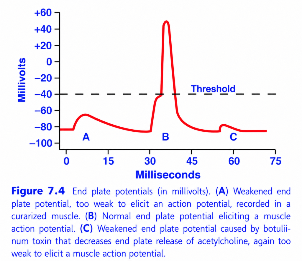
nd Plate Potentials (EPPs) and Muscle Action Potential (Figure 7.4) – Easy Self learning Summary for MBBS
This figure explains what happens at the neuromuscular junction (NMJ) after a motor nerve stimulates a skeletal muscle.
It compares three different end plate potentials (EPPs):
- A: Weak end plate potential (Curare poisoning)
- B: Normal end plate potential
- C: Weak end plate potential (Botulinum toxin)
The figure demonstrates one important principle:
Only an end plate potential that reaches the threshold can generate a muscle action potential and produce muscle contraction.
Basic Concept
When a motor nerve reaches the neuromuscular junction:
- The nerve releases acetylcholine (ACh).
- ACh binds to nicotinic receptors on the motor end plate.
- Sodium (Na⁺) enters the muscle fiber.
- The muscle membrane becomes less negative (depolarization).
- This local depolarization is called the End Plate Potential (EPP).
If the EPP reaches the threshold, it triggers a:
➡️ Muscle Action Potential
which spreads over the muscle membrane and causes contraction.
Understanding the Axes
X-axis (Milliseconds)
Shows time after nerve stimulation.
Moving to the right means:
➡️ More time has passed.
Y-axis (Millivolts, mV)
Shows the membrane potential of the muscle fiber.
- Resting membrane potential ≈ −90 mV
- Threshold ≈ −40 mV
The dashed horizontal line represents the:
Threshold Potential
Only if the membrane reaches this level,
a muscle action potential is produced.
Understanding Each Curve
A – Weak End Plate Potential (Curare Poisoning)
What happens?
A small depolarization occurs.
However,
It does not reach the threshold.
Therefore,
No muscle action potential is generated.
Why?
Curare blocks:
Nicotinic acetylcholine receptors (Nm receptors).
Since fewer receptors are available,
less sodium enters the muscle fiber.
Therefore,
The end plate potential is too small.
Easy Concept
Imagine trying to ring a doorbell,
but your finger only presses the button halfway.
The bell never rings.
Similarly,
The depolarization is insufficient to trigger an action potential.
Key Point
Curare:
- Blocks ACh receptors.
- Produces a weak EPP.
- No muscle action potential.
- No muscle contraction.
B – Normal End Plate Potential
This is the normal neuromuscular response.
What happens?
The motor nerve releases a normal amount of acetylcholine.
The end plate depolarizes.
The membrane reaches the threshold (−40 mV).
Immediately,
a muscle action potential is generated.
The membrane rapidly depolarizes to about +50 mV, then repolarizes back toward the resting membrane potential.
Why?
A sufficient number of nicotinic receptors are activated.
Enough sodium enters the muscle fiber.
The threshold is reached.
Voltage-gated sodium channels open.
This produces the muscle action potential.
Easy Concept
Imagine pressing a doorbell completely.
The bell rings immediately.
Similarly,
The threshold is reached,
and an action potential is generated.
Key Point
Normal ACh release → Threshold reached → Muscle action potential → Muscle contraction.
C – Weak End Plate Potential (Botulinum Toxin)
What happens?
A small end plate potential is produced.
It again fails to reach the threshold.
No muscle action potential occurs.
Why?
Botulinum toxin prevents:
Acetylcholine release from the motor nerve terminal.
Since very little acetylcholine is released,
only a few receptors are activated.
Therefore,
the end plate potential remains too small.
Easy Concept
Imagine the doorbell works normally,
but no one presses it.
Since no signal is sent,
the bell never rings.
Similarly,
very little acetylcholine is released,
so no action potential is generated.
Key Point
Botulinum toxin:
- Prevents ACh release.
- Produces a weak EPP.
- No muscle action potential.
- Muscle paralysis occurs.
Why is Curve B So Much Larger?
Curve B includes two electrical events:
1. End Plate Potential (EPP)
A small local depolarization produced by acetylcholine.
2. Muscle Action Potential
Once the threshold is reached,
voltage-gated sodium channels open.
This produces the large spike.
Why Don’t A and C Produce the Large Spike?
Because their end plate potentials never reach the threshold.
Without reaching threshold,
voltage-gated sodium channels remain closed.
Therefore,
no muscle action potential occurs.
Difference Between End Plate Potential and Muscle Action Potential
| End Plate Potential (EPP) | Muscle Action Potential |
|---|---|
| Local depolarization | Propagated electrical impulse |
| Produced by acetylcholine | Produced by voltage-gated Na⁺ channels |
| Graded response | All-or-none response |
| Cannot spread far | Travels along the entire muscle fiber |
| May or may not reach threshold | Occurs only if threshold is reached |
Comparison of A, B, and C
| Feature | A (Curare) | B (Normal) | C (Botulinum Toxin) |
|---|---|---|---|
| ACh release | Normal | Normal | ↓ Decreased |
| Nicotinic receptors | Blocked | Normal | Normal |
| End plate potential | Weak | Normal | Weak |
| Threshold reached? | ✘ No | ✔ Yes | ✘ No |
| Muscle action potential | ✘ No | ✔ Yes | ✘ No |
| Muscle contraction | ✘ No | ✔ Yes | ✘ No |
Clinical Importance
Curare Poisoning
Mechanism
Blocks nicotinic acetylcholine receptors at the neuromuscular junction.
Result
- Weak EPP
- No muscle action potential
- Flaccid paralysis
Botulism
Mechanism
Blocks acetylcholine release from motor nerve terminals.
Result
- Weak EPP
- No muscle action potential
- Flaccid paralysis
Normal Neuromuscular Transmission
- Adequate ACh release
- Normal receptor activation
- Threshold reached
- Muscle action potential generated
- Normal contraction
Quick Memory Table
| Condition | Main Defect | Result |
|---|---|---|
| Normal | Normal ACh release and receptors | Muscle action potential and contraction |
| Curare | Blocks nicotinic ACh receptors | Weak EPP, no action potential |
| Botulinum toxin | Blocks ACh release | Weak EPP, no action potential |
Easy Memory Trick
Curare = “Can’t Catch the Signal”
- ACh is released.
- Receptors are blocked.
- Signal cannot be received.
Botulinum = “No Bullet Sent”
- Nerve fails to release ACh.
- No signal reaches the muscle.
Normal = “Message Sent and Received”
- ACh released.
- Receptors activated.
- Threshold reached.
- Muscle contracts.
Key Concept
This figure illustrates that an end plate potential (EPP) is a local, graded depolarization produced when acetylcholine (ACh) binds to nicotinic receptors at the neuromuscular junction. An EPP must reach the threshold potential (about −40 mV) to activate voltage-gated sodium channels and generate a muscle action potential, which then spreads along the muscle fiber and initiates contraction. In normal neuromuscular transmission (B), sufficient ACh is released, the threshold is reached, and a muscle action potential is produced. In curare poisoning (A), ACh release is normal but nicotinic receptors are blocked, producing an EPP that is too small to reach threshold. In botulinum toxin poisoning (C), ACh release is markedly reduced, so only a small EPP develops, which also fails to reach threshold. Thus, both curare and botulinum toxin prevent muscle contraction, but by different mechanisms: curare blocks ACh receptors, whereas botulinum toxin blocks ACh release.
SAFETY FACTOR FOR TRANSMISSION AT THE NEUROMUSCULAR JUNCTION—FATIGUE OF THE JUNCTION
Safety Factor of the Neuromuscular Junction
- Under normal conditions, every nerve impulse reaching the neuromuscular junction generates a large end plate potential (EPP).
- The end plate potential is about three times greater than the minimum required to produce a muscle action potential.
- This extra margin of depolarization is called the safety factor.
- Because of this high safety factor:
- Every normal nerve impulse reliably produces a muscle action potential.
- Muscle contraction occurs consistently, even if there are small changes in neuromuscular transmission.
Fatigue of the Neuromuscular Junction
- If a motor nerve is stimulated continuously at a very high frequency (more than 100 impulses per second) for several minutes:
- The stored acetylcholine-containing synaptic vesicles gradually become depleted.
- Less acetylcholine is released with each nerve impulse.
- As a result:
- The end plate potential becomes progressively smaller.
- Eventually, it may not reach the threshold needed to generate a muscle action potential.
- When this occurs, the nerve impulse fails to activate the muscle fiber.
- This condition is called fatigue of the neuromuscular junction.
Cause of Neuromuscular Junction Fatigue
- The main cause is the depletion of acetylcholine synaptic vesicles in the presynaptic nerve terminal.
- With fewer vesicles available:
- Less acetylcholine is released into the synaptic cleft.
- Fewer nicotinic receptors are activated.
- The end plate potential decreases.
Comparison with Central Nervous System Synapses
- Fatigue of the neuromuscular junction occurs by the same basic mechanism as fatigue at synapses in the central nervous system (CNS).
- In both cases:
- Repeated excessive stimulation depletes neurotransmitter stores, reducing synaptic transmission.
Occurrence Under Normal Conditions
- During normal daily activities, fatigue of the neuromuscular junction is very uncommon.
- It occurs only during extremely prolonged and exhausting muscle activity.
- Therefore, under normal physiological conditions, the high safety factor ensures reliable transmission from nerve to muscle.
KEY CONCEPT
- The neuromuscular junction has a high safety factor because each nerve impulse produces an end plate potential about three times greater than the threshold needed to trigger a muscle action potential.
- This safety factor ensures dependable nerve-to-muscle transmission under normal conditions.
- Prolonged stimulation at frequencies greater than 100 impulses per second for several minutes can deplete acetylcholine vesicles.
- Reduced acetylcholine release decreases the end plate potential, and transmission may fail, causing neuromuscular junction fatigue.
- This mechanism is similar to fatigue at central nervous system synapses.
- Neuromuscular junction fatigue is rare in normal physiology and occurs only during extremely strenuous muscle activity.
ACETYLCHOLINE FORMATION AND RELEASE
1. Formation and Transport of Synaptic Vesicles
- Small synaptic vesicles, about 40 nanometers in diameter, are formed by the Golgi apparatus in the cell body of the motor neuron located in the anterior horn of the spinal cord.
- These vesicles are transported through the axon by axoplasmic flow from the spinal cord to the nerve terminal at the neuromuscular junction.
- About 300,000 synaptic vesicles are normally stored in the nerve terminal of one motor end plate.
2. Synthesis and Storage of Acetylcholine
- Acetylcholine (ACh) is synthesized in the cytosol of the nerve terminal.
- Immediately after synthesis, acetylcholine is transported into the synaptic vesicles.
- Inside the vesicles, acetylcholine is stored in a highly concentrated form.
- Each synaptic vesicle contains about 10,000 acetylcholine molecules.
3. Release of Acetylcholine (Exocytosis)
- When an action potential reaches the nerve terminal, it opens numerous voltage-gated Ca²⁺ channels.
- Because the nerve terminal contains many of these channels, Ca²⁺ rapidly enters the terminal.
- The intracellular Ca²⁺ concentration increases about 100-fold.
- The increased Ca²⁺ concentration:
- Greatly accelerates fusion of synaptic vesicles with the presynaptic membrane.
- Increases the rate of vesicle fusion about 10,000-fold.
- The fused vesicles rupture and release acetylcholine into the synaptic cleft by exocytosis.
- About 125 synaptic vesicles release acetylcholine with each action potential.
4. Breakdown and Recycling of Acetylcholine
- Within a few milliseconds, acetylcholinesterase breaks down acetylcholine into:
- Acetate ion
- Choline
- Choline is actively transported back into the nerve terminal.
- The recycled choline is used to synthesize new acetylcholine molecules.
- The entire process—from acetylcholine release to its breakdown and choline recycling—takes only about 5–10 milliseconds.
5. Recycling of Synaptic Vesicles
- The nerve terminal contains only enough synaptic vesicles for a few thousand nerve impulses.
- Therefore, new vesicles must be continuously formed to maintain neuromuscular transmission.
- Within a few seconds after acetylcholine release:
- Coated pits appear in the presynaptic membrane.
- These coated pits are produced by contractile proteins, especially clathrin, which remains attached to the membrane where the original vesicles fused.
- Within about 20 seconds:
- Clathrin contracts, causing the coated pits to pinch off from the membrane.
- This forms new synaptic vesicles.
- Within another few seconds:
- Acetylcholine is transported into these newly formed vesicles.
- The vesicles are then ready for the next cycle of neurotransmitter release.
Sequence of Acetylcholine Formation and Release
- Golgi apparatus forms synaptic vesicles in the motor neuron cell body.
- Vesicles are transported by axoplasmic flow to the nerve terminal.
- Acetylcholine is synthesized in the cytosol and stored inside vesicles.
- An action potential opens voltage-gated Ca²⁺ channels.
- Ca²⁺ enters the nerve terminal and triggers vesicle fusion.
- About 125 vesicles release acetylcholine by exocytosis.
- Acetylcholinesterase breaks acetylcholine into acetate and choline.
- Choline is reabsorbed and reused to synthesize new acetylcholine.
- Clathrin-mediated endocytosis forms new synaptic vesicles.
- The recycled vesicles are refilled with acetylcholine for the next nerve impulse.
KEY CONCEPT
- Synaptic vesicles (≈40 nm) are formed by the Golgi apparatus in the motor neuron cell body and transported to the neuromuscular junction.
- About 300,000 vesicles are stored in one motor nerve terminal, and each vesicle contains about 10,000 acetylcholine molecules.
- An action potential opens voltage-gated Ca²⁺ channels, increasing intracellular Ca²⁺ about 100-fold and enhancing vesicle fusion about 10,000-fold.
- Approximately 125 vesicles release acetylcholine by exocytosis with each nerve impulse.
- Acetylcholinesterase rapidly breaks acetylcholine into acetate and choline; choline is actively reabsorbed and recycled to synthesize new acetylcholine within 5–10 milliseconds.
- New synaptic vesicles are continuously regenerated by clathrin-mediated endocytosis, refilled with acetylcholine, and reused for subsequent neurotransmission.
DRUGS THAT STIMULATE THE MUSCLE FIBER BY ACETYLCHOLINE-LIKE ACTION
Acetylcholine-Like Drugs
- Some drugs produce effects similar to acetylcholine (ACh) at the neuromuscular junction.
- These drugs bind to nicotinic acetylcholine receptors on the motor end plate and stimulate the muscle fiber.
- Examples include:
- Methacholine
- Carbachol
- Nicotine
Difference Between These Drugs and Acetylcholine
- Although these drugs act like acetylcholine, they differ in one important way:
- Acetylcholine:
- Is rapidly broken down by acetylcholinesterase.
- Its action lasts only a few milliseconds.
- Methacholine, carbachol, and nicotine:
- Are not destroyed by acetylcholinesterase, or
- Are broken down very slowly.
- Therefore, their effects last much longer, often from several minutes to several hours.
Mechanism of Action
- These drugs bind to nicotinic acetylcholine receptors at the motor end plate.
- This opens acetylcholine-gated ion channels.
- As a result:
- Na⁺ enters the muscle fiber.
- The motor end plate becomes depolarized.
- This creates localized areas of persistent depolarization at the motor end plate.
Repeated Action Potential Generation
- After each muscle contraction:
- The muscle fiber repolarizes and recovers.
- However, because the drug continues to stimulate the acetylcholine receptors, the motor end plate remains partially depolarized.
- This persistent depolarization causes continuous leakage of ions.
- The ion leakage repeatedly reaches threshold, generating new muscle action potentials.
- Consequently, the muscle contracts repeatedly.
Effect on the Muscle
- Continuous stimulation produces repeated muscle contractions.
- This results in a state of sustained muscle spasm.
KEY CONCEPT
- Methacholine, carbachol, and nicotine mimic the action of acetylcholine at the neuromuscular junction.
- Unlike acetylcholine, these drugs are not destroyed by acetylcholinesterase or are degraded very slowly, so their effects persist for minutes to hours.
- They activate nicotinic acetylcholine receptors at the motor end plate, causing persistent local depolarization.
- This persistent depolarization repeatedly initiates muscle action potentials after each recovery, producing continuous muscle contractions and muscle spasm.
Drugs That Stimulate the Neuromuscular Junction By Inactivating Acetylcholinesterase
- Some drugs stimulate the neuromuscular junction by inactivating acetylcholinesterase.
- These drugs include:
- Neostigmine
- Physostigmine
- Diisopropyl fluorophosphate
- These drugs inactivate acetylcholinesterase, so the enzyme can no longer break down acetylcholine.
- As a result, acetylcholine remains in the synaptic cleft for a longer time.
- With each new nerve impulse, more acetylcholine accumulates in the synaptic cleft.
- The accumulated acetylcholine repeatedly stimulates the muscle fiber.
- This repeated stimulation causes muscle spasm, even when only a few nerve impulses reach the muscle.
- Severe muscle spasm can involve the larynx (voice box).
- Laryngeal spasm can block the airway, leading to asphyxiation (death due to inability to breathe).
- Neostigmine and physostigmine bind to acetylcholinesterase and inactivate it for several hours.
- After several hours, these drugs separate from the enzyme, allowing acetylcholinesterase to become active again.
- Diisopropyl fluorophosphate inactivates acetylcholinesterase for several weeks.
- Because its effect lasts for a very long time, diisopropyl fluorophosphate is a powerful nerve gas poison and is particularly lethal.
KEY CONCEPT
- Neostigmine, physostigmine, and diisopropyl fluorophosphate stimulate the neuromuscular junction by inactivating acetylcholinesterase.
- Inactivation of acetylcholinesterase prevents the breakdown of acetylcholine, causing it to accumulate in the synaptic cleft.
- Accumulated acetylcholine repeatedly stimulates the muscle fiber, producing muscle spasm.
- Severe stimulation may cause laryngeal spasm, leading to asphyxiation.
- Neostigmine and physostigmine act for several hours, whereas diisopropyl fluorophosphate acts for several weeks and is highly lethal.
Drugs That Block Transmission at the Neuromuscular Junction
- A group of drugs called curariform drugs blocks the transmission of nerve impulses from the motor nerve to the skeletal muscle.
- These drugs prevent nerve impulses from producing muscle contraction.
- d-Tubocurarine is an example of a curariform drug.
- d-Tubocurarine blocks the action of acetylcholine on nicotinic acetylcholine receptors of the muscle fiber.
- Because acetylcholine cannot bind to its receptors, the acetylcholine-gated ion channels do not open properly.
- As a result, not enough Na⁺ enters the muscle fiber.
- Therefore, the muscle membrane does not become sufficiently permeable to Na⁺.
- The end plate potential remains too small to reach threshold.
- Consequently, a muscle action potential is not generated.
- Since no muscle action potential is produced, the nerve impulse cannot pass from the nerve ending to the muscle fiber.
- Therefore, muscle contraction is prevented.
KEY CONCEPT
- Curariform drugs block transmission at the neuromuscular junction.
- d-Tubocurarine blocks the action of acetylcholine on nicotinic acetylcholine receptors of the muscle fiber.
- Blocking these receptors prevents sufficient Na⁺ entry into the muscle fiber.
- The end plate potential does not reach threshold, so no muscle action potential is generated.
- As a result, nerve impulses fail to stimulate the muscle, and muscle contraction is prevented.
Myasthenia Gravis Causes Muscle Weakness
- Myasthenia gravis is a disease that causes muscle weakness.
- It occurs in about 1 out of every 20,000 people.
- The weakness occurs because the neuromuscular junction cannot transmit enough nerve signals to the muscle fibers.
- Myasthenia gravis is believed to be an autoimmune disease.
- In this disease, the body produces antibodies against its own nicotinic acetylcholine receptors at the postsynaptic neuromuscular junction.
- These antibodies block or destroy the acetylcholine receptors.
- As a result, fewer acetylcholine receptors are available for acetylcholine to bind.
- Therefore, the end plate potential (EPP) becomes too small.
- The weak end plate potential cannot open enough voltage-gated Na⁺ channels.
- Because of this, the muscle fiber does not depolarize.
- Without sufficient depolarization, a muscle action potential is not generated.
- As a result, the muscle contracts weakly, causing muscle weakness.
- If the disease becomes severe, the respiratory muscles become very weak.
- Severe weakness of the respiratory muscles can cause respiratory failure and may lead to death.
- The symptoms can usually be improved for several hours by giving neostigmine or another anticholinesterase drug.
- These drugs inhibit acetylcholinesterase, allowing more acetylcholine to accumulate in the synaptic cleft.
- The increased amount of acetylcholine improves stimulation of the remaining acetylcholine receptors.
- Within a few minutes, many patients recover almost normal muscle function.
- After a few hours, the effect wears off, and another dose of neostigmine is needed.
KEY CONCEPT
- Myasthenia gravis is an autoimmune disease that causes muscle weakness by impairing neuromuscular transmission.
- Autoantibodies block or destroy nicotinic acetylcholine receptors at the postsynaptic neuromuscular junction.
- Reduced receptor number produces a weak end plate potential that cannot adequately open voltage-gated Na⁺ channels.
- The muscle fiber fails to depolarize sufficiently, resulting in weak muscle contraction.
- Severe disease may cause respiratory failure due to weakness of the respiratory muscles.
- Neostigmine and other anticholinesterase drugs improve symptoms by increasing acetylcholine concentration in the synaptic cleft for several hours.
Made by Self learning Dr sheen
