


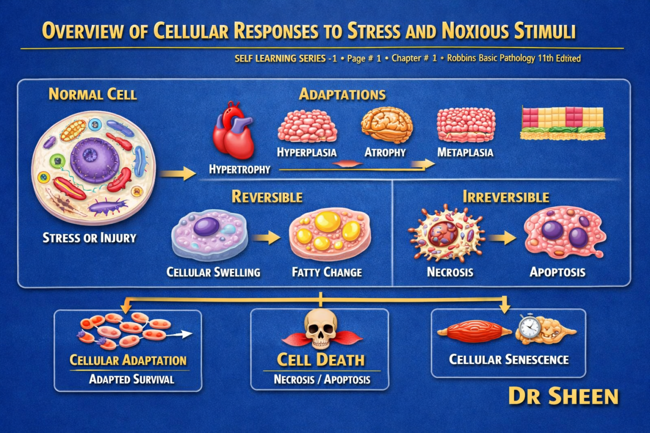
- Cells constantly interact with their surroundings.
- They continuously adjust their structure and function.
- These changes help them meet new demands and respond to outside stresses.
- The main goal is to keep the body’s internal condition stable (homeostasis).
- When cells face normal stress or harmful stimuli, they can adapt.
- Adaptation helps the cell reach a new stable state.
- This allows the cell to stay alive and continue working normally.
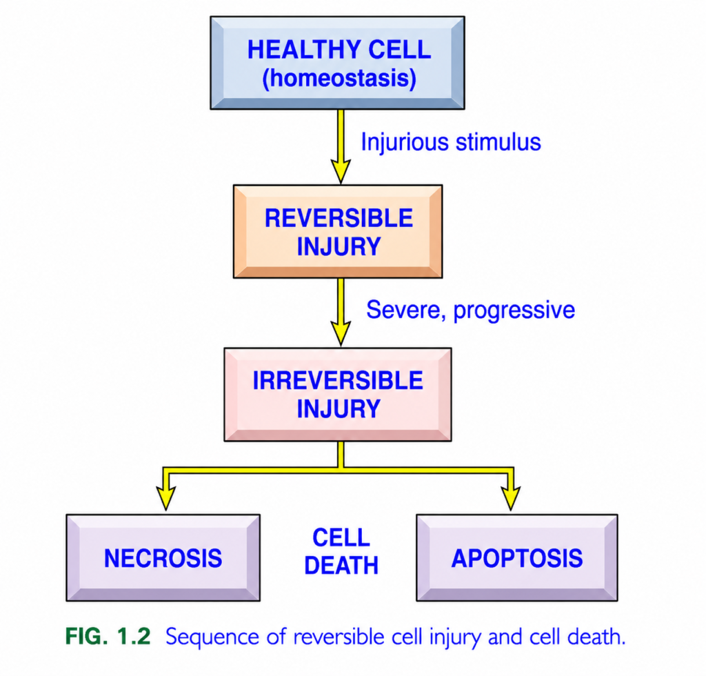
- If the stress is greater than the cell’s ability to adapt, or if the harmful stimulus is too severe, cell injury occurs (Fig. 1.2).
- Mild or short-term cell injury is reversible.
- If the harmful stimulus is removed, the cell returns to normal.
- Homeostasis is restored.
- Severe or long-lasting injury causes irreversible cell injury.
- Irreversible injury leads to cell death.
- Cell death is an important event in the development of many diseases.
- Cell injury is the basic cause of all diseases.
- This chapter explains:
- The causes of cell injury.
- The mechanisms of reversible cell injury and cell death.
- The consequences of reversible injury and cell death.
- Cellular adaptations to stress.
- Deposition of abnormal substances in cells and tissues.
- Cell aging.
KEY CONCEPT
- Cells maintain a stable internal environment by homeostasis.
- Cells can adapt to normal stress and remain functional.
- If stress is too severe, cell injury occurs (Fig. 1.2).
- Reversible injury allows recovery and restoration of homeostasis.
- Irreversible injury leads to cell death.
- Cell injury is the fundamental basis of all diseases.
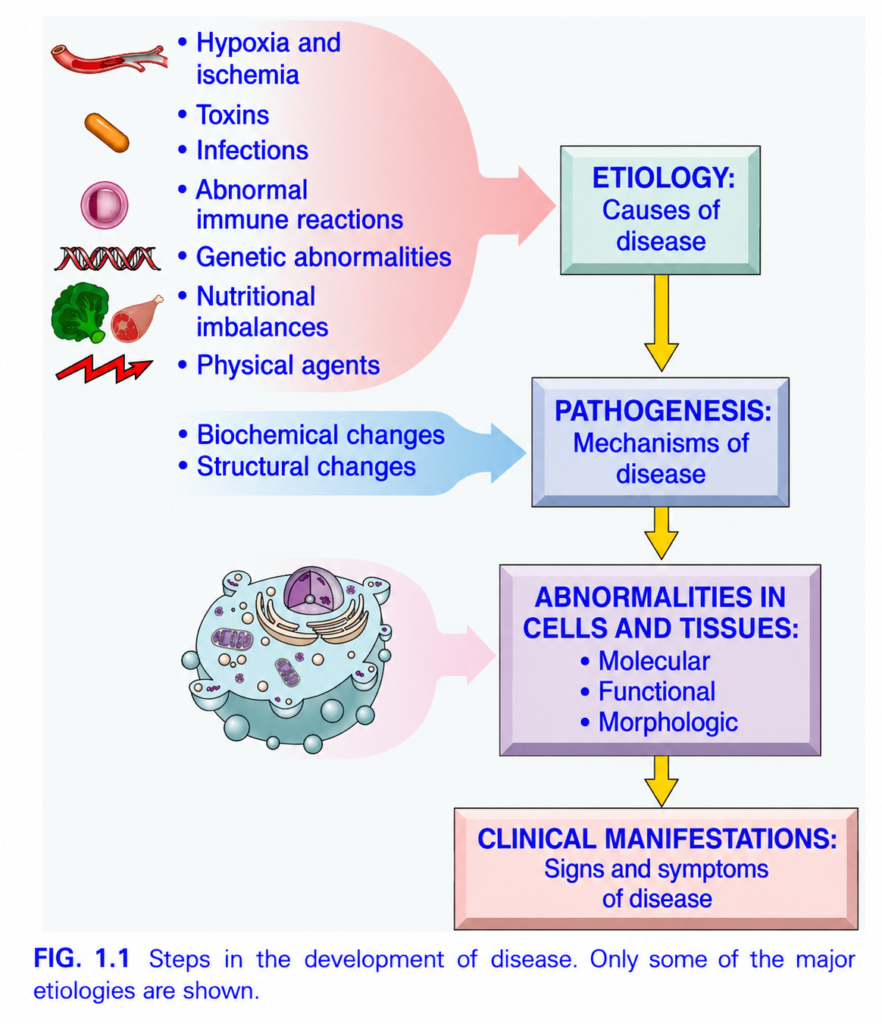
CAUSES OF CELL INJURY
- The major causes of cell injury are grouped into different categories.
- Hypoxia and ischemia
- Hypoxia means a lack of oxygen.
- Ischemia means reduced blood supply.
- These are the most common causes of cell injury.
- Both reduce the oxygen needed to produce energy for cell survival.
- Ischemia also decreases the supply of nutrients.
- The most common cause of hypoxia is ischemia due to blockage of an artery.
- Hypoxia can also occur because:
- Lung diseases reduce oxygen in the blood.
- Anemia reduces the oxygen-carrying capacity of blood.
- Toxins
- Harmful substances are encountered every day.
- Examples include:
- Air pollutants
- Insecticides
- Carbon monoxide
- Asbestos
- Cigarette smoke
- Ethanol (alcohol)
- Drugs
- Some medicines can also injure cells if:
- The patient is susceptible.
- They are used in excessive amounts.
- They are used incorrectly.
- Infectious agents
- Many infectious organisms can damage cells.
- These include:
- Viruses
- Bacteria
- Fungi
- Parasites
- They injure cells by:
- Releasing toxins.
- Triggering harmful immune responses.
- Immunologic reactions
- The immune system normally protects the body from infections.
- Sometimes immune reactions damage normal cells and tissues.
- Examples include:
- Autoimmune diseases (attack on the body’s own tissues)
- Allergic reactions to environmental substances
- Excessive or long-lasting immune responses to microbes
- These immune reactions cause inflammation.
- Inflammation is often responsible for cell and tissue damage.
- Genetic abnormalities
- Chromosomal abnormalities and gene mutations can cause disease.
- They may produce:
- Major birth defects, such as Down syndrome.
- Small genetic changes, such as sickle cell anemia.
- Mutations may injure cells by:
- Decreasing protein function.
- Increasing protein function.
- Causing damaged DNA to accumulate.
- Causing misfolded proteins to accumulate.
- These changes can lead to cell death.
- Mutations also play an important role in the development of cancer.
- Nutritional imbalances
- Protein-calorie deficiency is a major cause of cell injury.
- Vitamin deficiencies are also common.
- Excessive food intake can cause obesity.
- Obesity increases the risk of diseases such as:
- Type 2 diabetes
- Atherosclerosis
- Physical agents
- Physical factors can also damage cells.
- Examples include:
- Trauma
- Extremely high or low temperatures
- Radiation
- Electric shock
- Sudden changes in atmospheric pressure
- After understanding the causes of cell injury, the next topics include:
- The process of cell injury.
- The structural (morphologic) changes caused by cell injury.
- The biochemical mechanisms by which harmful stimuli damage cells.
KEY CONCEPT
- Hypoxia = Lack of oxygen.
- Ischemia = Reduced blood supply.
- The major causes of cell injury are:
- Hypoxia and ischemia
- Toxins
- Infectious agents
- Immunologic reactions
- Genetic abnormalities
- Nutritional imbalances
- Physical agents
- All these factors damage cells through different mechanisms and can lead to cell injury or cell death.
SEQUENCE OF EVENTS IN CELL INJURY AND CELL DEATH
- Although different harmful stimuli damage cells in different ways, they produce a similar sequence of structural and morphological changes in most cells.
Reversible Cell Injury
- Reversible cell injury means the cell can recover if the harmful stimulus is removed (Fig. 1.3).
- During reversible injury:
- The cell swells.
- Cell organelles also swell.
- Water enters the cell because energy-dependent ion pumps in the plasma membrane stop working properly.
- In some types of injury:
- Damaged organelles accumulate inside the cell.
- Lipids (fat) also accumulate inside the injured cell.
- Some harmful stimuli produce specific changes in cell organelles.
- One example is the smooth endoplasmic reticulum (smooth ER).
- The smooth ER helps metabolize:
- Alcohol
- Drugs such as barbiturates
- When cells are repeatedly exposed to these chemicals:
- The smooth ER becomes enlarged (hypertrophy).
- This is an adaptive response.
- It can change how the cell processes other drugs.
- Cells adapted to one drug can metabolize other drugs more rapidly if they use the same metabolic system.
- Example:
- A patient taking phenobarbital for epilepsy who drinks more alcohol may develop increased smooth ER activity.
- This increases metabolism of phenobarbital.
- Blood levels of the antiseizure drug fall below the therapeutic range.
- If cell injury becomes severe or continues for a long time:
- The cell reaches the “point of no return.”
- Recovery is no longer possible.
- The cell undergoes death, usually by necrosis.
- There is no single definite structural or biochemical marker of irreversible injury.
- However, irreversible injury is consistently characterized by:
- Failure to restore mitochondrial function.
- Failure to restart oxidative phosphorylation and ATP production.
- Damage to the plasma membrane and intracellular membranes, causing loss of their normal function.
- Loss of the normal structure of DNA and chromatin.
- Damage to lysosomal membranes releases digestive enzymes.
- These enzymes digest the injured cell.
- This enzymatic digestion is the final step of necrosis.
KEY CONCEPT
- Different harmful stimuli produce a similar sequence of cell injury.
- Reversible cell injury allows recovery if the harmful stimulus is removed (Fig. 1.3).
- Reversible injury causes:
- Cell swelling
- Organelle swelling
- Accumulation of damaged organelles and lipids
- The smooth ER enlarges in response to alcohol and certain drugs, increasing drug metabolism.
- Severe or persistent injury reaches the “point of no return” and causes necrosis.
- Irreversible injury is characterized by:
- Failure of mitochondrial ATP production
- Membrane damage
- DNA and chromatin damage
- Lysosomal enzyme release leading to cell digestion (necrosis)
- Fig. 1.3: Reversible cell injury.
- Fig. 1.2: Sequence of reversible cell injury and cell death.
- Fig. 1.1: Steps in the development of disease.
MORPHOLOGY
- The two most common structural (morphologic) changes seen in reversible cell injury are:
- Cellular swelling
- Fatty change
- Cellular swelling (Fig. 1.4B)
- Commonly occurs when cells are injured by:
- Hypoxia
- Toxins
- Other harmful causes
- It may be difficult to see with a light microscope because water is removed during tissue processing.
- It is often easier to recognize when the whole organ is examined.
- If many cells are affected, the organ shows:
- Pale appearance
- Increased firmness (turgor)
- Increased weight
- Under the microscope, small clear vacuoles may be seen in the cytoplasm.
- These vacuoles are enlarged and pinched-off parts of the endoplasmic reticulum (ER).
- This reversible change is also called:
- Hydropic change
- Vacuolar degeneration
- Commonly occurs when cells are injured by:
- Fatty change
- Fatty change appears as lipid (fat) vacuoles inside the cytoplasm.
- It mainly occurs in organs involved in fat metabolism.
- The liver is the most common organ affected.
- During cell injury:
- The cytoplasm becomes more eosinophilic (red staining) with H&E stain.
- This red staining becomes more obvious as the cell progresses toward necrosis.
- Other changes best seen with electron microscopy (eFig. 1.1) include:
- Plasma membrane changes
- Blebbing
- Blunting or distortion of microvilli
- Loosening of connections between adjacent cells
- Mitochondrial changes
- Swelling
- Appearance of phospholipid-rich amorphous densities
- Endoplasmic reticulum (ER) changes
- Dilation of the ER
- Ribosomes detach from the ER
- Polysomes separate into individual ribosomes
- Nuclear changes
- Clumping of chromatin
- Plasma membrane changes
- The cytoplasm may also contain myelin figures.
- Myelin figures are collections of phospholipids.
- They resemble myelin sheaths.
- They are formed from damaged cell membranes.
KEY CONCEPT
- The two major morphologic features of reversible cell injury are:
- Cellular swelling (Fig. 1.4B)
- Fatty change
- Cellular swelling causes:
- Cell enlargement
- Organ pallor
- Increased organ weight
- Hydropic change (vacuolar degeneration)
- Fatty change is the accumulation of lipid vacuoles, mainly in the liver.
- Injured cells also show:
- Increased eosinophilic (red) cytoplasm
- Plasma membrane damage
- Mitochondrial swelling
- ER dilation with ribosome loss
- Chromatin clumping
- Formation of myelin figures from damaged cell membranes.
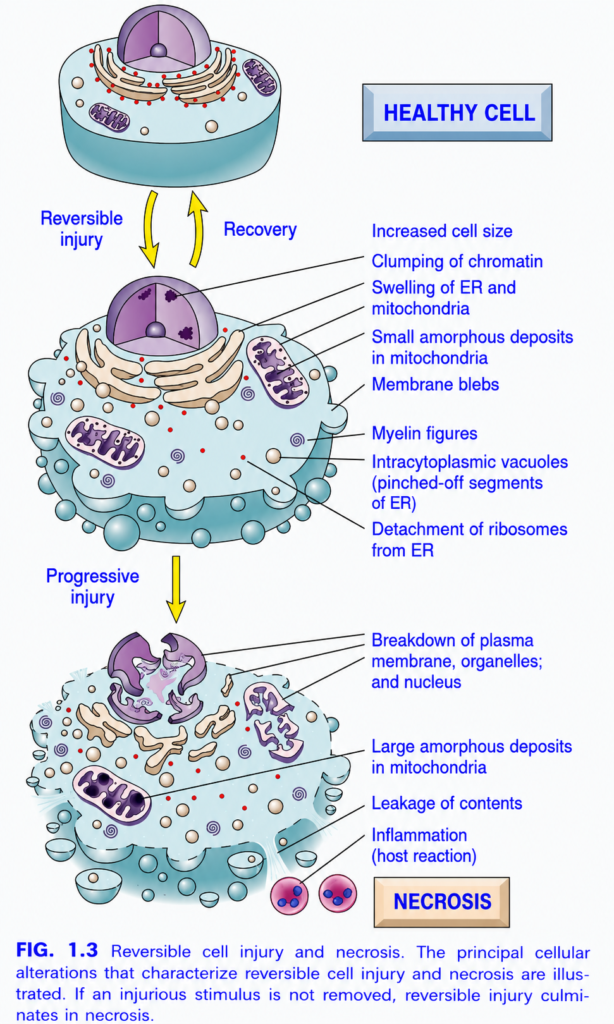
Reversible Cell Injury and Necrosis (Figure 1.3) — Easiest SELF LEARNING Summary
This figure shows the journey of a cell after injury.
A cell has two possible outcomes:
- The injury is mild and temporary → The cell recovers (Reversible injury).
- The injury is severe or continues → The cell dies (Necrosis).
Overall Flow of the Figure
Healthy Cell
      ↓
Cell Injury
      ↓
Reversible Cell Injury
      ↓
 ┌───────────────┐
 │ Injury stops  │────────► Recovery (Normal Cell)
 └───────────────┘
      │
      │ Injury continues
      ▼
Irreversible Cell Injury
      ▼
Necrosis (Cell Death)
Stage 1: Healthy Cell
The top cell is completely normal.
Features
- Normal cell size
- Intact plasma membrane
- Healthy nucleus
- Normal mitochondria
- Rough endoplasmic reticulum (RER) covered with ribosomes
- Normal protein synthesis
- Normal ATP production
Concept
Everything inside the cell is working efficiently.
Stage 2: Reversible Cell Injury
The middle cell has been injured, but it is still alive.
If the cause of injury is removed (e.g., oxygen returns or toxin is removed), the cell can recover completely.
Changes Seen During Reversible Injury
1. Increased Cell Size (Cell Swelling)
The cell becomes larger because water enters the cell.
Why?
- ATP production decreases.
- Sodium–potassium pump fails.
- Sodium accumulates inside the cell.
- Water follows sodium into the cell.
Easy Concept
Pump fails → Sodium stays inside → Water enters → Cell swells
This is the earliest and most common sign of reversible injury.
2. Clumping of Chromatin
Inside the nucleus, chromatin becomes darker and clumps together.
Why?
- Reduced ATP
- Altered nuclear metabolism
Important Point
The nucleus is still intact.
3. Swelling of Endoplasmic Reticulum (ER)
The ER becomes swollen because of water accumulation.
Effect
- Protein production slows.
- Cell functions become less efficient.
4. Swelling of Mitochondria
Mitochondria become enlarged.
Why?
- Water enters mitochondria.
- ATP production decreases.
Important Point
Mitochondria are damaged but still functional.
5. Small Amorphous Deposits in Mitochondria
Small dense deposits appear inside mitochondria.
Meaning
These indicate early mitochondrial injury, but recovery is still possible.6. Membrane Blebs
Small balloon-like bulges form on the plasma membrane.
Why?
- Cell membrane loses stability.
- Cytoskeleton becomes weak.
Important Point
The membrane is still intact, so the cell remains alive.
7. Myelin Figures
Whorled, spiral structures appear in the cytoplasm.
Why?
They are formed from damaged membrane phospholipids.
Meaning
Early membrane injury.
8. Intracytoplasmic Vacuoles
Small clear vacuoles appear in the cytoplasm.
Why?
They are pinched-off segments of the swollen endoplasmic reticulum.
9. Detachment of Ribosomes from ER
Normally, ribosomes are attached to the rough ER.
During injury, they detach.
Result
- Protein synthesis decreases.
- Cell function slows.
Can the Cell Recover?
Yes.
If oxygen supply is restored or the harmful stimulus is removed,
the cell returns to normal.
The arrow labeled Recovery shows this.
Stage 3: Progressive Injury
If the damaging stimulus continues,
the injury becomes more severe.
The cell reaches a point of no return.
Now recovery is no longer possible.
Stage 4: Necrosis (Irreversible Cell Injury)
The bottom cell is dead.
This is called necrosis.
The cell loses its structure completely.
Changes Seen in Necrosis
1. Breakdown of Plasma Membrane
The cell membrane ruptures.
Result
The contents of the cell leak outside.
2. Breakdown of Organelles
Organelles lose their structure.
Examples
- Mitochondria
- ER
- Lysosomes
They can no longer function.
3. Breakdown of the Nucleus
The nucleus is destroyed.
DNA is permanently damaged.
This confirms irreversible cell death.
4. Large Amorphous Deposits in Mitochondria
Large dense deposits appear inside mitochondria.
Meaning
Mitochondria have permanently failed.
ATP production stops completely.
5. Leakage of Cellular Contents
Because the membrane is ruptured,
proteins, enzymes, and other intracellular substances escape into the surrounding tissue and blood.
Clinical Importance
These leaked enzymes are often measured in blood tests.
Examples:
- Troponin → Heart muscle injury
- ALT/AST → Liver injury
- Amylase/Lipase → Pancreatic injury
6. Inflammation (Host Reaction)
Leaked cellular contents trigger inflammation.
White blood cells migrate to the damaged area to remove dead cells.
Important Point
Inflammation is a hallmark of necrosis.
Comparison: Reversible Injury vs. Necrosis
| Feature | Reversible Cell Injury | Necrosis (Irreversible Injury) |
|---|---|---|
| Cell status | Alive | Dead |
| Recovery | Possible | Impossible |
| Cell size | Increased (swelling) | Severe damage and rupture |
| Plasma membrane | Intact with blebs | Broken |
| Mitochondria | Swollen with small deposits | Destroyed with large deposits |
| Endoplasmic reticulum | Swollen | Fragmented |
| Ribosomes | Detached | Lost |
| Nucleus | Chromatin clumping, intact | Broken down |
| Cell contents | Remain inside | Leak outside |
| Inflammation | Absent | Present |
Easy Memory Trick
Reversible Injury = “SWELL”
- S = Swollen cell
- W = Water enters
- E = ER swelling
- L = Little mitochondrial deposits
- L = Life preserved (recovery possible)
Necrosis = “BREAK”
- B = Broken membrane
- R = Ruptured organelles
- E = Enzymes leak out
- A = Acute inflammation
- K = Killed cell (irreversible)
Clinical Examples
Reversible Injury
- Brief ischemia (temporary reduction in blood flow)
- Mild hypoxia
- Early toxin exposure
- Mild thermal injury
➡️ Remove the cause → Cell recovers.
Necrosis
- Prolonged myocardial infarction
- Severe burns
- Major trauma
- Severe bacterial infection
- Long-lasting ischemia
➡️ Cell dies permanently.
Important Points from the Figure
- A healthy cell becomes injured when exposed to harmful stimuli.
- Reversible injury is characterized by cell swelling, chromatin clumping, swelling of the ER and mitochondria, membrane blebs, myelin figures, cytoplasmic vacuoles, and ribosome detachment.
- If the injury is removed at this stage, the cell recovers completely.
- Continued injury leads to irreversible injury (necrosis).
- Necrosis is characterized by rupture of the plasma membrane, destruction of organelles and nucleus, large mitochondrial deposits, leakage of cellular contents, and inflammation.
- The key difference is that reversible injury is recoverable, whereas necrosis is permanent cell death with inflammation.
KEY CONCEPT (Figure 1.3)
This figure demonstrates the progression from a healthy cell to reversible cell injury and finally to necrosis. In reversible injury, the cell remains alive and shows changes such as cell swelling, chromatin clumping, swollen ER and mitochondria, membrane blebs, vacuoles, and ribosome detachment. If the damaging stimulus is removed, the cell can recover completely. If the injury persists, the damage becomes irreversible, leading to necrosis, characterized by membrane rupture, destruction of organelles and nucleus, leakage of intracellular contents, and inflammation. The presence of membrane integrity and the ability to recover distinguish reversible injury from necrosis.
MADE BY SELF LEARNING DR SHEEN.
