


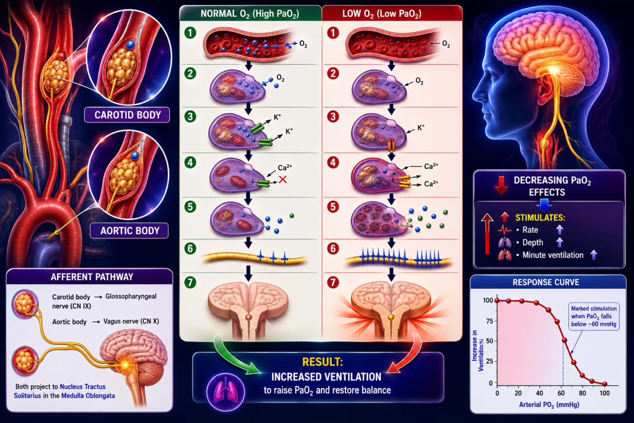
- In addition to the brain respiratory center, the body has another mechanism to control breathing called the peripheral chemoreceptor system.
- Figure 42.4 shows the peripheral chemoreceptor system.
- The peripheral chemoreceptors are special sensory receptors located outside the brain.
- Their main function is to detect changes in blood oxygen (O₂).
- They also respond to:
- Changes in CO₂
- Changes in H⁺ (blood acidity)
- However, their response to CO₂ and H⁺ is much weaker than their response to O₂.
- When these chemoreceptors detect changes in the blood:
- They send nerve signals to the respiratory center in the brain.
- This helps adjust the rate and depth of breathing.
- Most peripheral chemoreceptors are located in the carotid bodies.
- A few chemoreceptors are located in the aortic bodies (Figure 42.4).
- A very small number are also present near other arteries in the thorax and abdomen.
- The carotid bodies are located on both sides at the bifurcation (division) of the common carotid arteries.
- Nerve signals from the carotid bodies travel:
- Through the Hering nerves
- Into the glossopharyngeal nerves (cranial nerve IX)
- Then to the dorsal respiratory area of the medulla.
- The aortic bodies are located along the arch of the aorta.
- Their nerve signals travel:
- Through the vagus nerves (cranial nerve X)
- To the dorsal respiratory area of the medulla.
- Each chemoreceptor body has its own small artery supplying blood directly from a nearby artery.
- Blood flow through these chemoreceptors is extremely high, about 20 times their own weight each minute.
- Because the blood flow is so high:
- Almost no oxygen is removed from the blood.
- Therefore, the chemoreceptors are always exposed to fresh arterial blood.
- As a result:
- They measure arterial PO₂, not venous PO₂.
Figure 42.4
Shows:
- Carotid bodies at the bifurcation of the common carotid arteries.
- Aortic bodies along the aortic arch.
- Carotid body signals travel via the Hering nerve → Glossopharyngeal nerve (CN IX) → Dorsal respiratory center.
- Aortic body signals travel via the Vagus nerve (CN X) → Dorsal respiratory center.
Easy Concept
Think of the peripheral chemoreceptors as oxygen alarm sensors.
When Oxygen Falls
↓ Arterial PO₂

↓

Peripheral Chemoreceptors detect it

↓

Send nerve signals

↓

Respiratory Center (Medulla)

↓

Breathing increases
Locations
Carotid Bodies

↓

Bifurcation of
Common Carotid Arteries

↓

Hering Nerve

↓

Glossopharyngeal Nerve (CN IX)

↓

Medulla
Aortic Bodies
↓
Aortic Arch
↓
Vagus Nerve (CN X)
↓
MedullaBlood Supply
Very High Blood Flow

↓

Almost No O₂ Removed

↓

Chemoreceptors always receive

Arterial Blood

↓

Measure Arterial PO₂
Easy Memory Trick
Peripheral Chemoreceptors

Main Job

↓

Detect LOW O₂
Carotid Body
↓
CN IX
(Glossopharyngeal)Aortic Body
↓
CN X
(Vagus)KEY CONCEPT
- The peripheral chemoreceptor system (Figure 42.4) helps regulate breathing by detecting changes in arterial blood, especially decreases in PO₂. Most chemoreceptors are located in the carotid bodies, while a few are found in the aortic bodies. Signals from the carotid bodies travel through the Hering nerve and glossopharyngeal nerve (CN IX), whereas signals from the aortic bodies travel through the vagus nerve (CN X) to the dorsal respiratory center of the medulla. Because these receptors have an exceptionally high blood flow, they continuously monitor arterial—not venous—PO₂.
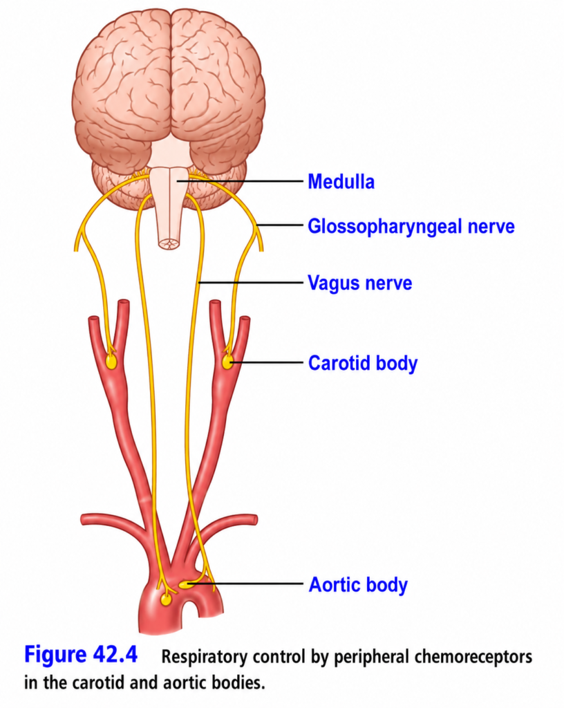
STIMULATION OF THE CHEMORECEPTORS BY DECREASED ARTERIAL PO₂
- When the oxygen (PO₂) level in arterial blood falls below normal, the peripheral chemoreceptors become strongly stimulated.
- This response is shown in Figure 42.5.
- The nerve impulse rate increases greatly when arterial PO₂ falls from 60 mmHg to 30 mmHg.
- In this PO₂ range, hemoglobin loses oxygen rapidly because its oxygen saturation decreases quickly.
- The carotid bodies and aortic bodies contain many specialized gland-like cells called glomus cells.
- These glomus cells are connected directly or indirectly to nerve endings.
- Current evidence suggests that glomus cells act as the chemoreceptors and then stimulate the nerve endings.
- This process is shown in Figure 42.6.
- Glomus cells are sensitive to oxygen (O₂).
- When blood PO₂ decreases markedly, the potassium (K⁺) channels become inactivated (close).
- Closing of K⁺ channels causes the glomus cell to depolarize.
- Depolarization opens voltage-gated calcium (Ca²⁺) channels.
- Calcium enters the glomus cell, increasing the intracellular calcium concentration.
- The increased calcium causes the release of neurotransmitters.
- These neurotransmitters activate the afferent sensory neurons.
- The afferent neurons send signals to the central nervous system.
- These signals increase respiration.
- Adenosine triphosphate (ATP) appears to be the main excitatory neurotransmitter released by carotid glomus cells during hypoxia.
- Dopamine or acetylcholine may also act as neurotransmitters released by the glomus cells.
- KEY CONCEPT
- A decrease in arterial PO₂ strongly stimulates the peripheral chemoreceptors, especially when PO₂ falls between 60 and 30 mmHg. Glomus cells detect the low oxygen, K⁺ channels close, the cells depolarize, Ca²⁺ enters, neurotransmitters (mainly ATP) are released, and afferent nerves stimulate the respiratory center to increase breathing.
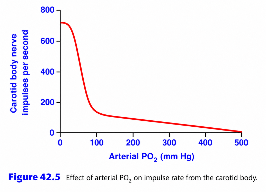
This is Figure 42.5 from Guyton Physiology, and it explains one of the most important concepts in the control of breathing:
⭐ Effect of Arterial PO₂ on the Carotid Body (Peripheral Chemoreceptors)
This graph answers the question:
How does low oxygen (hypoxia) stimulate breathing?
The answer is:
Low arterial PO₂ stimulates the carotid bodies, causing them to send many nerve impulses to the respiratory center, which increases ventilation.
🎯 One-Line Concept
The lower the arterial PO₂, the higher the firing rate of the carotid body chemoreceptors.
Simply,
- ↓ PO₂ → ↑ Carotid body impulses → ↑ Breathing
- ↑ PO₂ → ↓ Carotid body impulses → Normal breathing
First Understand What the Carotid Body Is
The carotid body is a small chemoreceptor organ located at the bifurcation of the common carotid artery.
Its job is to continuously monitor:
- ✅ Arterial PO₂
- ✅ Arterial PCO₂
- ✅ Arterial pH
It is the most important peripheral oxygen sensor.
Simple Flow Chart
Low Arterial PO₂
        ↓
Carotid Body Stimulated
        ↓
More Nerve Impulses
        ↓
Glossopharyngeal Nerve (CN IX)
        ↓
Medulla (Respiratory Center)
        ↓
↑ Rate and Depth of Breathing
        ↓
More Oxygen Enters Lungs
Step 1: Understand the Axes
X-Axis (Horizontal)
Arterial PO₂ (mmHg)
This shows the
partial pressure of oxygen in arterial blood.
Starts from
0 mmHg
↓
500 mmHgEasy Memory
Move right
↓
More oxygen in blood
Move left
↓
Less oxygen in blood
Y-Axis (Vertical)
Carotid Body Nerve Impulses/Second
This shows
how frequently the carotid body sends signals to the brain.
Higher value
↓
More stimulation
↓
More breathing
Lower value
↓
Less stimulation
↓
Less effect on breathing
Understanding the Curve
Look carefully.
The curve has three regions.
Region 1
Severe Hypoxia
PO₂ = 0–60 mmHg
Look at the left side.
The curve is very high.
What Happens?
Oxygen is dangerously low.
The carotid body becomes
strongly stimulated.
It sends
hundreds of nerve impulses every second.
Why?
The body is trying to save itself.
The brain receives the message:
“Oxygen is falling—breathe faster!”
Result
- Respiratory rate ↑
- Depth of breathing ↑
- Alveolar ventilation ↑
Easy Memory
Very low oxygen
↓
Alarm rings
↓
Carotid body fires rapidly
↓
Breathing increases
Region 2
Normal Physiological Range
PO₂ ≈60–100 mmHg
This is the
most important region.
Notice
The curve drops very steeply.
Why?
Small decreases in PO₂ below about 60 mmHg cause a large increase in carotid body firing.
Above about 60 mmHg, changes in PO₂ have much less effect.
Normal Arterial PO₂
≈95–100 mmHg
At this level,
the carotid body sends only a small number of impulses.
Breathing is mainly controlled by CO₂, not oxygen.
Why Is 60 mmHg So Important?
This is the critical threshold.
Above 60 mmHg
The carotid body is only mildly stimulated.
Breathing changes very little.
Below 60 mmHg
The carotid body becomes strongly activated.
Breathing increases rapidly.
MBBS Golden Number
60 mmHg is the critical PO₂ at which hypoxia begins to strongly stimulate peripheral chemoreceptors.
Region 3
Very High PO₂
PO₂ = 100–500 mmHg
Look at the far right.
The curve becomes almost flat.
Why?
The carotid body already has plenty of oxygen.
Extra oxygen
does not stimulate it.
Instead,
its firing rate becomes very low.
Result
Very little chemoreceptor stimulation.
Why Doesn’t High Oxygen Increase Impulses?
Because
the carotid body is designed to detect
oxygen deficiency, not oxygen excess.
Think of it as a smoke detector.
A smoke detector
responds to smoke,
not to clean air.
Similarly,
the carotid body responds to low oxygen, not high oxygen.
Why Does the Curve Fall So Steeply?
Imagine
Arterial PO₂ falls from
100
↓
60 mmHg.
The carotid body suddenly senses that oxygen delivery to tissues may become inadequate.
It rapidly increases its nerve firing.
This provides an early warning system.
Clinical Correlation
1. High Altitude
Atmospheric oxygen decreases.
↓
Arterial PO₂ falls.
↓
Carotid body firing increases.
↓
Hyperventilation develops.
This is the body’s first adaptation to altitude.
2. COPD
Some patients have chronically high CO₂.
Over time,
central chemoreceptors become less responsive.
Breathing depends more on
low PO₂ stimulating the carotid bodies (hypoxic drive).
3. Pneumonia
Poor oxygen exchange
↓
PO₂ falls
↓
Carotid body stimulation
↓
Rapid breathing (tachypnea)
4. Severe Blood Loss
Reduced oxygen delivery
↓
Carotid body activation
↓
Increased ventilation
High-Yield MBBS Viva Points
Carotid Body
- Located at the bifurcation of the common carotid artery.
- Main peripheral chemoreceptor.
- Afferent nerve: Glossopharyngeal nerve (CN IX).
- Sends signals to the medullary respiratory center.
Stimulated By
- ↓ PO₂ (most important when PO₂ < 60 mmHg)
- ↑ PCO₂
- ↓ pH
Response
- ↑ Respiratory rate
- ↑ Tidal volume
- ↑ Alveolar ventilation
Compare Different PO₂ Levels
| Arterial PO₂ | Carotid Body Activity | Effect on Breathing |
|---|---|---|
| 100 mmHg | Low | Normal breathing |
| 80 mmHg | Slightly increased | Little change |
| 60 mmHg | Rapid increase | Ventilation begins to rise markedly |
| 40 mmHg | Very high | Strong hyperventilation |
| 20 mmHg | Maximum | Emergency response |
Easy Story
Imagine
The carotid body is a security guard.
Plenty of oxygen (PO₂ = 100 mmHg)
Everything is normal.
The guard stays calm.
Oxygen begins to fall (PO₂ ≈60 mmHg)
The guard notices danger.
He starts calling for help.
Very low oxygen (PO₂ = 30–40 mmHg)
The guard activates the emergency alarm.
The brain immediately tells the lungs:
“Breathe faster!”
Exactly what this graph shows.
Difference Between Central and Peripheral Chemoreceptors
| Central Chemoreceptors | Peripheral Chemoreceptors (Carotid Bodies) |
|---|---|
| Located in medulla | Located in carotid bodies |
| Respond mainly to CO₂/H⁺ | Respond mainly to low PO₂ (also ↑CO₂ and ↓pH) |
| Not directly stimulated by low PO₂ | Strongly stimulated when PO₂ < 60 mmHg |
🎯 MBBS Golden Rule
The Carotid Body Is an Oxygen Alarm
- PO₂ > 60 mmHg → Minimal stimulation.
- PO₂ < 60 mmHg → Rapid increase in nerve firing.
- Lower PO₂ → More impulses → Faster and deeper breathing.
Final Concept to Never Forget
The key message of Figure 42.5 is that the carotid body is the body’s emergency oxygen sensor.
- When arterial PO₂ is normal (≈95–100 mmHg), it sends only a few impulses.
- As PO₂ falls below about 60 mmHg, carotid body activity increases dramatically.
- These impulses travel through the glossopharyngeal nerve (cranial nerve IX) to the medullary respiratory center, causing rapid, deep breathing to restore oxygen levels.
This mechanism protects the body during hypoxia, such as at high altitude, pneumonia, severe lung disease, or shock, making the carotid body the primary peripheral chemoreceptor for oxygen sensing.
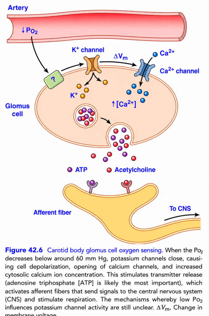
Carotid Body Oxygen Sensing (Guyton Fig. 42.6) – Easiest & Most Conceptual Explanation
🎯 One-Line Concept
When arterial PO₂ falls below about 60 mm Hg, the carotid body detects the low oxygen and immediately tells the brain to breathe faster and deeper.
💡 Golden Rule
Low O₂ → Glomus Cell Activated → Brain Stimulated → Breathing Increases
Think of the carotid body as the body’s oxygen alarm system.
- 🚨 Normal oxygen = Alarm OFF
- 🚨 Low oxygen = Alarm ON
The Big Picture
This figure explains
How the carotid body detects low oxygen (hypoxia).
The main cell responsible is the
⭐ Glomus Cell (Type I Cell)
This cell acts like an oxygen sensor.
Where is the Carotid Body?
📍 Located at the bifurcation (division) of the common carotid artery in the neck.
Its job is to continuously monitor:
- ✅ PO₂ (oxygen)
- ✅ PCO₂ (carbon dioxide)
- ✅ H⁺ (pH)
But this figure focuses on
Low Oxygen (Low PO₂).
Step-by-Step Story
Step 1. Normal Oxygen
Normally,
Arterial PO₂ is about
95–100 mm Hg
At this level,
the glomus cell is quiet.
Normal PO₂
      ↓
K⁺ channels open
      ↓
Cell remains at resting membrane potential
      ↓
No neurotransmitter release
      ↓
Normal breathing
Step 2. Oxygen Falls Below 60 mm Hg ⭐
This is the most important number.
When arterial PO₂ falls below about 60 mm Hg
the carotid body becomes strongly stimulated.
PO₂ < 60 mm Hg
        ↓
Glomus cell activated
This is why the caption mentions 60 mm Hg.
Step 3. Potassium (K⁺) Channels Close
Normally,
potassium leaves the glomus cell through K⁺ channels.
When oxygen falls,
these channels close.
Low PO₂
      ↓
K⁺ channels close
      ↓
K⁺ cannot leave cell
Why Is This Important?
Normally,
K⁺ leaving the cell helps keep the inside of the cell negative.
When K⁺ cannot leave,
positive charge accumulates inside.
Step 4. Cell Depolarizes
Because K⁺ remains inside,
the membrane becomes less negative.
This is called
Depolarization
K⁺ retained
      ↓
Inside becomes positive
      ↓
Depolarization
This is shown in the figure as
ΔVm
(Change in membrane potential)
Step 5. Calcium Channels Open
Depolarization opens
Voltage-Gated Ca²⁺ Channels
Depolarization
      ↓
Ca²⁺ channels open
Now,
calcium enters the glomus cell.
Step 6. Intracellular Calcium Increases
Large amounts of calcium enter.
Ca²⁺ enters
      ↓
Intracellular Ca²⁺ ↑
Calcium is the signal that tells the cell:
“Release neurotransmitters!”
Step 7. Neurotransmitters Are Released
The glomus cell releases neurotransmitters onto the nearby sensory nerve ending.
The figure shows
- 🟣 ATP ⭐ (most important)
- 🔴 Acetylcholine (ACh)
Other transmitters (not shown) include dopamine in some situations.
Step 8. Sensory Nerve Is Activated
The neurotransmitters stimulate the
Afferent Nerve Fiber
ATP + ACh
      ↓
Afferent nerve stimulated
This nerve carries the signal to the brainstem through the glossopharyngeal nerve (cranial nerve IX).
Step 9. Signal Reaches the Brain
The nerve sends signals to the
Respiratory Center (Medulla)
Carotid Body
      ↓
Glossopharyngeal Nerve
      ↓
Medulla
      ↓
Respiratory Center
Step 10. Breathing Increases
The respiratory center stimulates
- Diaphragm
- External intercostal muscles
Result
Ventilation ↑Breathing becomes
- Faster
- Deeper
More oxygen enters the lungs.
Complete Flow Chart
Arterial PO₂ Falls
(<60 mm Hg)
        │
        ▼
Glomus Cell Detects Low O₂
        │
        ▼
K⁺ Channels Close
        │
        ▼
Depolarization
(ΔVm)
        │
        ▼
Voltage-Gated Ca²⁺ Channels Open
        │
        ▼
Ca²⁺ Enters Cell
        │
        ▼
ATP & Acetylcholine Released
        │
        ▼
Afferent Nerve Activated
        │
        ▼
Brainstem Respiratory Center
        │
        ▼
Breathing Increases
        │
        ▼
Blood Oxygen Rises
Understanding Each Label in the Figure
🔵 ↓ PO₂
Means
Oxygen level has fallen.
🟠 K⁺ Channel
Normally open.
Low oxygen causes it to close.🔷 ΔVm
Means
Change in membrane voltage
(Depolarization).
🔵 Ca²⁺ Channel
Opens after depolarization.
Allows calcium to enter.
🔵 ↑ [Ca²⁺]
Means
Intracellular calcium concentration increases.
🟣 ATP
The most important neurotransmitter released by the glomus cell.Acetylcholine
Another neurotransmitter that helps stimulate the sensory nerve.
🟡 Afferent Fiber
Carries information from the carotid body to the brain.
Everyday Analogy
Imagine a building with an oxygen detector.
- 🏢 Carotid body = Oxygen detector
- 🔋 Glomus cell = Sensor
- 🚪 K⁺ channel = Exit door
- ⚡ Ca²⁺ = Electrical switch
- 📞 ATP & ACh = Emergency phone call
- 🧠 Brain = Control room
- 🫁 Lungs = Ventilation system
When oxygen falls,
the detector sends an emergency message,
and the ventilation system works harder.
High-Yield MBBS Points
| Event | Result |
|---|---|
| PO₂ < 60 mm Hg | Carotid body strongly stimulated |
| K⁺ channels close | Depolarization |
| Depolarization | Voltage-gated Ca²⁺ channels open |
| Ca²⁺ enters | Neurotransmitter release |
| ATP (mainly) + ACh released | Afferent nerve stimulated |
| Brainstem activated | Ventilation increases |
Important Viva Question
Why does the carotid body respond mainly when PO₂ falls below 60 mm Hg?
Because above 60 mm Hg, hemoglobin remains highly saturated with oxygen, so oxygen delivery is usually adequate. When PO₂ falls below this level, oxygen content drops more rapidly, making hypoxia a significant threat. The carotid body therefore becomes strongly activated to increase ventilation and restore oxygen levels.🌟 Super Memory Summary
Low PO₂
(<60 mm Hg)
        │
K⁺ Channels Close
        │
Depolarization
        │
Ca²⁺ Channels Open
        │
Ca²⁺ Enters Cell
        │
ATP + ACh Released
        │
Afferent Nerve
        │
Brainstem
        │
Breathing ↑
        │
O₂ Restored
🧠 Easy Mnemonic
“Low O₂ → K⁺ Closes → Ca²⁺ Opens → ATP Released → Breathe More”
Or even shorter:
O₂ ↓ → K⁺ ↓ → Ca²⁺ ↑ → ATP ↑ → Ventilation ↑
💎 Golden Rule
The carotid body is the body’s primary oxygen sensor. When arterial PO₂ falls below about 60 mm Hg, glomus cells close K⁺ channels, depolarize, open voltage-gated Ca²⁺ channels, release ATP (and acetylcholine), activate afferent fibers to the brainstem, and rapidly increase ventilation to restore oxygen levels.
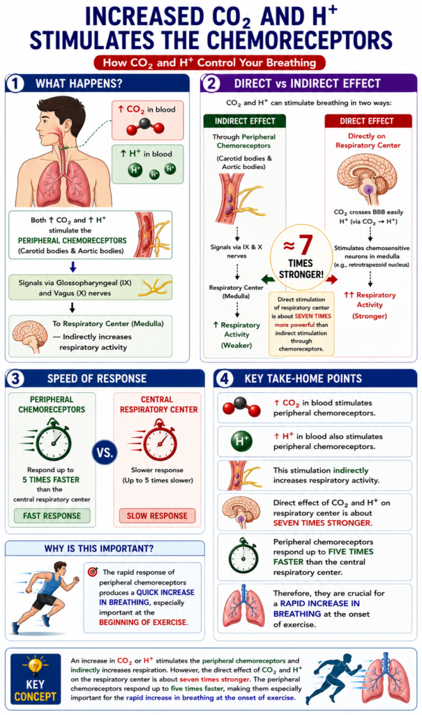
INCREASED CO₂ AND H⁺ CONCENTRATION STIMULATES THE CHEMORECEPTORS
- An increase in blood CO₂ concentration stimulates the peripheral chemoreceptors.
- An increase in H⁺ concentration also stimulates the peripheral chemoreceptors.
- Stimulation of the chemoreceptors indirectly increases respiratory activity.
- However, the direct effects of CO₂ and H⁺ on the respiratory center are much stronger than their effects through the peripheral chemoreceptors.
- The direct stimulation of the respiratory center is about seven times more powerful than the indirect stimulation through the chemoreceptors.
- One important difference between the peripheral and central effects of CO₂ is the speed of response.
- Peripheral chemoreceptors respond up to five times faster than the central respiratory center.
- Therefore, the peripheral chemoreceptors are especially important for producing a rapid increase in breathing.
- This rapid response is particularly important at the beginning of exercise.
KEY CONCEPT
- An increase in CO₂ or H⁺ stimulates the peripheral chemoreceptors and indirectly increases respiration. However, the direct effect of CO₂ and H⁺ on the respiratory center is about seven times stronger. The peripheral chemoreceptors respond up to five times faster, making them especially important for the rapid increase in breathing at the onset of exercise.
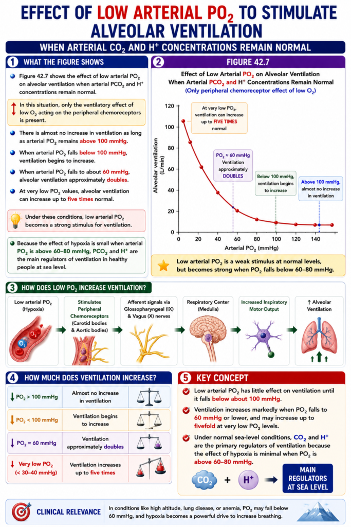
EFFECT OF LOW ARTERIAL PO₂ TO STIMULATE ALVEOLAR VENTILATION WHEN ARTERIAL CO₂ AND H⁺ CONCENTRATIONS REMAIN NORMAL
- Figure 42.7 shows the effect of low arterial PO₂ on alveolar ventilation when arterial PCO₂ and H⁺ concentrations remain normal.
- In this situation, only the ventilatory effect of low O₂ acting on the peripheral chemoreceptors is present.
- There is almost no increase in ventilation as long as arterial PO₂ remains above 100 mmHg.
- When arterial PO₂ falls below 100 mmHg, ventilation begins to increase.
- When arterial PO₂ falls to about 60 mmHg, alveolar ventilation approximately doubles.
- At very low PO₂ values, alveolar ventilation can increase up to five times normal.
- Under these conditions, low arterial PO₂ becomes a strong stimulus for ventilation.
- Because the effect of hypoxia is small when arterial PO₂ is above 60–80 mmHg, PCO₂ and H⁺ are the main regulators of ventilation in healthy people at sea level.
KEY CONCEPT
- Low arterial PO₂ has little effect on ventilation until it falls below about 100 mmHg. Ventilation increases markedly when PO₂ falls to 60 mmHg or lower, and may increase up to fivefold at very low PO₂ levels. Under normal sea-level conditions, CO₂ and H⁺ are the primary regulators of ventilation because the effect of hypoxia is minimal when PO₂ is above 60–80 mmHg.
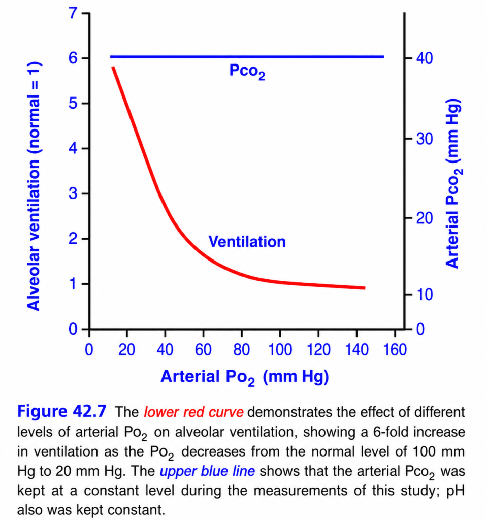
This is Figure 42.7 from Guyton Physiology, and it explains how low oxygen (hypoxia) alone stimulates breathing when carbon dioxide (PCO₂) and pH are kept constant.
This is one of the highest-yield MBBS physiology graphs because it proves an important fact:
Oxygen is a relatively weak stimulus for breathing until arterial PO₂ falls below about 60 mmHg.
The graph also shows that this response is produced only by the peripheral chemoreceptors (mainly the carotid bodies) because CO₂ and pH are held constant.
⭐ Figure 42.7: Effect of Low Arterial PO₂ on Alveolar Ventilation (PCO₂ and pH Constant)
🎯 One-Line Concept
When arterial PO₂ falls below about 60 mmHg, alveolar ventilation increases rapidly because the carotid bodies strongly stimulate the respiratory center.
Simply,
Normal PO₂ (100 mmHg)
        ↓
Little effect on breathing

PO₂ < 60 mmHg
        ↓
Carotid body strongly stimulated
        ↓
Breathing increases rapidly
First Understand Why This Experiment Was Done
Normally,
When PO₂ falls,
two things happen:
- Breathing increases.
- CO₂ falls because of hyperventilation.
Since CO₂ is itself a powerful stimulator of breathing,
scientists wanted to know:
“Does low oxygen alone stimulate breathing?”
So in this experiment:
✔ PCO₂ was kept constant at 40 mmHg
✔ pH was kept constant.
Therefore,
only the effect of oxygen was studied.tep 1: Understand the Axes
X-Axis (Horizontal)
Arterial PO₂ (mmHg)
This is
the oxygen pressure in arterial blood.
Starts from
20 mmHg
↓
140 mmHg
Easy Memory
Move right
↓
More oxygen
Move left
↓
Less oxygen
Left Y-Axis
Alveolar Ventilation
Normal ventilation
=
1
If ventilation becomes
2
↓
Breathing doubles.
If ventilation becomes
6
↓
Breathing becomes six times normal.
Right Y-Axis
Arterial PCO₂
Notice
The blue scale.
It stays
40 mmHg
throughout the graph.
Understanding the Two Lines
🔵 Blue Horizontal Line
PCO₂ = 40 mmHg
This line never changes.
Why?
Because the experiment kept CO₂ constant.
This means
CO₂ is not responsible for any change in breathing.
Only oxygen is affecting ventilation.
Easy Memory
Blue line
↓
No CO₂ change
🔴 Red Curve
This is the
most important line.
It shows
how ventilation changes
as PO₂ changes.
Region 1
PO₂ = 100–140 mmHg
Look at the far right.
The curve is almost flat.
Ventilation
≈1
(normal).
Why?
There is plenty of oxygen.
The carotid body is almost silent.
Very little stimulation reaches the respiratory center.
Therefore,
breathing remains normal.
Easy Memory
High oxygen
↓
No alarm
↓
Normal breathing
Region 2
PO₂ = 60–100 mmHg
Notice
The curve changes only slightly.
Why?
The carotid body is only mildly stimulated.
Even though oxygen falls,
the respiratory center hardly changes ventilation.
Important Point
Above
60 mmHg
oxygen has
very little effect
on breathing.
Region 3
PO₂ Below 60 mmHg
This is
the most important part.
Look carefully.
The curve suddenly rises steeply.
What Happens?
Once PO₂ falls below
60 mmHg,
the carotid body becomes strongly stimulated.
It sends many impulses to the medulla.
Breathing increases rapidly.
Result
Ventilation rises dramatically.
Example
PO₂
100 mmHg
↓
Ventilation = 1×
Normal
PO₂
20 mmHg
↓
Ventilation ≈6×
Normal
That means
breathing becomes
about six times faster/deeper.
Why Does the Curve Rise So Rapidly?
Because
the carotid body is an
oxygen emergency sensor.
Above
60 mmHg
↓
No emergency.
Below
60 mmHg
↓
Emergency alarm.
The respiratory center immediately increases ventilation.
Why Is 60 mmHg So Important?
This is the
critical threshold.
Above
60 mmHg
↓
Hemoglobin remains almost fully saturated.
The tissues still receive enough oxygen.
Below
60 mmHg
↓
Hemoglobin saturation begins to fall rapidly.
Tissues may become hypoxic.
Therefore,
the carotid body reacts strongly.
Link This with Figure 41.8
Remember the
oxygen-hemoglobin dissociation curve.
At
PO₂
≈60 mmHg,
hemoglobin saturation is still about
90%.
This is why
little ventilatory response occurs above this value.
Below
60 mmHg,
oxygen content begins to fall rapidly.
The body responds with hyperventilation.
Step-by-Step Mechanism
Suppose
Arterial PO₂ falls to
40 mmHg.
Step 1
Carotid body detects low oxygen.
↓
Step 2
Carotid body firing increases.
↓
Step 3
Signals travel via
Glossopharyngeal nerve (CN IX)
↓
Step 4
Respiratory center in the medulla is stimulated.
↓
Step 5
Breathing becomes faster and deeper.
↓
Step 6
More oxygen enters alveoli.
↓
PO₂ begins to improve.
Why Doesn’t Ventilation Continue Increasing Forever?
Because
once oxygen improves,
carotid body stimulation decreases.
Negative feedback occurs.Clinical Correlation
1. High Altitude
Atmospheric oxygen falls.
↓
Arterial PO₂ decreases.
↓
Carotid body stimulation.
↓
Hyperventilation develops.
This is the first adaptation to high altitude.
2. Severe Pneumonia
Poor gas exchange.
↓
PO₂ falls below
60 mmHg.
↓
Rapid breathing develops.
3. COPD
Some patients have chronic CO₂ retention.
Their central chemoreceptors become less responsive.
Breathing depends more on
low PO₂ stimulating the carotid body (hypoxic drive).
4. Pulmonary Edema
Poor oxygen diffusion.
↓
PO₂ falls.
↓
Hyperventilation occurs.
High-Yield MBBS Viva Points
Blue Line
- Arterial PCO₂ = 40 mmHg
- Kept constant.
- pH also kept constant.
Red Curve
Shows
the effect of
oxygen alone
on ventilation.
Critical PO₂
60 mmHg
Below this,
ventilation rises sharply.
Maximum Response
PO₂
20 mmHg
↓
Ventilation
≈6×
Normal.
Compare Different PO₂ Levels
| Arterial PO₂ | Carotid Body Activity | Ventilation |
|---|---|---|
| 100 mmHg | Minimal | Normal (≈1×) |
| 80 mmHg | Slight | Nearly normal |
| 60 mmHg | Begins to increase markedly | Slight increase |
| 40 mmHg | High | Markedly increased |
| 20 mmHg | Maximum | ≈6× normal |
Super Easy Memory Story
Imagine
The carotid body is a
🚨 Fire Alarm.
Oxygen = 100 mmHg
No smoke.
Alarm is silent.
People breathe normally.
Oxygen = 70 mmHg
A little smoke.
Alarm barely notices.
Oxygen = 50 mmHg
Lots of smoke.
The alarm rings loudly.
Everyone runs.
Breathing becomes rapid.
Exactly like the graph.
Difference Between CO₂ and O₂ Control of Breathing
| Carbon Dioxide | Oxygen |
|---|---|
| Main regulator of breathing under normal conditions | Secondary regulator |
| Acts mainly through central chemoreceptors | Acts mainly through carotid bodies |
| Small rise causes strong increase in ventilation | Significant effect mainly when PO₂ < 60 mmHg |
🎯 MBBS Golden Rule
Oxygen Alone Stimulates Breathing Only When
PO₂ < 60 mmHg
Above
60 mmHg
↓
Little effect.
Below
60 mmHg
↓
Rapid hyperventilation.
Final Concept to Never Forget
The key message of Figure 42.7 is:
- When CO₂ and pH are kept constant, oxygen has very little effect on breathing until arterial PO₂ falls below about 60 mmHg.
- Below 60 mmHg, the carotid bodies are strongly stimulated, causing a rapid increase in ventilation.
- As PO₂ falls from 100 mmHg to about 20 mmHg, alveolar ventilation increases to approximately six times the normal level.
This graph demonstrates that oxygen is an emergency regulator of breathing, whereas carbon dioxide is the primary regulator under normal physiological conditions.
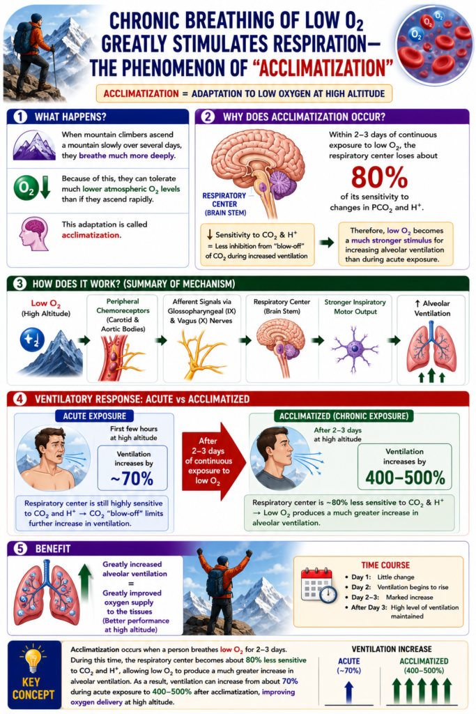
CHRONIC BREATHING OF LOW O₂ GREATLY STIMULATES RESPIRATION—THE PHENOMENON OF “ACCLIMATIZATION”
- When mountain climbers ascend a mountain slowly over several days, they breathe much more deeply.
- Because of this, they can tolerate much lower atmospheric O₂ levels than if they ascend rapidly.
- This adaptation is called acclimatization.
- The reason for acclimatization is that within 2–3 days, the respiratory center in the brain stem loses about 80% of its sensitivity to changes in PCO₂ and H⁺.
- As a result, the excess loss (blow-off) of CO₂ during increased ventilation no longer strongly inhibits respiration.
- Therefore, low O₂ becomes a much stronger stimulus for increasing alveolar ventilation than it is during acute exposure.
- During acute exposure to low O₂, ventilation may increase by about 70%.
- After 2–3 days of continuous exposure to low O₂, alveolar ventilation can increase by 400% to 500%.
- This large increase in ventilation greatly improves oxygen supply to the mountain climber.
KEY CONCEPT
- Acclimatization occurs when a person breathes low O₂ for 2–3 days. During this time, the respiratory center becomes about 80% less sensitive to CO₂ and H⁺, allowing low O₂ to produce a much greater increase in alveolar ventilation. As a result, ventilation can increase from about 70% during acute exposure to 400–500% after acclimatization, improving oxygen delivery at high altitude.
COMPOSITE EFFECTS OF PCO₂, pH, AND PO₂ ON ALVEOLAR VENTILATION
- Figure 42.8 shows the combined effects of PO₂, PCO₂, and pH on alveolar ventilation.
- The four red curves were recorded at different arterial PO₂ values:
- 40 mmHg
- 50 mmHg
- 60 mmHg
- 100 mmHg
- For each red curve, the PCO₂ was changed from low to high values.
- Therefore, the red curves show the combined effects of PO₂ and PCO₂ on alveolar ventilation.
- The blue curves were recorded at a blood pH of 7.3.
- The red curves were recorded at a blood pH of 7.4.
- Thus, the red and blue curves together show the combined effects of PCO₂ and PO₂ at two different blood pH values.
- If the blood pH becomes higher than 7.4, the family of curves shifts to the right.
- If the blood pH becomes lower than 7.3, the family of curves shifts to the left.
- Using this diagram, the level of alveolar ventilation can be predicted from the combined values of alveolar PCO₂, alveolar PO₂, and arterial pH.
KEY CONCEPT
- Figure 42.8 shows the combined effects of PCO₂, PO₂, and pH on alveolar ventilation. The red curves represent different arterial PO₂ levels at pH 7.4, while the blue curves represent the same relationship at pH 7.3. Higher pH shifts the curves to the right, lower pH shifts them to the left, allowing prediction of alveolar ventilation from the combined values of PCO₂, PO₂, and pH.
his is Figure 42.8 from Guyton Physiology, and it is one of the most important and most confusing graphs in respiratory physiology.
It combines the effects of three variables on breathing:
- PCO₂ (Carbon dioxide) ✅
- PO₂ (Oxygen) ✅
- pH (Hydrogen ion concentration) ✅
This graph answers the question:
How do CO₂, O₂, and pH work together to control alveolar ventilation?
⭐ Figure 42.8: Combined Effects of PCO₂, PO₂, and pH on Alveolar Ventilation
🎯 One-Line Concept
CO₂ is the strongest stimulus for breathing, but low PO₂ and low pH greatly increase the ventilatory response to CO₂.
In simple words,
- ↑ PCO₂ → ↑↑ Ventilation
- ↓ PO₂ → Even greater increase in ventilation
- ↓ pH → Even greater increase in ventilation
STEP 1: Understand the Axes
X-Axis (Horizontal)
Alveolar PCO₂ (mmHg)
This is
Carbon dioxide pressure inside the alveoli.
Normally,
Alveolar PCO₂
≈ 40 mmHg
Easy Memory
Move right
↓
More CO₂
Move left
↓
Less CO₂
Y-Axis (Vertical)
Alveolar Ventilation (L/min)
This shows
How much air enters and leaves the alveoli each minute.
Higher value
↓
More breathing
Lower value
↓
Less breathing
STEP 2: Understand the Colors
The graph has two groups of curves.
🔴 Red Curves
pH = 7.4 (Normal pH)
These curves show
normal blood acidity.
🔵 Blue Curves
pH = 7.3
This represents
Acidosis
(Higher H⁺ concentration)
Easy Memory
Blue
↓
Acidosis
↓
Breathing increases more
STEP 3: Understand the Numbers on Each Curve
Each curve has
40
50
60
100
These numbers represent
Arterial PO₂ (mmHg)
NOT PCO₂.
Therefore
Each curve is drawn at
a different oxygen level.Red Curves (Normal pH)
There are four curves.
Curve 1
PO₂ = 100 mmHg
Normal oxygen.
Curve 2
PO₂ = 60 mmHg
Mild hypoxia.
Curve 3
PO₂ = 50 mmHg
Moderate hypoxia.
Curve 4
PO₂ = 40 mmHg
Severe hypoxia.
Exactly the same four oxygen levels are shown by the blue curves, but at pH = 7.3.
STEP 4: Understand Each Red Curve
Red Curve (PO₂ = 100 mmHg)
This is
the normal person.
Normal oxygen.
Normal pH.
Suppose
PCO₂ rises
40
↓
50 mmHg.
Ventilation increases.
This is
the normal CO₂ response.
Red Curve (PO₂ = 60 mmHg)
Now
oxygen falls.
Everything else remains normal.Look carefully.
The whole curve shifts
to the left.
Why?
Because
hypoxia stimulates the carotid body.
Now
even a small rise in CO₂
produces more ventilation.
Red Curve (PO₂ = 50 mmHg)
Now
oxygen falls even more.Breathing becomes even more sensitive
to CO₂.
Red Curve (PO₂ = 40 mmHg)
Severe hypoxia.
Now
very small increases in CO₂
produce
massive hyperventilation.
Golden Rule
Lower PO₂
↓
Curve shifts LEFT
↓
Ventilation increases more.
STEP 5: Understand the Blue Curves
Blue means
pH
=
7.3
(acidosis)
Compare
Red
↓
Normal pH
Blue
↓
Low pH
Notice
Every blue curve
lies
to the
LEFT
of its corresponding red curve.
Why?
Acidosis itself
stimulates breathing.
Now
CO₂ becomes
even more effective.
Therefore
Acidosis
↓
Curve shifts LEFT.
What Does “Shift to the Left” Mean?
Suppose
PCO₂
=
40 mmHg.
Normal pH
↓
Ventilation
≈12 L/min
Acidosis
↓
Ventilation
≈18 L/min
Same CO₂
Different pH
Much more breathing.
Why?
Hydrogen ions stimulate
peripheral chemoreceptors.
Therefore,
ventilation increases.
Which Stimulus Is Strongest?
This graph answers that too.
① Carbon Dioxide
Strongest stimulus.
Even
small increases
produce
large increases in ventilation.
② Low Oxygen
Weak stimulus
until
PO₂ falls below
≈60 mmHg.
After that,
it greatly enhances
the CO₂ response.
③ Acidosis
Also increases
ventilation.
Especially
when CO₂ is elevated.
How Do All Three Work Together?
Imagine
A patient with
Pneumonia.
PO₂ falls.
↓
PCO₂ rises.
↓
Acidosis develops.
Now
all three stimuli
act together.
Result
↓
Very rapid breathing.
Clinical Correlation
1. High Altitude
PO₂ decreases.
↓
Red curves shift left.
↓
Hyperventilation.
2. COPD Exacerbation
CO₂ rises.
↓
Ventilation increases.
If hypoxemia is also present,
the response becomes even stronger through the peripheral chemoreceptors.
3. Diabetic Ketoacidosis
pH falls.
↓
Blue curves apply.
↓
Kussmaul breathing develops
(deep, rapid breathing).
4. Exercise
Muscles produce
CO₂.
↓
CO₂ rises.
↓
Ventilation increases.
If lactic acid accumulates,
pH falls,
making ventilation increase even further.
High-Yield MBBS Viva Points
Red Curves
- pH = 7.4
- Normal acid-base balance.
Blue Curves
- pH = 7.3
- Acidosis.
Numbers
40
50
60
100
=
Arterial PO₂
Lower PO₂
↓
Curve shifts LEFT
↓
More ventilation.
Lower pH
↓
Curve shifts LEFT
↓
More ventilation.
Higher PCO₂
↓
Moves upward on the curve
↓
More ventilation.
Compare the Effects
| Variable | Effect on Ventilation |
|---|---|
| ↑ PCO₂ | Strongly increases ventilation |
| ↓ PO₂ | Enhances the ventilatory response, especially when PO₂ < 60 mmHg |
| ↓ pH | Enhances ventilation by stimulating chemoreceptors |
Super Easy Memory Story
Imagine
The respiratory center is a car accelerator.
Three people can press the accelerator.
🚗 Person 1 = CO₂
Presses hardest.
The car speeds up immediately.
🚗 Person 2 = Low Oxygen
Usually presses lightly.
But
below
60 mmHg,
pushes much harder.
🚗 Person 3 = Acidosis
Also presses the accelerator.
Together with CO₂,
the car moves even faster.
When
CO₂
Low O₂
Acidosis
occur together,
the accelerator is pushed the hardest,
causing marked hyperventilation.
Quick Summary Table
| Condition | Effect on Graph |
|---|---|
| ↑ PCO₂ | Ventilation increases |
| ↓ PO₂ | Curves shift left (greater response to CO₂) |
| ↓ pH | Curves shift further left (greater response to CO₂) |
| PO₂ = 100 mmHg | Least ventilatory response |
| PO₂ = 40 mmHg | Greatest ventilatory response |
🎯 MBBS Golden Rule
Remember This Order
CO₂ > H⁺ > O₂
Meaning:
- CO₂ is the primary regulator of breathing under normal conditions.
- Acidosis (↑H⁺) significantly augments ventilation.
- Low O₂ becomes a powerful stimulus mainly when PO₂ falls below about 60 mmHg.
Final Concept to Never Forget
The key message of Figure 42.8 is that breathing is controlled by the combined action of carbon dioxide, oxygen, and pH—not by any one factor alone.
- Carbon dioxide is the most powerful physiological stimulus for ventilation.
- Low oxygen (especially PO₂ < 60 mmHg) makes the respiratory center much more responsive to CO₂ by activating the peripheral chemoreceptors.
- Acidosis (low pH) also enhances the ventilatory response, shifting the curves to the left.
Therefore:
The greatest increase in ventilation occurs when all three are present together: high CO₂, low PO₂, and low pH. This coordinated response helps restore normal gas exchange and acid-base balance.
MADE BY SELF LEARNING CEO AND FOUNDER DR SHEEN
