


CHRONIC PULMONARY EMPHYSEMA
Definition
Major Pathophysiological Changes
1. Chronic Infection and Airway Damage
- Long-term inhalation of tobacco smoke or other irritants causes chronic infection of the bronchi and bronchioles.
- This infection damages the normal protective mechanisms of the airways.
- The cilia of the respiratory epithelium become partially paralyzed and eventually destroyed.
- This damage is caused by toxic substances present in tobacco smoke and air pollution.
- Because the cilia cannot function properly, mucus cannot be cleared effectively from the airways.
- At the same time, mucus secretion increases, causing further blockage of the airways.
- Alveolar macrophages are also inhibited, making them less effective at fighting infection.
2. Chronic Airway Obstruction
- Chronic infection, excess mucus, and inflammatory swelling (edema) of the bronchiolar lining together produce chronic obstruction of many small airways.
3. Air Trapping and Alveolar Destruction
- Because the airways are narrowed, expiration becomes especially difficult.
- Air becomes trapped inside the alveoli.
- The trapped air overstretches the alveoli.
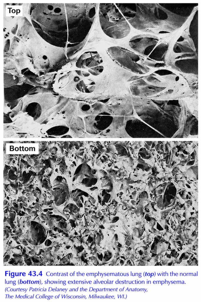
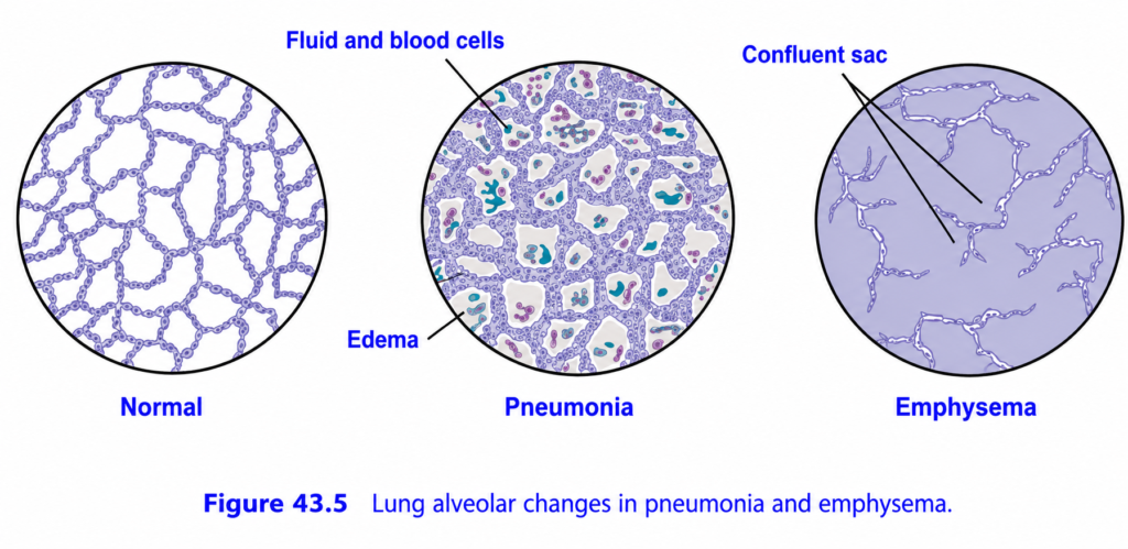
- Overstretching, together with chronic infection, destroys about 50–80% of the alveolar walls.
- The emphysematous lung therefore contains:
- Large, overdistended air spaces.
- Marked loss of normal alveolar walls. (Figs. 43.4 and 43.5)
Physiological Effects of Chronic Emphysema
1. Increased Airway Resistance
- Bronchiolar obstruction increases airway resistance.
- As a result, the work of breathing increases greatly.
- Expiration is especially difficult.
- During expiration:
- Pressure outside the lungs compresses both the alveoli and bronchioles.
- This further narrows the bronchioles.
- Airflow resistance increases even more.
2. Decreased Diffusing Capacity
- Destruction of alveolar walls greatly reduces the lung’s diffusing capacity.
- Consequently:
- Less oxygen diffuses into the blood.
- Less carbon dioxide diffuses out of the blood.
3. Ventilation–Perfusion (V̇A/Q̇) Mismatch
- Airway obstruction is more severe in some lung regions than in others.
- Therefore:
- Some lung regions are well ventilated.
- Other regions are poorly ventilated.
- This produces markedly abnormal ventilation–perfusion (V̇A/Q̇) ratios.
- In poorly ventilated regions:
- V̇A/Q̇ is very low.
- This creates a physiological shunt.
- Blood remains poorly oxygenated.
- In well-ventilated but poorly perfused regions:
- V̇A/Q̇ is very high.
- This creates a physiological dead space.
- Ventilation is wasted because little blood is available for gas exchange.
- Both physiological shunt and physiological dead space can occur simultaneously in the same lungs.
4. Pulmonary Hypertension and Right-Sided Heart Failure
- Destruction of the alveolar walls also destroys many pulmonary capillaries.
- Consequently:
- Pulmonary vascular resistance increases.
- Pulmonary hypertension develops.
- The right ventricle must pump against a higher resistance.
- This places an increased workload on the right side of the heart.
- Over time, right-sided heart failure may develop.
Progression of Disease
- Chronic emphysema usually progresses slowly over many years.
- As the disease worsens:
- Hypoxia develops because many alveoli are poorly ventilated and many alveolar walls are destroyed.
- Hypercapnia develops because carbon dioxide cannot be removed efficiently.
- The combination of hypoxia and hypercapnia produces severe, prolonged shortness of breath (dyspnea).
- This air hunger may continue for years.
- Eventually, severe hypoxia and hypercapnia can lead to death.
KEY CONCEPT
- Chronic pulmonary emphysema is a chronic obstructive and destructive lung disease, most commonly caused by long-term smoking.
- Chronic infection damages cilia, increases mucus secretion, and weakens alveolar macrophage function.
- Inflammation and mucus obstruct small airways, making expiration difficult and causing air trapping.
- Air trapping and infection destroy 50–80% of the alveolar walls, producing enlarged air spaces.
- Airway obstruction increases the work of breathing, especially during expiration.
- Loss of alveolar walls decreases the lung’s diffusing capacity, reducing oxygen uptake and carbon dioxide removal.
- Uneven airway obstruction produces severe ventilation–perfusion mismatch, causing both physiological shunt (low V̇A/Q̇) and physiological dead space (high V̇A/Q̇).
- Loss of pulmonary capillaries increases pulmonary vascular resistance, leading to pulmonary hypertension and right-sided heart failure.
- The disease progresses slowly, causing chronic hypoxia, hypercapnia, severe dyspnea, and eventually death if advanced.
PREPARE AND MADE BY SELF LEARNING DR SHEEN.
