


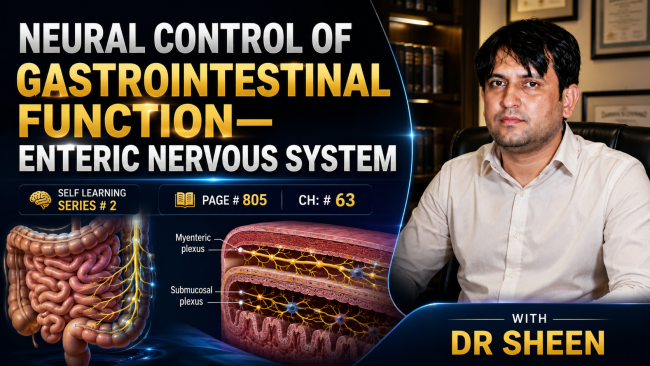
- The gastrointestinal (GI) tract has its own nervous system, called the enteric nervous system (ENS).
- The ENS is located entirely within the wall of the gastrointestinal tract.
- It extends from the esophagus to the anus.
- The ENS contains more than 100 million neurons.
- This is more neurons than the entire spinal cord.
- The ENS is highly developed and is especially important for controlling gastrointestinal movement and secretion.
Two Plexuses of the Enteric Nervous System
- The enteric nervous system consists mainly of two nerve plexuses (Fig. 63.4).
1. Myenteric Plexus (Auerbach Plexus)
- It is the outer plexus.
- It lies between the longitudinal and circular muscle layers.
- It mainly controls gastrointestinal movements (motility).
2. Submucosal Plexus (Meissner Plexus)
- It is the inner plexus.
- It lies in the submucosa.
- It mainly controls gastrointestinal secretion and local blood flow.
- The myenteric plexus and submucosal plexus are connected to each other by nerve connections (Fig. 63.4).
Connection With the Autonomic Nervous System
- The enteric nervous system is connected to:
- Parasympathetic nerve fibers
- Sympathetic nerve fibers
- These extrinsic nerve fibers connect with both the:
- Myenteric plexus
- Submucosal plexus
- The ENS can function independently, even without these external nerves.
- However:
- Parasympathetic stimulation usually enhances gastrointestinal activity.
- Sympathetic stimulation usually inhibits gastrointestinal activity.
Sensory Nerve Endings
- Sensory nerve endings are present in:
- The gastrointestinal epithelium
- The gut wall
- These sensory nerves send afferent (sensory) fibers to:
- The myenteric plexus
- The submucosal plexus
- The prevertebral sympathetic ganglia
- The spinal cord
- The brainstem through the vagus nerves
Functions of These Sensory Nerves
- These sensory nerves can produce local reflexes within the wall of the gut.
- They can also trigger long reflexes, which travel through:
- The prevertebral sympathetic ganglia, or
- The brainstem
- These reflexes then return to the gut and help regulate gastrointestinal function.
KEY CONCEPT
- The enteric nervous system (ENS) is the intrinsic nervous system of the GI tract.
- It extends from the esophagus to the anus.
- It contains more than 100 million neurons.
- The ENS has two major plexuses (Fig. 63.4):
- Myenteric (Auerbach) plexus → Controls GI motility.
- Submucosal (Meissner) plexus → Controls GI secretion and local blood flow.
- The ENS can work independently, but parasympathetic and sympathetic nerves modify its activity.
- Sensory nerves produce both:
- Local gut reflexes
- Long reflexes involving the spinal cord, sympathetic ganglia, and brainstem
Conceptual Examples
Example 1: Food Enters the Intestine
Food stretches the intestinal wall.
⬇
Sensory nerves detect the stretch.
⬇
Signals enter the enteric nervous system.
⬇
The myenteric plexus increases intestinal movement.
⬇
Food is pushed forward by peristalsis.
Example 2: Acid Enters the Small Intestine
Acid reaches the intestinal lining.
⬇
Sensory nerves detect the acid.
⬇
The submucosal plexus stimulates secretion of protective mucus and increases local blood flow.le 3: Parasympathetic Stimulation
Eating food activates the parasympathetic nervous system.
⬇
The parasympathetic nerves stimulate the ENS.
⬇
The ENS increases:
- Gastrointestinal movement
- Digestive secretions
Example 4: Sympathetic Stimulation
During stress or fear:
⬇
The sympathetic nervous system is activated.
⬇
It inhibits the ENS.
⬇
Gastrointestinal movement and secretion decrease.
Easy Flow Chart
Food enters gut
⬇
Sensory receptors detect stretch or chemicals
⬇
Signals reach the Enteric Nervous System (ENS)
⬇
Myenteric plexus → Controls movement
Submucosal plexus → Controls secretion & blood flow
⬇
Food is mixed, digested, and moved forward
Easy Comparison Table
| Myenteric Plexus (Auerbach) | Submucosal Plexus (Meissner) |
|---|---|
| Outer plexus | Inner plexus |
| Between longitudinal and circular muscle layers | Located in the submucosa |
| Controls gastrointestinal movement (motility) | Controls secretion |
| Coordinates peristalsis | Regulates local blood flow |
| Main target = Muscle | Main target = Mucosa and glands |
Easy Memory Trick
A = Auerbach = Action
- Action = Movement (Motility)
M = Meissner = Mucosa
- Mucosa = Secretion + Blood flow
DIFFERENCES BETWEEN THE MYENTERIC AND SUBMUCOSAL PLEXUSES
- The myenteric plexus is mainly a long chain of interconnected neurons.
- This chain extends throughout the entire gastrointestinal tract.
- A part of this chain is shown in Fig. 63.4.
- Because the myenteric plexus extends along the whole intestine and lies between the longitudinal and circular smooth muscle layers, it is especially important for controlling muscle activity along the gut.
- When the myenteric plexus is stimulated, it:
- Increases the resting tone (tonic contraction) of the gut wall.
- Increases the strength of rhythmic contractions.
- Slightly increases the rate of rhythmic contractions.
- Increases the speed of conduction of excitatory waves, so peristaltic waves move faster.
- The excitatory motor neurons contain neurotransmitters:
- Acetylcholine
- Substance P
- Glutamate
- The myenteric plexus is not completely excitatory.
- Some of its neurons are inhibitory.
- These inhibitory neurons release:
- Vasoactive intestinal polypeptide (VIP)
- Nitric oxide (NO)
- Adenosine triphosphate (ATP)
- These inhibitory signals are especially important for relaxing intestinal sphincters that normally slow the movement of food.
- Examples include:
- Pyloric sphincter, which controls the movement of food from the stomach to the duodenum.
- Ileocecal sphincter, which controls the movement of food from the small intestine to the cecum.
- The submucosal plexus, unlike the myenteric plexus, is mainly important for controlling the inner wall of each small segment of the intestine.
- Many sensory signals begin in the gastrointestinal epithelium.
- These signals are processed in the submucosal plexus.
- The submucosal plexus helps control:
- Local intestinal secretion
- Local absorption
- Local contraction of the submucosal muscle
- These contractions produce different degrees of folding (infolding) of the gastrointestinal mucosa.
KEY CONCEPT
- The myenteric plexus mainly controls gastrointestinal muscle movement.
- It increases gut tone, strength and rate of contractions, and the speed of peristalsis.
- It contains both excitatory and inhibitory neurons.
- The inhibitory neurons relax sphincters, allowing food to move forward.
- The submucosal plexus mainly controls the inner wall of the intestine.
- It regulates local secretion, absorption, and mucosal folding.
Conceptual Examples
- Myenteric plexus activated → Stronger and faster peristalsis → Food moves forward more quickly.
- Inhibitory neurons of the myenteric plexus activated → Pyloric sphincter relaxes → Food passes from the stomach into the duodenum.
- Submucosal plexus activated → More secretion and absorption in that small intestinal area.
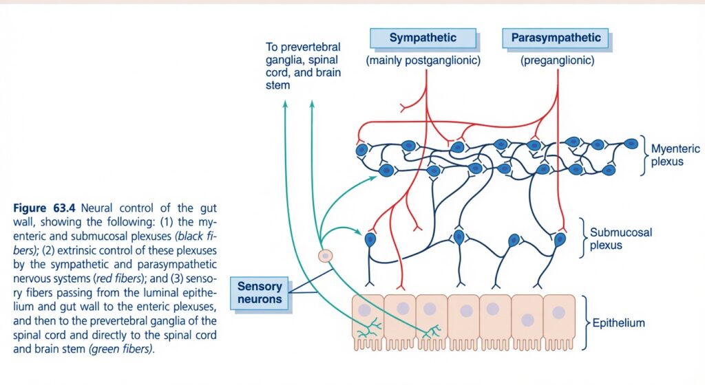
Figure 63.4: Neural Control of the Gut Wall
Easiest and Most Conceptual SELF LEARNING Explanation
⭐ One-Line Concept
The gastrointestinal (GI) tract has its own nervous system called the Enteric Nervous System (ENS). It contains two plexuses (myenteric and submucosal), receives control from the sympathetic and parasympathetic nervous systems, and continuously receives sensory information from the gut wall.
First Understand the Whole Story
Imagine the intestine is a smart factory.
It has:
- Its own manager → Enteric Nervous System (ENS)
- A boss from outside → Autonomic Nervous System
- Workers that sense everything happening inside → Sensory neurons
So, the intestine can:
- Work by itself.
- Receive instructions from the brain.
- Send information back to the brain.
Color Guide (Most Important Part of the Figure)
The figure uses three different colored nerve fibers.
| Color | Represents | Function |
|---|---|---|
| Black (dark blue) | Enteric plexuses | Local control inside the gut wall |
| Red | Sympathetic and parasympathetic nerves | External autonomic control |
| Green | Sensory nerve fibers | Carry sensory information from the gut |
Understanding Every Structure
1. Epithelium (Bottom Pink Cells)
These pink cells represent the intestinal epithelium.
This is the innermost lining of the intestine.
What does the epithelium do?
It:
- Absorbs nutrients.
- Secretes digestive fluids.
- Detects food, stretch, chemicals, and irritation.
Easy Concept
Think of the epithelium as the floor of the intestine where food touches first.
2. Sensory Neurons (Green Fibers)
The green fibers begin near the epithelium.
These are sensory neurons.
What do they detect?
They detect:
- Stretch
- Food
- Chemicals
- Pain
- Irritation
What happens next?
They send this information to:
- Enteric plexuses
- Prevertebral ganglia
- Spinal cord
- Brainstem
Easy Concept
They are the intestine’s information reporters.
3. Myenteric Plexus
The upper network of dark blue/black neurons is the myenteric (Auerbach’s) plexus.
It lies:
Between the longitudinal and circular muscle layers.
Main Function
Controls:
- Gut movement (motility)
- Muscle tone
- Strength of contraction
- Speed of peristalsis
Easy Concept
The myenteric plexus is the movement controller of the intestine.
Think:
Myenteric = Muscle Movement
4. Submucosal Plexus
The lower network of dark blue/black neurons is the submucosal (Meissner’s) plexus.
It lies in the submucosa.
Main Function
Controls:
- Intestinal secretion
- Local blood flow
- Absorption
Easy Concept
The submucosal plexus is the secretion controller.
Think:
Submucosal = Secretion and Absorption
5. Black (Dark Blue) Solid Lines
These represent the enteric nervous system fibers.
They connect:
- Myenteric neurons
- Submucosal neurons
Why?
Because both plexuses communicate continuously.
Easy Concept
These are the intestine’s internal telephone wires.
6. Red Fibers (Autonomic Nerves)
The red fibers come from outside the gut.
They represent:
- Sympathetic nerves
- Parasympathetic nerves
These fibers modify the activity of the enteric nervous system.
Sympathetic Fibers (Left Upper Red)
Labeled:
Sympathetic (mainly postganglionic)
Main Effects
Usually:
- Decrease motility
- Decrease secretion
- Constrict blood vessels
Easy Concept
Sympathetic nerves tell the intestine:
“Slow down.”
Parasympathetic Fibers (Right Upper Red)
Labeled:
Parasympathetic (preganglionic)
Main Effects
Usually:
- Increase motility
- Increase secretion
- Stimulate digestion
Easy Concept
Parasympathetic nerves tell the intestine:
“Work harder.”
Why Do Both Red Fibers End in the Plexuses?
Notice:
The red fibers do not directly stimulate the muscle or epithelium.
Instead,
they first synapse within:
- Myenteric plexus
- Submucosal plexus
Why?
Because the enteric nervous system is the final local controller.
The autonomic nervous system simply adjusts its activity.
Easy Concept
The brain is the boss,
but the enteric plexuses are the local managers.
7. Green Fibers Going Upward
These arrows represent sensory signals leaving the gut.
They travel to:
- Prevertebral ganglia
- Spinal cord
- Brainstem
Why?
To inform the central nervous system about:
- Stretch
- Pain
- Distension
- Chemical irritation
Easy Concept
The intestine continuously reports its condition to the brain.
8. Communication Between Plexuses
Notice that the myenteric and submucosal plexuses are connected.
This allows coordination between:
Movement
↓
Secretion
↓
Blood flow
↓
Digestion
Example
Food enters the intestine.
↓
Sensory neurons detect stretch.
↓
Submucosal plexus increases secretion.
↓
Myenteric plexus increases peristalsis.
↓
Food moves forward while digestive juices are released.What Happens When Food Enters the Intestine?
Food enters
↓
Epithelium detects food and stretch
↓
Sensory neurons become activated (green fibers)
↓
Signals enter the enteric plexuses (black fibers)
↓
Myenteric plexus increases motility
↓
Submucosal plexus increases secretion
↓
Sympathetic or parasympathetic nerves (red fibers) adjust the response if needed
↓
Food is mixed, digested, and propelled forward
Difference Between the Two Plexuses
| Myenteric Plexus | Submucosal Plexus |
|---|---|
| Located between muscle layers | Located in submucosa |
| Controls movement | Controls secretion |
| Controls muscle tone | Controls absorption |
| Controls peristalsis | Controls blood flow |
Meaning of Every Colored Line
| Color | Represents | Main Function |
|---|---|---|
| Black (dark blue) | Enteric plexuses | Local communication and control within the gut wall |
| Red | Sympathetic and parasympathetic autonomic fibers | External regulation of the enteric plexuses |
| Green | Sensory neurons | Carry sensory information from the gut to the enteric plexuses and central nervous system |
Easy Story
Imagine a large shopping mall.
- The shops = intestinal cells.
- The local manager = enteric nervous system.
- The company headquarters = brain.
- The security cameras = sensory neurons.
The cameras detect a problem.
↓
The local manager immediately solves most problems.
↓
If needed, the manager also informs headquarters.
↓
Headquarters can send instructions back.
This is exactly how the intestine works.
Simple Flow Diagram
Food enters intestine
↓
Epithelium detects food and stretch
↓
Sensory neurons (green) carry information
↓
Enteric plexuses (black) process the information
↓
Myenteric plexus
→ Controls motility
Submucosal plexus
→ Controls secretion and blood flow
↓
Sympathetic nerves (red)
→ Usually decrease activity
OR
Parasympathetic nerves (red)
→ Usually increase activity
↓
Coordinated digestion
High-Yield Summary Table
| Structure | Function | Easy Concept |
|---|---|---|
| Epithelium | Detects food, chemicals, and stretch | Inner lining that senses the gut contents |
| Sensory neurons (green) | Carry sensory information | Reporters sending information to the nervous system |
| Myenteric plexus | Controls motility and peristalsis | Movement controller |
| Submucosal plexus | Controls secretion, absorption, and blood flow | Secretion controller |
| Black fibers | Internal enteric nerve network | Local communication lines |
| Sympathetic fibers (red) | Usually inhibit GI activity | Slow-down signal |
| Parasympathetic fibers (red) | Usually stimulate GI activity | Work-harder signal |
Key Concept (Figure 63.4)
Figure 63.4 illustrates the neural control of the gut wall. The black (dark blue) fibers represent the enteric nervous system, consisting of the myenteric (Auerbach’s) plexus, which primarily controls gut motility, and the submucosal (Meissner’s) plexus, which primarily regulates secretion, absorption, and local blood flow. The red fibers represent extrinsic autonomic nerves. Parasympathetic (preganglionic) fibers generally stimulate enteric activity, whereas sympathetic (mainly postganglionic) fibers generally inhibit motility and secretion and constrict blood vessels. The green fibers are sensory neurons that detect stretch, chemical composition, and other stimuli in the gut wall and carry this information to the enteric plexuses, prevertebral ganglia, spinal cord, and brainstem. Together, these three neural systems coordinate intestinal movement, secretion, blood flow, and digestive function.✕
TYPES OF NEUROTRANSMITTERS SECRETED BY ENTERIC NEURONS
- More than 25 neurotransmitter substances have been identified from the nerve endings of different enteric neurons.
- These include:
- Acetylcholine
- Norepinephrine
- Adenosine triphosphate (ATP)
- Serotonin
- Dopamine
- Cholecystokinin (CCK)
- Substance P
- Vasoactive intestinal polypeptide (VIP)
- Somatostatin
- Leu-enkephalin
- Met-enkephalin
- Bombesin
- Neuropeptide Y
- Nitric oxide (NO)
- The functions of many of these neurotransmitters are not yet fully understood.
- Therefore, only their main characteristics are discussed here.
- Acetylcholine usually stimulates (excites) gastrointestinal activity.
- Norepinephrine usually inhibits gastrointestinal activity.
- Epinephrine also inhibits gastrointestinal activity.
- Epinephrine reaches the gastrointestinal tract mainly through the blood after being released from the adrenal medulla into the circulation.
- The other neurotransmitters may be either excitatory or inhibitory, depending on the situation.
- Some of these neurotransmitters are discussed further in Chapter 64.
KEY CONCEPT
- Enteric neurons release more than 25 neurotransmitters.
- Acetylcholine usually increases gastrointestinal activity.
- Norepinephrine usually decreases gastrointestinal activity.
- Epinephrine also decreases gastrointestinal activity.
- The remaining neurotransmitters may either stimulate or inhibit gastrointestinal function.
Conceptual Examples
- Acetylcholine released → Gut movement and secretion increase.
- Norepinephrine released → Gut movement and secretion decrease.
- Epinephrine released during stress → Gastrointestinal activity decreases.
- Different neurotransmitters allow the enteric nervous system to precisely regulate gastrointestinal function.
AUTONOMIC CONTROL OF THE GASTROINTESTINAL TRACT
Parasympathetic Stimulation Increases Activity of the Enteric Nervous System
- The parasympathetic nerve supply to the gastrointestinal tract is divided into two parts:
- Cranial division
- Sacral division
- Except for a few parasympathetic fibers supplying the mouth and pharynx, almost all cranial parasympathetic fibers travel through the vagus nerves.
- The vagus nerves provide extensive nerve supply to:
- Esophagus
- Stomach
- Pancreas
- They also supply the intestines up to the first half of the large intestine, but to a lesser extent.
- The sacral parasympathetic fibers arise from the 2nd, 3rd, and 4th sacral segments (S2, S3, S4) of the spinal cord.
- These fibers pass through the pelvic nerves.
- They supply:
- Distal half of the large intestine
- Rectum
- Anal canal
- Up to the anus
- The sigmoid colon, rectum, and anus receive more parasympathetic fibers than other intestinal regions.
- These fibers are especially important for carrying out the defecation reflexes.
- The postganglionic parasympathetic neurons are located mainly in:
- Myenteric plexus
- Submucosal plexus
- Stimulation of the parasympathetic nerves generally increases the activity of the entire enteric nervous system.
- As a result, most gastrointestinal functions become more active.
KEY CONCEPT
- The parasympathetic supply of the GI tract has cranial and sacral divisions.
- Vagus nerves supply the esophagus, stomach, pancreas, and up to the first half of the large intestine.
- Pelvic nerves (S2–S4) supply the distal large intestine, rectum, and anus.
- Sigmoid colon, rectum, and anus receive the greatest parasympathetic supply.
- Parasympathetic postganglionic neurons are mainly located in the myenteric and submucosal plexuses.
- Parasympathetic stimulation increases enteric nervous system activity, thereby enhancing most gastrointestinal functions.
Conceptual Examples
- Eating food → Vagus nerve is activated → Enteric nervous system becomes more active → Digestion and gut movement increase.
- Rectum fills with feces → Pelvic nerves (S2–S4) are activated → Defecation reflex occurs.
- Parasympathetic stimulation → More gut movement, more secretion, and better digestion.
AUTONOMIC CONTROL OF THE GASTROINTESTINAL TRACT
Sympathetic Stimulation Usually Inhibits Gastrointestinal Tract Activity
- The sympathetic nerve fibers supplying the gastrointestinal tract arise from the T5 to L2 segments of the spinal cord.
- Most preganglionic sympathetic fibers leave the spinal cord and enter the sympathetic chains located beside the vertebral column.
- Many of these fibers then pass through the sympathetic chains to prevertebral ganglia, including:
- Celiac ganglion
- Mesenteric ganglia
- Most postganglionic sympathetic neuron cell bodies are located in these ganglia.
- The postganglionic fibers then travel to all parts of the gastrointestinal tract.
- Unlike the parasympathetic nerves, the sympathetic nerves supply almost the entire gastrointestinal tract equally.
- The sympathetic nerve endings mainly release norepinephrine.
- In general, sympathetic stimulation inhibits gastrointestinal activity.
- Its effects are mostly opposite to those of the parasympathetic nervous system.
- It produces its effects in two ways:
- Directly, norepinephrine slightly inhibits the smooth muscle of the gastrointestinal tract except the mucosal muscle, which it stimulates.
- Indirectly, norepinephrine strongly inhibits the neurons of the enteric nervous system.
- The indirect inhibitory effect on the enteric nervous system is the major mechanism.
- Strong sympathetic stimulation can greatly reduce gastrointestinal movements.
- It can almost completely stop the movement of food through the gastrointestinal tract.
KEY CONCEPT
- The sympathetic supply to the gastrointestinal tract originates from T5–L2.
- Preganglionic fibers synapse mainly in the celiac and mesenteric ganglia.
- Postganglionic fibers distribute to the entire gastrointestinal tract.
- Sympathetic nerve endings mainly release norepinephrine.
- Norepinephrine inhibits gastrointestinal activity mainly by suppressing the enteric nervous system.
- Strong sympathetic stimulation can almost stop gastrointestinal motility.
Conceptual Examples
- Stress or fear → Sympathetic nervous system is activated → Norepinephrine is released → Gut movement and secretion decrease.
- Norepinephrine inhibits the enteric nervous system → Peristalsis slows down.
- Very strong sympathetic stimulation → Food movement through the intestine may almost stop.
Afferent Sensory Nerve Fibers From the Gut
- The gastrointestinal tract contains many afferent (sensory) nerve fibers.
- Some of these sensory nerve fibers have their cell bodies in the enteric nervous system.
- Other sensory nerve fibers have their cell bodies in the dorsal root ganglia of the spinal cord.
- These sensory nerves are stimulated by:
- Irritation of the gut mucosa
- Excessive stretching (distention) of the gut
- Specific chemical substances present in the gut
- The sensory signals carried by these nerve fibers can:
- Increase (excite) intestinal movement or secretion, or
- Decrease (inhibit) intestinal movement or secretion, depending on the situation.
- Some sensory signals from the gut travel to the spinal cord and even to the brainstem.
- About 80% of the nerve fibers in the vagus nerves are afferent (sensory) rather than efferent (motor).
- These afferent vagal fibers carry sensory information from the gastrointestinal tract to the medulla of the brain.
- The medulla then sends vagal reflex signals back to the gastrointestinal tract.
- These reflexes help control many gastrointestinal functions.
KEY CONCEPT
- The gastrointestinal tract has many afferent (sensory) nerve fibers.
- Their cell bodies are located in the enteric nervous system or the dorsal root ganglia.
- These sensory nerves respond to:
- Mucosal irritation
- Gut distention
- Chemical substances
- The sensory signals can either stimulate or inhibit intestinal movement and secretion.
- Many sensory signals travel to the brainstem through the vagus nerve.
- About 80% of vagus nerve fibers are sensory (afferent).
- The brainstem sends reflex signals back to the gut, helping regulate gastrointestinal functions.
Conceptual Examples
- Food stretches the stomach → Sensory nerves are activated → Reflexes increase gut movement.
- Irritating substances in the intestine → Sensory nerves send signals → Gut secretion or movement changes.
- Sensory signals travel through the vagus nerve to the medulla → The medulla sends reflex signals back → Normal digestion is maintained.
Gastrointestinal Reflexes
- The enteric nervous system, together with its sympathetic and parasympathetic connections, forms three types of gastrointestinal reflexes.
- These reflexes are essential for normal control of gastrointestinal function.
- 1. Reflexes integrated entirely within the enteric nervous system (gut wall)
- These reflexes occur completely within the wall of the gastrointestinal tract.
- They control:
- Gastrointestinal secretion
- Peristalsis
- Mixing contractions
- Local inhibitory effects
- Other local gastrointestinal activities
- 2. Reflexes from the gut to the prevertebral sympathetic ganglia and back to the gastrointestinal tract
- These reflexes carry signals over long distances to different parts of the gastrointestinal tract.
- Examples include:
- Gastrocolic reflex → Signals from the stomach stimulate emptying of the colon.
- Enterogastric reflex → Signals from the small intestine and colon inhibit stomach movement and stomach secretion.
- Colonoileal reflex → Signals from the colon inhibit emptying of ileal contents into the colon.
- 3. Reflexes from the gut to the spinal cord or brainstem and back to the gastrointestinal tract
- These reflexes travel to the spinal cord or brainstem and then return to the gastrointestinal tract.
- They include:
- Vagovagal reflexes → Signals from the stomach and duodenum travel to the brainstem and return through the vagus nerves to control gastric movement and secretion.
- Pain reflexes → Pain signals cause general inhibition of the entire gastrointestinal tract.
- Defecation reflexes → Signals travel from the colon and rectum to the spinal cord and back, producing strong contractions of the colon, rectum, and abdominal muscles required for defecation.
KEY CONCEPT
- Gastrointestinal function is controlled by three main reflex pathways:
- Local enteric reflexes → Control secretion, peristalsis, mixing, and local inhibition.
- Prevertebral reflexes → Carry signals between different parts of the gastrointestinal tract.
- Spinal cord and brainstem reflexes → Control gastric activity, pain responses, and defecation.
Conceptual Examples
- Food stretches the intestine → Local enteric reflex → Peristalsis increases.
- Food enters the stomach → Gastrocolic reflex → Colon begins emptying.
- Acid enters the small intestine → Enterogastric reflex → Stomach movement slows down.
- Rectum fills with feces → Defecation reflex → Strong contractions help expel feces.
MADE BY EASIEST SELF LEARNING CEO AND FOUNDER DSR SHEEN.
