


Physiologic Anatomy of Smooth Muscle Neuromuscular Junctions
- Smooth muscle does not have the highly organized neuromuscular junctions seen in skeletal muscle.
- Instead, autonomic nerve fibers branch widely over a sheet of smooth muscle fibers (Fig. 8.6).
- In most cases, these nerve fibers do not make direct contact with the smooth muscle cell membrane.
- Instead, they form diffuse junctions.
- At diffuse junctions, the nerve releases its transmitter substance into the matrix surrounding the smooth muscle.
- The transmitter is released a few nanometers to a few micrometers away from the muscle cells.
- The transmitter substance then diffuses to the smooth muscle cells.
- When smooth muscle has many layers, the nerve fibers usually supply only the outer layer.
- Muscle excitation then spreads from the outer layer to the inner layers.
- This spread occurs by action potentials traveling through the muscle or by further diffusion of the transmitter substance.
- The axons supplying smooth muscle do not have motor end plates like skeletal muscle.
- Instead, the terminal axons contain many varicosities along their length.
- At the varicosities, the covering Schwann cells are interrupted.
- This allows the transmitter substance to be released through the wall of each varicosity.
- Each varicosity contains vesicles filled with transmitter substance.
- These vesicles are similar to those found at the skeletal muscle motor end plate.
- In skeletal muscle, the vesicles always contain acetylcholine.
- In smooth muscle autonomic nerve endings, the vesicles may contain:
- Acetylcholine
- Norepinephrine
- Occasionally other transmitter substances
- In some cases, especially in multi-unit smooth muscle, the varicosities lie only 20–30 nanometers from the smooth muscle cell membrane.
- This distance is similar to the synaptic cleft at the skeletal muscle neuromuscular junction.
- These are called contact junctions.
- Contact junctions work in almost the same way as skeletal muscle neuromuscular junctions.
- Smooth muscle fibers supplied by contact junctions contract much faster than fibers supplied by diffuse junctions.
KEY CONCEPT
- Smooth muscle does not have true motor end plates like skeletal muscle.
- Autonomic nerves usually form diffuse junctions (Fig. 8.6).
- The transmitter substance diffuses from the nerve ending to the smooth muscle cells.
- Varicosities are swellings along autonomic nerve fibers that release neurotransmitters.
- Smooth muscle neurotransmitters may be acetylcholine, norepinephrine, or other substances.
- Contact junctions are present mainly in multi-unit smooth muscle.
- Contact junctions produce faster contraction than diffuse junctions.
Conceptual Examples
- Skeletal muscle: Nerve → Motor end plate → Direct stimulation → Fast contraction.
- Most smooth muscle: Autonomic nerve → Diffuse junction → Neurotransmitter diffuses → Smooth muscle contracts.
- Varicosity acts like a small neurotransmitter release station along the nerve fiber.
- Contact junction (20–30 nm gap) → Faster response, similar to a skeletal muscle neuromuscular junction.
- Diffuse junction → Slower response because the neurotransmitter must diffuse before reaching the muscle cells.
KEEP IN MIND
- Distance between contact junction and smooth muscle cell membrane: 20–30 nanometers (nm).
- This is approximately the same width as the synaptic cleft in skeletal muscle.
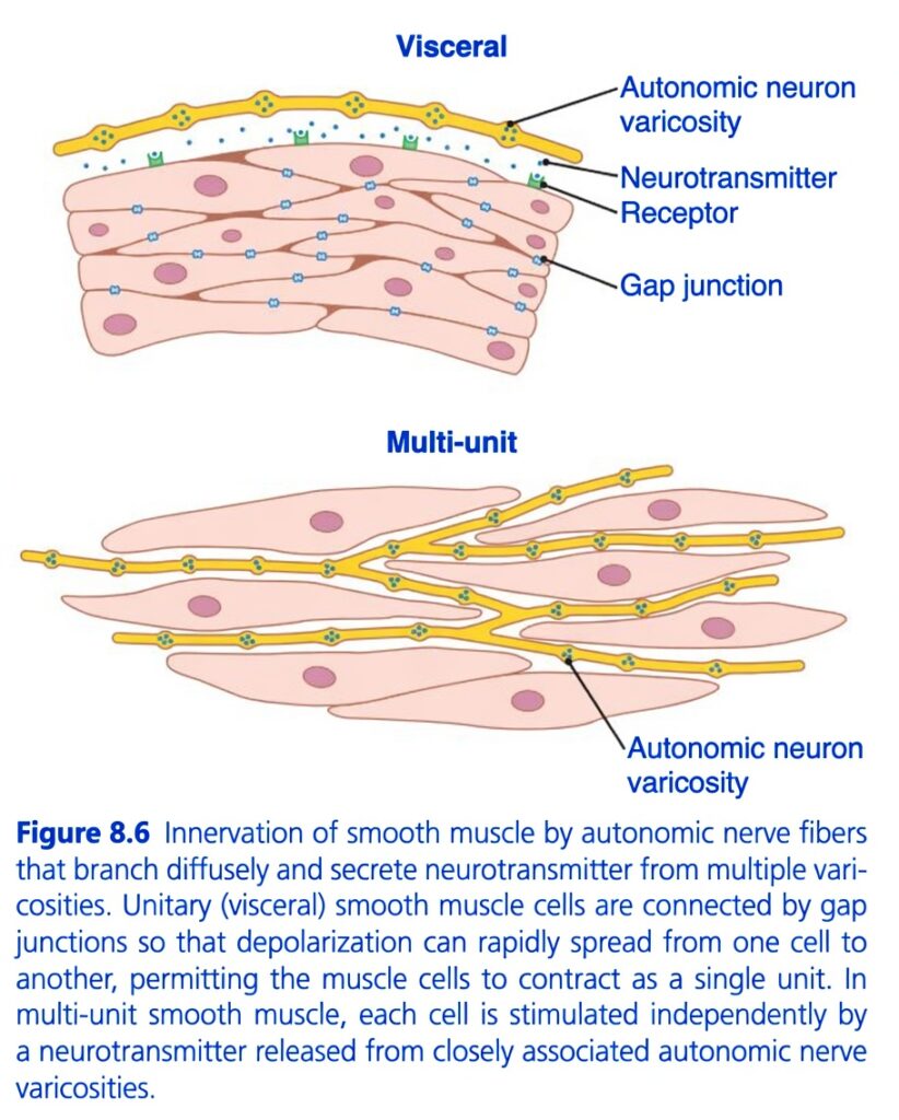
Figure 8.6: Innervation of Smooth Muscle
Easiest and Most Conceptual Explanation
⭐ One-Line Concept
Smooth muscle is controlled by autonomic nerves. There are two types of smooth muscle:
- Visceral (single-unit) smooth muscle → cells are connected by gap junctions and contract together.
- Multi-unit smooth muscle → cells are not well connected and each cell is stimulated separately.
First Understand the Whole Story
Imagine there are 100 workers.
Situation 1: Everyone holds hands
If you tell one worker to start working,
↓
The message quickly spreads to everyone.
↓
All workers work together.
This is Visceral (Single-Unit) Smooth Muscle.
Situation 2: Nobody holds hands
Each worker must receive his own instruction.
↓
One worker cannot tell another.
↓
Each works independently.
This is Multi-Unit Smooth Muscle.
Understanding Every Part of the Figure
The figure has two parts.
- Upper part = Visceral (Single-Unit) Smooth Muscle
- Lower part = Multi-Unit Smooth Muscle
Part A: Visceral (Single-Unit) Smooth Muscle
The upper figure shows visceral smooth muscle.
Examples include:
- Intestine
- Stomach
- Uterus
- Urinary bladder
- Ureter
These organs contract as one functional unit.
1. Autonomic Neuron
The yellow nerve fiber at the top is an autonomic nerve.
Unlike skeletal muscle,
it does not form a neuromuscular junction.
Instead,
it runs over the muscle surface.
Easy Concept
Think of it as a sprinkler pipe running above a garden.
2. Autonomic Neuron Varicosities
The small swellings along the yellow nerve are varicosities.
These are small enlargements containing neurotransmitters.
What do varicosities do?
They release neurotransmitters at many places along the nerve.
There is no single end plate like in skeletal muscle.
Easy Concept
Think of varicosities as many tiny spray nozzles.
Each nozzle sprays neurotransmitter onto nearby muscle cells.
3. Neurotransmitter
The blue dots represent neurotransmitter molecules.
Examples:
- Acetylcholine
- Norepinephrine
They diffuse through the extracellular fluid.
↓
Bind to receptors on smooth muscle.
↓
Start contraction or relaxation.
Easy Concept
These blue dots are the chemical messages sent by the autonomic nerve.
4. Receptors
The small green structures on the muscle cell membrane are receptors.
Neurotransmitters bind here.
Different receptors produce different effects.
Some receptors:
✅ Cause contraction.
Others:
✅ Cause relaxation.
Easy Concept
Receptors are like locks, and neurotransmitters are the keys.
5. Gap Junctions
The blue connections between neighboring cells are gap junctions.
These are tiny channels joining adjacent muscle cells.
Why are gap junctions important?
Electrical signals pass directly from one cell to another.
Therefore,
if one cell is stimulated,
↓
Neighboring cells are also stimulated.
↓
The entire muscle contracts together.
Easy Concept
Gap junctions are like bridges connecting neighboring houses.
A message crosses the bridge and reaches everyone.
Final Result of Visceral Smooth Muscle
One autonomic nerve releases neurotransmitter.
↓
One cell is stimulated.
↓
Gap junctions spread the electrical signal.
↓
All cells contract together.
Concept
One message → Whole muscle contracts
Part B: Multi-Unit Smooth Muscle
The lower figure shows multi-unit smooth muscle.
Examples include:
- Iris of the eye
- Ciliary muscle
- Piloerector muscles (hair follicles)
- Large airways
These muscles require very precise control.
1. Separate Muscle Cells
Notice the cells are not connected by many gap junctions.
Therefore,
signals cannot spread easily from one cell to another.
Easy Concept
Each cell behaves like an independent worker.
2. Numerous Autonomic Nerve Fibers
Many autonomic nerve fibers run between the muscle cells.
Each fiber has many varicosities.
Why?
Each muscle cell must receive its own nerve signal.
Concept
Instead of one nerve controlling many cells,
many nerves control many individual cells.
3. Autonomic Varicosities
Again, the yellow swellings are varicosities.
Each releases neurotransmitter very close to its target cell.
Easy Concept
Each muscle cell has its own personal messenger.
Final Result of Multi-Unit Smooth Muscle
Each muscle cell receives its own nerve signal.
↓
Each contracts independently.
↓
Produces very fine, precise movements.
Concept
One nerve → One muscle cell
Comparison of the Two Types
| Feature | Visceral (Single-Unit) | Multi-Unit |
|---|---|---|
| Gap junctions | Many | Few or absent |
| Electrical signal | Spreads from cell to cell | Does not spread significantly |
| Contraction | Whole muscle contracts together | Each cell contracts independently |
| Nerve supply | One nerve can activate many cells | Each cell requires its own nerve supply |
| Precision | Less precise | Very precise |
| Examples | Intestine, stomach, uterus, bladder | Iris, ciliary muscle, piloerector muscles |
Relationship to the Text
Unlike skeletal muscle, smooth muscle can be stimulated by:
- Autonomic nerves
- Hormones
- Stretch
- Other local chemical factors
This figure specifically illustrates the nervous (autonomic) control of smooth muscle.
Because smooth muscle has many receptor proteins, the same neurotransmitter may produce contraction or relaxation depending on the receptor type.
Easy Story
Imagine two classrooms.
Classroom 1 (Visceral Smooth Muscle)
Students are holding hands.
The teacher whispers to one student.
↓
The message passes through everyone.
↓
The whole class stands up together.
Classroom 2 (Multi-Unit Smooth Muscle)
No students are holding hands.
The teacher must tell each student individually.
↓
Each student stands up separately.
Simple Flow Diagram
Visceral (Single-Unit) Smooth Muscle
Autonomic nerve
↓
Varicosities release neurotransmitter
↓
One muscle cell is stimulated
↓
Electrical signal spreads through gap junctions
↓
All muscle cells contract together
Multi-Unit Smooth Muscle
Autonomic nerve
↓
Varicosities release neurotransmitter
↓
Each muscle cell receives its own signal
↓
Each cell contracts independently
↓
Precise muscle control
High-Yield Summary Table
| Structure | Function | Easy Concept |
|---|---|---|
| Autonomic nerve | Controls smooth muscle | The messenger |
| Varicosities | Release neurotransmitter at many sites | Tiny spray nozzles |
| Neurotransmitter | Chemical signal to muscle | Message sent by the nerve |
| Receptors | Bind neurotransmitter | Locks that receive the key |
| Gap junctions | Connect neighboring cells electrically | Bridges between cells |
| Visceral smooth muscle | Contracts as one unit | One message activates all cells |
| Multi-unit smooth muscle | Each cell contracts independently | One message for one cell |
Key Concept (Figure 8.6)
Figure 8.6 shows the two patterns of autonomic innervation in smooth muscle. In visceral (single-unit) smooth muscle, autonomic nerve fibers release neurotransmitters from multiple varicosities. The neurotransmitters bind to receptors on nearby muscle cells, and because the cells are connected by numerous gap junctions, the electrical signal spreads rapidly from one cell to another. As a result, the entire muscle contracts as a single functional unit. In multi-unit smooth muscle, there are few or no gap junctions, so electrical signals do not spread effectively between cells. Instead, each muscle cell receives its own autonomic nerve supply and contracts independently, allowing fine and precise control of movement.
Excitatory and Inhibitory Transmitter Substances Secreted at the Smooth Muscle Neuromuscular Junction
- The two most important neurotransmitters released by autonomic nerves supplying smooth muscle are:
- Acetylcholine (ACh)
- Norepinephrine (NE)
- The same nerve fiber never releases both acetylcholine and norepinephrine.
- In some organs, acetylcholine stimulates (excites) smooth muscle contraction.
- In other organs, acetylcholine inhibits smooth muscle contraction.
- When acetylcholine excites a smooth muscle, norepinephrine usually inhibits it.
- Conversely, when acetylcholine inhibits a smooth muscle, norepinephrine usually excites it.
- Why do these different responses occur?
- Both acetylcholine and norepinephrine first bind to receptor proteins on the surface of the smooth muscle cell membrane.
- Some receptor proteins are excitatory receptors.
- Other receptor proteins are inhibitory receptors.
- Therefore, the type of receptor, not just the neurotransmitter itself, determines whether the smooth muscle contracts or relaxes.
- The receptor type also determines whether acetylcholine or norepinephrine will produce excitation or inhibition.
- These receptors are discussed in more detail in Chapter 61, which explains the autonomic nervous system.
KEY CONCEPT
- The two main autonomic neurotransmitters for smooth muscle are:
- Acetylcholine (ACh)
- Norepinephrine (NE)
- One nerve fiber releases only one of these neurotransmitters.
- Acetylcholine may either excite or inhibit smooth muscle, depending on the organ.
- Norepinephrine usually has the opposite effect of acetylcholine in the same organ.
- The response depends on the receptor type:
- Excitatory receptor → Smooth muscle contracts.
- Inhibitory receptor → Smooth muscle relaxes.
Conceptual Examples
- Organ A
- Acetylcholine binds to an excitatory receptor → Smooth muscle contracts.
- Norepinephrine binds to an inhibitory receptor → Smooth muscle relaxes.
- Organ B
- Acetylcholine binds to an inhibitory receptor → Smooth muscle relaxes.
- Norepinephrine binds to an excitatory receptor → Smooth muscle contracts.
- Easy way to remember:
- Neurotransmitter = Messenger
- Receptor = Decision maker
- The receptor type decides whether the message causes contraction or relaxation.
Membrane Potentials in Smooth Muscle
- The membrane potential of smooth muscle changes depending on the condition of the muscle.
- In the normal resting state, the intracellular membrane potential is about −50 to −60 mV.
- This is about 30 mV less negative than the resting membrane potential of skeletal muscle.
Action Potentials in Unitary Smooth Muscle
- Action potentials occur in unitary (visceral) smooth muscle.
- They are produced in the same general way as in skeletal muscle.
- Most multi-unit smooth muscle normally does not produce action potentials.
- The action potentials of visceral smooth muscle occur in two forms:
- Spike potentials
- Action potentials with plateaus
Spike Potentials
- Spike potentials are the typical action potentials seen in most types of unitary smooth muscle.
- They are similar to the action potentials of skeletal muscle.
- The duration of a spike potential is 10–50 milliseconds (Fig. 8.7A).
- Spike action potentials can be produced by:
- Electrical stimulation
- Hormonal stimulation
- Neurotransmitters released from nerve fibers
- Stretch of the muscle
- Spontaneous generation within the smooth muscle fiber
KEY CONCEPT
- The resting membrane potential of smooth muscle is −50 to −60 mV.
- Smooth muscle is less negative than skeletal muscle by about 30 mV.
- Unitary (visceral) smooth muscle generates action potentials.
- Most multi-unit smooth muscle normally does not generate action potentials.
- Unitary smooth muscle has two types of action potentials:
- Spike potentials
- Plateau action potentials
- Spike potentials last 10–50 ms and can be triggered by electrical stimulation, hormones, neurotransmitters, stretch, or spontaneous activity (Fig. 8.7A).
Conceptual Examples
- Resting smooth muscle → Membrane potential ≈ −50 to −60 mV.
- Intestinal (visceral) smooth muscle receives a stimulus → Spike action potential develops → Muscle contracts.
- Stretching the intestine → Spike action potentials may occur → Smooth muscle contracts.
- Hormone or neurotransmitter binds to receptors → Spike action potential → Contraction.
KEEP IN MIND
- Resting membrane potential of smooth muscle: −50 to −60 mV
- Compared with skeletal muscle:
- Skeletal muscle ≈ −80 to −90 mV
- Smooth muscle ≈ 30 mV less negative than skeletal muscle.
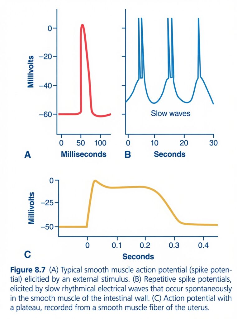
Figure 8.7: Types of Smooth Muscle Action Potentials
Easiest and Most Conceptual Explanation
⭐ One-Line Concept
Smooth muscle can produce three main electrical patterns:
- Single spike action potential (short, external stimulus)
- Repeated spike action potentials on slow waves (intestinal smooth muscle)
- Plateau action potential (long-lasting contraction, e.g., uterus)
First Understand the Whole Story
Think of smooth muscle like three different types of light switches.
Type 1 (Figure A)
Press the switch once.
↓
The light flashes quickly.
↓
Then turns off.
➡ Single spike action potential
Type 2 (Figure B)
The light slowly becomes brighter and dimmer.
Whenever it becomes bright enough,
↓
It flashes.
↓
Then repeats again.
➡ Slow waves with spike action potentials
Type 3 (Figure C)
Turn the light on,
↓
It stays on for a long time,
↓
Then slowly turns off.
➡ Plateau action potential
Understanding the Axes
Y-axis (Millivolts)
Shows the membrane potential.
- Around −60 mV = resting membrane potential.
- Near 0 mV = depolarization.
X-axis
Shows time.
Notice:
- Figure A = milliseconds
- Figure B = seconds
- Figure C = seconds
This means:
- Figure A is very fast.
- Figure B is slower and rhythmic.
- Figure C lasts the longest.
Figure A — Typical Spike Action Potential
This is the simplest smooth muscle action potential.
Step 1: Resting Membrane Potential
Starts at about −60 mV.
The muscle is relaxed.
Step 2: External Stimulus
A nerve,
or
A hormone,
or
Stretch,
stimulates the smooth muscle.
Step 3: Rapid Depolarization
Voltage-gated Ca²⁺ channels open.
Ca²⁺ enters the cell.
The membrane potential rises rapidly.
From:
−60 mV
↓
Almost 0 mV.
Concept
Ca²⁺ enters → electrical signal starts.
Step 4: Repolarization
Ca²⁺ channels close.
K⁺ leaves the cell.
The membrane potential returns to about −60 mV.
Final Result
One electrical spike
↓
One contraction
↓
Muscle relaxes.
Easy Concept
Think of it like ringing a doorbell once.
One press
↓
One ring.
Figure B — Slow Waves with Repeated Spike Potentials
This graph is commonly seen in the gastrointestinal (GI) tract.
What are Slow Waves?
The smooth muscle membrane slowly depolarizes and repolarizes by itself, even without a nerve impulse.
These are called slow waves.
Important Point
A slow wave is not an action potential.
It is only a rhythmic change in membrane potential.
What happens during each slow wave?
The membrane slowly rises.
If it does not reach threshold,
↓
No spike occurs.
↓
No strong contraction.
If it reaches threshold,
↓
Voltage-gated Ca²⁺ channels open.
↓
One or more spike action potentials appear on top of the slow wave.
Notice in the graph:
The blue curve rises slowly.
Then sharp spikes appear at the top.
Those spikes produce contraction.
Why are there several spikes?
The membrane remains above threshold for a short period.
Therefore,
more than one spike can occur.
Concept
The slow wave is like climbing a hill.
When you reach the top,
↓
You jump (spike).
↓
Each jump produces contraction.
Final Result
Slow waves
↓
Repeated spikes
↓
Repeated rhythmic contractions.
Where does this occur?
Mainly in:
- Small intestine
- Stomach
- Colon
Figure C — Plateau Action Potential
This graph is different.
Instead of coming down immediately,
it remains near the top for a long time.
This flat portion is called the plateau.
Step 1: Depolarization
Ca²⁺ enters.
The membrane depolarizes.
Step 2: Plateau Phase
Ca²⁺ channels remain open longer.
Ca²⁺ continues entering.
The membrane remains depolarized.
Why is this important?
More Ca²⁺ enters.
↓
Contraction lasts much longer.
Concept
Instead of a quick flash,
the muscle receives a continuous electrical signal.
Step 3: Repolarization
Eventually,
Ca²⁺ channels close.
K⁺ leaves the cell.
The membrane returns to resting potential.
Final Result
Long-lasting action potential
↓
Long-lasting contraction.
Where does this occur?
Commonly in:
- Uterus
- Some vascular smooth muscle
- Ureter (in certain regions)
Why Does Plateau Matter?
The uterus must contract for a prolonged period during labor.
A short spike would not be enough.
The plateau allows sustained contraction.
Comparing the Three Graphs
| Figure | Type | Duration | Main Feature | Example |
|---|---|---|---|---|
| A | Spike action potential | Very short (milliseconds) | Single spike after a stimulus | General smooth muscle |
| B | Slow waves with spikes | Rhythmic (seconds) | Slow waves trigger repeated spikes | Intestinal smooth muscle |
| C | Plateau action potential | Long (0.3–0.4 s or more) | Sustained depolarization | Uterus |
Easy Story
Imagine three kinds of fireworks.
Firework A
Boom!
Finished.
➡ One spike.
Firework B
Boom…
Boom…
Boom…
Repeated regularly.
➡ Slow waves with repeated spikes.
Firework C
Boom…
Keeps glowing.
Then slowly fades.
➡ Plateau action potential.
Simple Flow Diagram
Figure A
External stimulus
↓
Ca²⁺ channels open
↓
Single spike action potential
↓
One contraction
↓
Relaxation
Figure B
Slow wave develops
↓
Threshold reached
↓
Repeated spike action potentials
↓
Rhythmic contractions
↓
Repeat
Figure C
Depolarization
↓
Plateau phase
↓
Prolonged Ca²⁺ entry
↓
Long-lasting contraction
↓
RepolarizationHigh-Yield Summary Table
| Feature | Figure A | Figure B | Figure C |
|---|---|---|---|
| Action potential | Single spike | Repeated spikes on slow waves | Plateau action potential |
| Duration | Very short | Rhythmic | Long-lasting |
| Trigger | External stimulus | Slow waves reaching threshold | Sustained Ca²⁺ entry |
| Contraction | Brief | Rhythmic | Sustained |
| Typical location | General smooth muscle | Gastrointestinal tract | Uterus |
Key Concept (Figure 8.7)
Figure 8.7 illustrates the three major electrical patterns of smooth muscle. Figure A shows a typical spike action potential, produced by an external stimulus, resulting in a brief contraction. Figure B shows slow waves, which are spontaneous rhythmic fluctuations in membrane potential commonly found in intestinal smooth muscle. Slow waves themselves do not cause contraction unless they reach the threshold, at which point spike action potentials are generated, producing rhythmic contractions. Figure C shows a plateau action potential, in which prolonged Ca²⁺ entry keeps the membrane depolarized for a longer time, producing a sustained contraction. This pattern is especially important in organs such as the uterus, where prolonged contractions are required.
Action Potentials With Plateaus
- Some smooth muscle cells produce action potentials with a plateau (Fig. 8.7C).
- The beginning (onset) of this action potential is similar to a normal spike action potential.
- However, after the spike, the membrane does not repolarize quickly.
- Instead, repolarization is delayed.
- This delay lasts for several hundred milliseconds up to about 1000 milliseconds (1 second).
- This prolonged plateau phase causes the smooth muscle to remain contracted for a longer time.
- Plateau action potentials occur in some types of smooth muscle, including:
- Ureter
- Uterus (under some conditions)
- Certain types of vascular smooth muscle
- Cardiac muscle fibers also have plateau action potentials, which produce a prolonged period of contraction.
KEY CONCEPT
- Plateau action potentials begin like normal spike action potentials.
- The difference is that repolarization is delayed.
- The plateau lasts several hundred milliseconds to about 1 second (1000 ms).
- Because repolarization is delayed, the muscle stays contracted longer.
- Plateau action potentials are found in:
- Ureter
- Uterus (under some conditions)
- Certain vascular smooth muscles
- Cardiac muscle
Conceptual Examples
- Normal spike action potential → Quick repolarization → Short contraction.
- Plateau action potential → Delayed repolarization → Long-lasting contraction.
- Ureter uses a plateau action potential to maintain contraction long enough to push urine forward.
- Uterus can maintain a prolonged contraction during certain conditions because of plateau action potentials.
- Cardiac muscle also has a plateau action potential, allowing the heart to contract long enough to pump blood effectively.
Figure Number: Fig. 8.7C
Equation/Calculation
- Plateau duration:
- Several hundred milliseconds
- Maximum ≈ 1000 milliseconds = 1 second
Easy Comparison
| Feature | Spike Action Potential | Plateau Action Potential |
|---|---|---|
| Beginning | Same | Same |
| Repolarization | Rapid | Delayed |
| Duration | 10–50 ms | Several hundred ms to 1000 ms (1 s) |
| Muscle contraction | Short | Prolonged |
| Examples | Most unitary smooth muscle | Ureter, uterus, vascular smooth muscle, cardiac muscle |
Calcium Channels Are Important in Generating the Smooth Muscle Action Potential
- Smooth muscle cell membranes contain many more voltage-gated Ca²⁺ channels than skeletal muscle.
- They contain only a few voltage-gated Na⁺ (sodium) channels.
- Therefore, Na⁺ has only a small role in producing the action potential in most smooth muscle.
- Instead, Ca²⁺ entering the smooth muscle cell is mainly responsible for generating the action potential.
- This Ca²⁺ influx occurs by a self-regenerative (positive feedback) mechanism, similar to the way Na⁺ channels generate action potentials in nerve fibers and skeletal muscle.
- However, Ca²⁺ channels open much more slowly than Na⁺ channels.
- They also remain open for a much longer time.
- Because they stay open longer, Ca²⁺ continues to enter the cell for a longer period.
- This prolonged Ca²⁺ entry is the main reason for the long-lasting (plateau) action potentials seen in some smooth muscle fibers.
- Another important point is that the Ca²⁺ entering during the action potential has a second function.
- The same Ca²⁺ directly activates the smooth muscle contractile mechanism.
- Therefore, Ca²⁺ performs two functions at the same time:
- Generates the action potential.
- Directly causes smooth muscle contraction.
KEY CONCEPT
- Smooth muscle has many voltage-gated Ca²⁺ channels but very few voltage-gated Na⁺ channels.
- Therefore, Ca²⁺ is the main ion responsible for generating the smooth muscle action potential.
- Ca²⁺ channels open slowly and remain open longer than Na⁺ channels.
- This prolonged opening produces plateau action potentials in some smooth muscle fibers.
- Ca²⁺ has a dual role:
- Generates the action potential.
- Directly initiates smooth muscle contraction.
Conceptual Examples
Example 1: Skeletal Muscle
- Na⁺ enters the cell → Action potential is generated.
- Later, Ca²⁺ is released from the sarcoplasmic reticulum → Muscle contracts.
- Na⁺ and Ca²⁺ perform different jobs.
Example 2: Smooth Muscle
- Ca²⁺ enters through voltage-gated Ca²⁺ channels.
- The same Ca²⁺ generates the action potential.
- The same Ca²⁺ immediately activates contraction.
- One ion (Ca²⁺) performs two jobs.
Easy Memory Trick
Skeletal Muscle
- Na⁺ = Electrical signal
- Ca²⁺ = Contraction
Smooth Muscle
- Ca²⁺ = Electrical signal + Contraction
Easy Comparison Table
| Feature | Skeletal Muscle | Smooth Muscle |
|---|---|---|
| Main ion for action potential | Na⁺ | Ca²⁺ |
| Number of voltage-gated Ca²⁺ channels | Few | Many |
| Number of voltage-gated Na⁺ channels | Many | Few |
| Channel opening speed | Fast | Slow |
| Channel remains open | Short time | Long time |
| Plateau action potential | Usually absent | Present in some smooth muscles |
| Ca²⁺ function | Mainly causes contraction | Generates action potential + causes contraction |
Slow Wave Potentials in Unitary Smooth Muscle Can Lead to Spontaneous Generation of Action Potentials
- Some unitary (visceral) smooth muscles are self-excitatory.
- This means action potentials can develop without any external stimulus.
- This spontaneous activity is usually associated with a basic slow wave rhythm of the membrane potential (Fig. 8.7B).
- The slow wave is NOT an action potential.
- It is not a self-regenerating electrical signal that spreads from one cell to another.
- Instead, it is a local rhythmic change in the membrane potential of the smooth muscle fibers.
- The exact cause of slow waves is not known.
- One proposed explanation is that the Na⁺ (sodium) pump activity increases and decreases rhythmically.
- When the Na⁺ pump works faster, more positive ions leave the cell, so the membrane becomes more negative (hyperpolarized).
- When the Na⁺ pump slows down, fewer positive ions leave the cell, so the membrane becomes less negative (depolarized).
- Another proposed explanation is that ion channels rhythmically open and close, causing regular changes in membrane permeability.
- Slow waves alone do not produce muscle contraction.
- Their importance is that they can trigger action potentials if they become large enough.
- When the membrane potential rises from about −60 mV to approximately −35 mV, it reaches the threshold.
- At this threshold, an action potential is generated.
- The action potential then spreads throughout the smooth muscle, causing muscle contraction.
- As shown in Fig. 8.7B, one or more action potentials occur at the peak of each slow wave.
- These repeated action potentials produce rhythmic contractions of the smooth muscle.
- Because slow waves determine the rhythm of these contractions, they are called pacemaker waves.
- In the gastrointestinal tract, these pacemaker waves control the rhythmic contractions of the gut.
KEY CONCEPT
- Some unitary smooth muscles generate action potentials spontaneously.
- This spontaneous activity is associated with slow waves (Fig. 8.7B).
- Slow waves are not action potentials and cannot directly cause contraction.
- Slow waves are local rhythmic fluctuations in membrane potential.
- When a slow wave reaches the threshold of about −35 mV, it triggers an action potential.
- The action potential causes smooth muscle contraction.
- Repeated slow waves produce rhythmic contractions, so they are called pacemaker waves.
- Pacemaker waves control the rhythmic movements of the gut.
Conceptual Examples
Example 1: Weak Slow Wave
- Membrane potential changes from −60 mV to −45 mV.
- Threshold (−35 mV) is not reached.
- ❌ No action potential
- ❌ No contraction
Example 2: Strong Slow Wave
- Membrane potential changes from −60 mV to −35 mV.
- Threshold is reached.
- ✅ Action potential develops
- ✅ Smooth muscle contracts
Example 3: Intestinal Smooth Muscle
- Slow waves occur continuously.
- Each strong slow wave reaches threshold.
- Action potentials are generated repeatedly.
- The intestine contracts rhythmically, helping move food forward.
Easy Flow Chart
Slow wave develops
⬇
Membrane potential rises from −60 mV toward −35 mV
⬇
Threshold reached (≈ −35 mV)
⬇
Action potential generated
⬇
Action potential spreads through smooth muscle
⬇
Smooth muscle contracts
⬇
Repeated cycles → Rhythmic contractions (Pacemaker activity)
Easy Comparison
| Feature | Slow Wave | Action Potential |
|---|---|---|
| Electrical signal | Local rhythmic fluctuation | Self-regenerating electrical impulse |
| Spreads through muscle | No | Yes |
| Directly causes contraction | No | Yes |
| Main role | Reaches threshold | Produces contraction |
| Threshold | Must reach ≈ −35 mV | Already generated after threshold |
Figure Number: Fig. 8.7B
Equation/Calculation
- Resting membrane potential: ≈ −60 mV
- Threshold for action potential: ≈ −35 mV
- Required depolarization:
−35 − (−60) = +25 mV
Concept: The membrane must become about 25 mV less negative (depolarize) for a slow wave to trigger an action potential.
Excitation of Visceral Smooth Muscle by Muscle Stretch
- When visceral (unitary) smooth muscle is stretched enough, it usually generates spontaneous action potentials.
- These action potentials are produced by a combination of two factors:
- Normal slow wave potentials already present in the muscle.
- Stretch decreases the negativity of the membrane potential (the membrane becomes less negative, i.e., depolarized).
- Because of these two factors, the membrane reaches the threshold more easily.
- As a result, action potentials are generated spontaneously.
- These action potentials then cause smooth muscle contraction.
- This stretch response is very important in the gastrointestinal (GI) tract.
- When the gut wall is excessively stretched, it automatically contracts in a rhythmic manner.
- This helps move the intestinal contents forward without needing an external stimulus.
- For example, when the intestine becomes overfilled with food or intestinal contents:
- The intestinal wall stretches.
- Stretch triggers spontaneous action potentials.
- The smooth muscle contracts automatically.
- These contractions produce peristaltic waves.
- The peristaltic waves push the intestinal contents away from the overfilled area, usually toward the anus.
KEY CONCEPT
- Stretch can directly excite visceral (unitary) smooth muscle.
- Stretch produces spontaneous action potentials by combining:
- Normal slow waves
- Depolarization (less negative membrane potential) caused by stretch
- These action potentials cause rhythmic smooth muscle contraction.
- In the gut, this mechanism automatically produces peristalsis.
- Peristalsis moves intestinal contents toward the anus, preventing excessive distension.
Conceptual Examples
Example 1: Normal Intestine
- Normal stretch → Slow waves continue.
- Threshold is not always reached.
- Mild or no contraction occurs.
Example 2: Overfilled Intestine
- Too much food stretches the intestinal wall.
- Stretch makes the membrane less negative.
- Slow waves now reach the threshold.
- ✅ Action potentials develop.
- ✅ Smooth muscle contracts rhythmically.
- ✅ Peristaltic waves move food toward the anus.
Example 3: Easy Story
Imagine the intestine is like a rubber tube.
- Small amount of food → Tube stretches a little → No strong response.
- Too much food → Tube stretches a lot → Stretch triggers action potentials.
- The intestine automatically squeezes and pushes the food forward.
Easy Flow Chart
Intestine becomes overfilled
⬇
Gut wall stretches
⬇
Stretch makes membrane less negative (depolarization)
- Normal slow waves
⬇
Threshold is reached
⬇
Action potentials are generated
⬇
Smooth muscle contracts
⬇
Peristaltic waves develop
⬇
Food moves toward the anus
Easy Comparison
| Normal Gut | Overfilled Gut |
|---|---|
| Mild stretch | Excessive stretch |
| Slow waves may not reach threshold | Stretch + slow waves reach threshold |
| Few or no action potentials | Spontaneous action potentials develop |
| Minimal contraction | Strong rhythmic contractions |
| Little movement | Peristaltic waves move food toward the anus |
DEPOLARIZATION OF MULTI-UNIT SMOOTH MUSCLE WITHOUT ACTION POTENTIALS
- Multi-unit smooth muscle usually contracts in response to nerve stimulation.
- Examples of multi-unit smooth muscle include:
- Muscle of the iris of the eye
- Piloerector (arrector pili) muscle attached to each hair
- The autonomic nerve endings release different neurotransmitters:
- Acetylcholine (ACh) in some multi-unit smooth muscles.
- Norepinephrine (NE) in other multi-unit smooth muscles.
- These neurotransmitters depolarize the smooth muscle cell membrane.
- This depolarization directly causes muscle contraction.
- Action potentials usually do not develop in multi-unit smooth muscle.
- The reason is that the smooth muscle fibers are very small, so they cannot usually generate a self-propagating action potential.
- In visceral (unitary) smooth muscle, about 30–40 smooth muscle fibers must depolarize together before a self-propagating action potential can occur.
- In contrast, multi-unit smooth muscle does not require an action potential for contraction.
- Instead, the neurotransmitter produces a local depolarization, called the junctional potential.
- This junctional potential spreads electrotonically (passively) throughout the entire muscle fiber.
- This passive spread is sufficient to produce contraction, even though no action potential is generated.
KEY CONCEPT
- Multi-unit smooth muscle contracts mainly because of nerve stimulation.
- The neurotransmitter may be:
- Acetylcholine
- Norepinephrine
- These neurotransmitters depolarize the membrane.
- Most multi-unit smooth muscle does not generate action potentials because the fibers are too small.
- Instead, a junctional potential spreads electrotonically through the cell.
- This local depolarization alone is enough to cause contraction.
Conceptual Examples
Example 1: Iris Muscle
- Light stimulates autonomic nerves.
- Nerves release acetylcholine or norepinephrine.
- The membrane becomes depolarized.
- A junctional potential spreads through the muscle.
- The iris contracts without an action potential.
Example 2: Arrector Pili Muscle
- Cold temperature or fear activates autonomic nerves.
- Neurotransmitter is released.
- A junctional potential develops.
- The small smooth muscle contracts.
- Hair stands up (goosebumps).
Easy Story
Imagine a small room.
- In a large room (unitary smooth muscle), you need a loudspeaker (action potential) so everyone hears the message.
- In a small room (multi-unit smooth muscle), a person speaking normally (junctional potential) is enough for everyone to hear.
- Similarly, small smooth muscle cells do not need an action potential—the junctional potential alone causes contraction.
Easy Comparison
| Unitary Smooth Muscle | Multi-Unit Smooth Muscle |
|---|---|
| Usually generates action potentials | Usually does not generate action potentials |
| About 30–40 fibers must depolarize together | Individual small fibers respond directly |
| Contraction follows an action potential | Contraction follows a junctional potential |
| Seen in gut, uterus, ureter | Seen in iris and arrector pili muscles |
KEEP IN MIND
- Approximately 30–40 smooth muscle fibers must depolarize simultaneously in unitary smooth muscle before a self-propagating action potential develops.
- Multi-unit smooth muscle: No action potential is usually required; a junctional potential alone produces contraction.
Local Tissue Factors and Hormones Can Cause Smooth Muscle Contraction Without Action Potentials
- About half of all smooth muscle contractions are started without action potentials.
- Instead, these contractions occur because stimulatory factors act directly on the smooth muscle contractile machinery.
- Two important types of these stimulating factors are:
- Local tissue chemical factors
- Hormones
Smooth Muscle Contraction in Response to Local Tissue Chemical Factors
- The small arterioles, metarterioles, and precapillary sphincters have little or no nerve supply.
- Even without nerves, their smooth muscle contracts very effectively.
- These muscles respond quickly to:
- Changes in local chemical conditions in the surrounding interstitial fluid.
- Stretch caused by changes in blood pressure.
- Under normal resting conditions, many of these small blood vessels remain partially contracted.
- When a tissue needs more blood flow, several local chemical factors cause the smooth muscle to relax.
- Relaxation of the vascular smooth muscle causes vasodilation (widening of blood vessels).
- Vasodilation increases blood flow to the tissue.
- In this way, local feedback mechanisms automatically regulate blood flow according to the tissue’s needs.
Local Factors That Cause Vasodilation
- Low oxygen (O₂) in the tissue
- Causes smooth muscle relaxation.
- Results in vasodilation.
- High carbon dioxide (CO₂)
- Causes vasodilation.
- High hydrogen ion (H⁺) concentration (low pH)
- Causes vasodilation.
- Other substances that also cause local vasodilation include:
- Adenosine
- Lactic acid
- Increased potassium ions (K⁺)
- Nitric oxide (NO)
- Increased body temperature
- Decreased blood pressure
- Reduces stretch of the vascular smooth muscle.
- This also causes the small blood vessels to dilate.
KEY CONCEPT
- About 50% of smooth muscle contractions occur without action potentials.
- Local tissue chemicals and hormones can directly activate or regulate smooth muscle.
- Small blood vessels do not need nerve stimulation to respond.
- Local chemical changes automatically adjust blood vessel diameter according to tissue needs.
- Most local metabolic factors relax vascular smooth muscle, causing vasodilation and increased blood flow.
Conceptual Examples
Example 1: Exercising Muscle
- A muscle is working hard.
- Oxygen decreases, while CO₂, H⁺, lactic acid, and adenosine increase.
- These chemicals relax the smooth muscle of nearby arterioles.
- Blood vessels dilate.
- More oxygen-rich blood reaches the active muscle.
Example 2: Warm Skin
- Body temperature rises.
- Increased temperature causes vasodilation.
- More blood flows to the skin.
- Heat is lost from the body.
Example 3: Low Blood Pressure
- Blood pressure falls.
- Blood vessels are stretched less.
- Reduced stretch causes vasodilation.
- The vessel adjusts automatically without needing nerve stimulation.
Easy Flow Chart
Local tissue needs more blood
⬇
Low O₂ / High CO₂ / High H⁺ / Adenosine / Lactic acid / K⁺ / NO / Heat
⬇
Smooth muscle relaxes
⬇
Vasodilation
⬇
Blood flow increases
⬇
More oxygen and nutrients reach the tissue
Easy Summary Table
| Local Factor | Effect on Smooth Muscle | Blood Vessel Response |
|---|---|---|
| ↓ Oxygen (O₂) | Relaxes | Vasodilation |
| ↑ Carbon dioxide (CO₂) | Relaxes | Vasodilation |
| ↑ Hydrogen ions (H⁺) | Relaxes | Vasodilation |
| Adenosine | Relaxes | Vasodilation |
| Lactic acid | Relaxes | Vasodilation |
| ↑ Potassium (K⁺) | Relaxes | Vasodilation |
| Nitric oxide (NO) | Relaxes | Vasodilation |
| ↑ Temperature | Relaxes | Vasodilation |
| ↓ Blood pressure (↓ Stretch) | Relaxes | Vasodilation |
Approximate proportion of smooth muscle contractions without action potentials:
- ≈ 50% (about half)
Effects of Hormones on Smooth Muscle Contraction
- Many hormones circulating in the blood affect smooth muscle contraction.
- Some hormones have very strong (profound) effects on smooth muscle.
- Important hormones include:
- Norepinephrine
- Epinephrine
- Angiotensin II
- Endothelin
- Vasopressin
- Oxytocin
- Serotonin
- Histamine
- A hormone causes contraction if the smooth muscle cell membrane has excitatory receptors for that hormone.
- A hormone causes relaxation (inhibition) if the membrane has inhibitory receptors instead.
- Therefore, the type of receptor determines the response, not the hormone alone.
Mechanisms of Smooth Muscle Excitation or Inhibition by Hormones or Local Tissue Factors
1. Excitation by Opening Na⁺ or Ca²⁺ Channels
- Some hormone receptors open Na⁺ or Ca²⁺ channels in the smooth muscle membrane.
- Positive ions enter the cell.
- The membrane becomes depolarized.
- This is similar to what happens after nerve stimulation.
- Depolarization may:
- Produce action potentials, or
- Increase existing action potentials.
- Sometimes no action potential develops, but the depolarization still allows Ca²⁺ to enter the cell.
- The increased intracellular Ca²⁺ causes smooth muscle contraction.
2. Inhibition by Closing Na⁺ and Ca²⁺ Channels or Opening K⁺ Channels
- Hormones or local tissue factors can close Na⁺ and Ca²⁺ channels.
- This prevents positive ions from entering the cell.
- They can also open K⁺ (potassium) channels.
- K⁺ leaves the cell.
- Loss of positive ions makes the inside of the cell more negative.
- This state is called hyperpolarization.
- Hyperpolarization strongly inhibits smooth muscle contraction.
3. Hormones Can Act Without Changing the Membrane Potential
- Some hormones do not change the membrane potential.
- Instead, they activate membrane receptors that do not open ion channels.
- These receptors produce internal changes inside the smooth muscle cell.
Example: Ca²⁺ Release
- A receptor stimulates the sarcoplasmic reticulum.
- The sarcoplasmic reticulum releases Ca²⁺ into the cytoplasm.
- The released Ca²⁺ causes smooth muscle contraction.
4. Inhibition Through Second Messengers (cAMP and cGMP)
- Some receptors activate the enzymes:
- Adenylate cyclase
- Guanylate cyclase
- These enzymes produce second messengers:
- cAMP (cyclic adenosine monophosphate)
- cGMP (cyclic guanosine monophosphate)
- These second messengers change the phosphorylation of several enzymes.
- As a result:
- The Ca²⁺ pump of the sarcoplasmic reticulum becomes more active.
- The Ca²⁺ pump of the cell membrane also becomes more active.
- More Ca²⁺ is removed from the cytoplasm.
- The intracellular Ca²⁺ concentration falls.
- This inhibits smooth muscle contraction.
Different Smooth Muscles Respond Differently
- Smooth muscles in different organs do not all respond the same way.
- The response depends on the type of receptors present.
- Therefore, the same hormone can produce opposite effects in different organs.
Example
- Norepinephrine
- Relaxes (inhibits) smooth muscle in the intestine.
- Contracts (stimulates) smooth muscle in blood vessels.
KEY CONCEPT
- Many hormones regulate smooth muscle activity.
- The effect of a hormone depends on the type of receptor:
- Excitatory receptor → Contraction
- Inhibitory receptor → Relaxation
- Hormones can cause contraction by:
- Opening Na⁺ or Ca²⁺ channels
- Increasing Ca²⁺ entry
- Releasing Ca²⁺ from the sarcoplasmic reticulum
- Hormones can cause relaxation by:
- Closing Na⁺ and Ca²⁺ channels
- Opening K⁺ channels (hyperpolarization)
- Increasing cAMP or cGMP, which remove Ca²⁺ from the cytoplasm
- The same hormone may cause contraction in one organ and relaxation in another, depending on the receptors present.
Conceptual Examples
Example 1: Excitation
Hormone binds to an excitatory receptor
⬇
Na⁺ or Ca²⁺ channels open
⬇
Ca²⁺ enters the cell
⬇
Smooth muscle contracts
Example 2: Inhibition
Hormone binds to an inhibitory receptor
⬇
Na⁺ and Ca²⁺ channels close or K⁺ channels open
⬇
Cell becomes hyperpolarized
⬇
Smooth muscle relaxes
Example 3: Internal Ca²⁺ Release
Hormone binds to receptor
⬇
Sarcoplasmic reticulum releases Ca²⁺
⬇
Smooth muscle contracts
Example 4: cAMP / cGMP Pathway
Hormone binds to receptor
⬇
Adenylate cyclase or guanylate cyclase activated
⬇
cAMP or cGMP formed
⬇
Ca²⁺ pumps become more active
⬇
Ca²⁺ leaves the cytoplasm
⬇
Smooth muscle relaxes
Easy Comparison Table
| Mechanism | Effect on Ca²⁺ | Result |
|---|---|---|
| Open Na⁺/Ca²⁺ channels | Ca²⁺ increases | Contraction |
| Release Ca²⁺ from sarcoplasmic reticulum | Ca²⁺ increases | Contraction |
| Close Na⁺/Ca²⁺ channels | Ca²⁺ decreases | Relaxation |
| Open K⁺ channels | Hyperpolarization | Relaxation |
| cAMP / cGMP activation | Ca²⁺ removed from cytoplasm | Relaxation |
Easy Memory Trick
To Contract:
- Ca²⁺ goes IN or is released → Contract
To Relax:
- Ca²⁺ goes OUT or is stored back → Relax
MADE BY SELF LEARNING CEO AND FOUNDER DR SHEEN
