


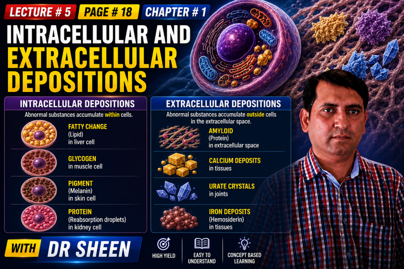
- Sometimes, cells or tissues accumulate abnormal amounts of different substances.
- These accumulated substances may be harmless or may cause cell injury.
Intracellular Accumulations
- The main ways abnormal substances accumulate inside cells are:
- The cell cannot remove or break down a substance properly.
- The cell produces too much of a substance normally made by the body.
- An abnormal substance from outside the body is deposited inside the cell (Fig. 1.24).
- These intracellular deposits may be found:
- In the cytoplasm
- Inside organelles, especially lysosomes
- In the nucleus
- Examples of these different types are described below.
Fatty Change (Steatosis)
- Fatty change, or steatosis, means an abnormal accumulation of triglycerides inside parenchymal cells.
- It occurs most commonly in the liver because the liver is the major organ involved in fat metabolism.
- It can also occur in the heart, skeletal muscle, kidney, and other organs.
- Steatosis can be caused by:
- Toxins
- Protein malnutrition
- Diabetes
- Obesity
- Anoxia
- In higher-income nations, the most common causes of fatty liver are alcohol abuse and diabetes associated with obesity.
- This process is discussed in more detail in Chapter 14.
Cholesterol and Cholesteryl Esters
- Cellular cholesterol metabolism is tightly controlled so that cells can normally make their cell membranes, in which cholesterol is an important component, without significant cholesterol accumulation inside the cell.
- However, phagocytic cells can become overloaded with lipids, including:
- Triglycerides
- Cholesterol
- Cholesteryl esters
- This can happen in several pathologic conditions in which there is increased lipid intake or decreased lipid breakdown.
- Among these conditions, atherosclerosis is the most important.
- The role of lipid and cholesterol deposition in atherosclerosis is discussed in Chapter 8.
Proteins
- Visible protein accumulations inside cells are less common than lipid accumulations.
- They may occur because of increased uptake or increased protein synthesis.
- In the kidney, small amounts of albumin normally pass through the glomerulus and are reabsorbed by pinocytosis in the proximal convoluted tubules.
- In conditions with heavy protein leakage through the glomerular filter, such as nephrotic syndrome (Chapter 12), much more protein enters the urine.
- The excessive albumin is reabsorbed and accumulates in vesicles inside tubular epithelial cells.
- These accumulated proteins appear as pink, hyaline cytoplasmic droplets.
- This process is reversible.
- If proteinuria decreases, the protein is degraded and the hyaline droplets disappear.
- Another example is marked accumulation of immunoglobulins in the rough ER of some plasma cells.
- These accumulated immunoglobulins form rounded, eosinophilic Russell bodies.
- Other examples of protein aggregation include alcoholic hyaline in the liver (Chapter 14) and neurofibrillary tangles in neurons (Chapter 21).
Glycogen
- Intracellular glycogen deposits are associated with abnormal metabolism of glucose or glycogen.
- In poorly controlled diabetes, glycogen accumulates in:
- Renal tubular epithelium
- Cardiac myocytes
- β cells of the islets of Langerhans
- Glycogen also accumulates inside cells in a group of genetic disorders called glycogen storage diseases (Chapter 4).
Pigments
- Pigments are colored substances.
- They may be:
- Exogenous — coming from outside the body
- Endogenous — produced within the body
- Carbon is the most common exogenous pigment.
- It is a common pollutant in urban air.
- When inhaled, carbon is taken up by alveolar macrophages.
- These macrophages transport the carbon through lymphatic channels to the regional tracheobronchial lymph nodes.
- Accumulated carbon makes the draining lymph nodes and pulmonary tissue black.
- This condition is called anthracosis (Chapter 11).
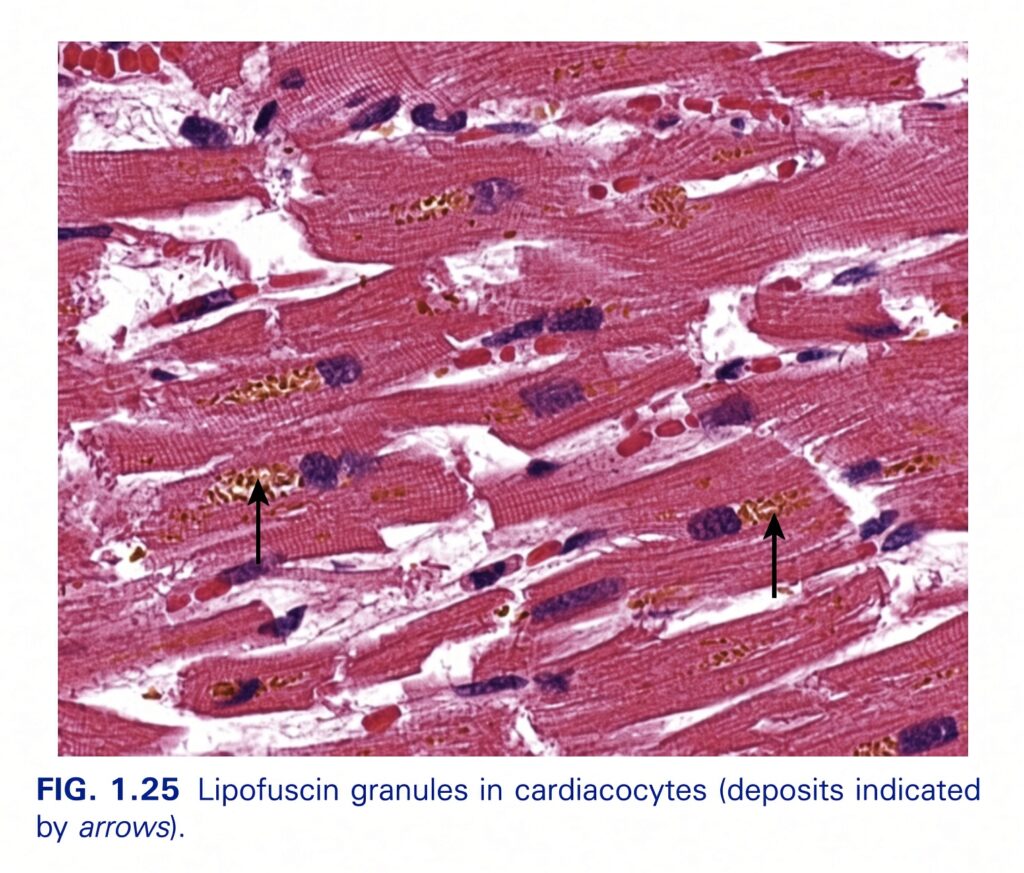
- Lipofuscin, also called “wear-and-tear pigment,” is an insoluble brownish-yellow granular material inside cells.
- It accumulates in many tissues, especially the heart, liver, and brain, with aging or atrophy.
- Lipofuscin consists of lipid-protein complexes produced by free-radical-mediated peroxidation of polyunsaturated lipids in intracellular membranes.
- It is a marker of previous free-radical injury, but it does not injure the cell.
- When large amounts accumulate, the brown pigment (Fig. 1.25) gives atrophic tissue, especially the heart, a brown appearance called brown atrophy.
- Melanin is an endogenous brown-black pigment.
- It is produced by melanocytes in the epidermis.
- Melanin acts as a screen against harmful ultraviolet radiation.
- Although melanocytes are the only cells that produce melanin, nearby basal keratinocytes can accumulate it, such as in freckles.
- Dermal macrophages can also accumulate melanin.
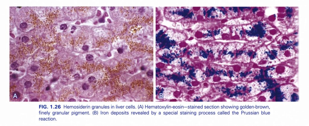
- Hemosiderin is a golden-yellow to brown, granular pigment derived from hemoglobin.
- It accumulates in tissues when there is a local or systemic excess of iron.
- Normally, iron is stored inside cells attached to the protein apoferritin, forming ferritin micelles.
- Hemosiderin consists of large aggregates of ferritin micelles.
- These aggregates can be easily seen by light and electron microscopy.
- The iron associated with hemosiderin can be specifically identified by the Prussian blue histochemical reaction (Fig. 1.26).
- Hemosiderin accumulation is usually pathologic.
- However, small amounts are normally present in bone marrow, spleen, and liver mononuclear phagocytes.
- These cells phagocytose and break down old red blood cells and recycle their iron for the production of new red blood cells (Chapter 10).
- Excessive hemosiderin deposition is called hemosiderosis.
- More extensive iron accumulation occurs in hereditary hemochromatosis, which is described in Chapter 14.
KEY CONCEPT
Intracellular accumulation = abnormal substance builds up inside a cell because it is not removed/degraded properly, is produced excessively, or comes from outside the body.
Conceptual examples:
- Triglycerides → accumulate in liver cells → fatty change (steatosis)
- Cholesterol/lipids → accumulate in phagocytic cells → important in atherosclerosis
- Albumin → excessive protein leakage → tubular cells reabsorb it → hyaline droplets
- Glucose metabolism abnormality → glycogen accumulation → poorly controlled diabetes
- Inhaled carbon → alveolar macrophages → lymph nodes/lung → anthracosis
- Aging/atrophy → lipofuscin accumulation → brown atrophy
- Melanocytes → melanin → protection against ultraviolet radiation
- Excess iron → ferritin aggregates → hemosiderin → hemosiderosis when excessive
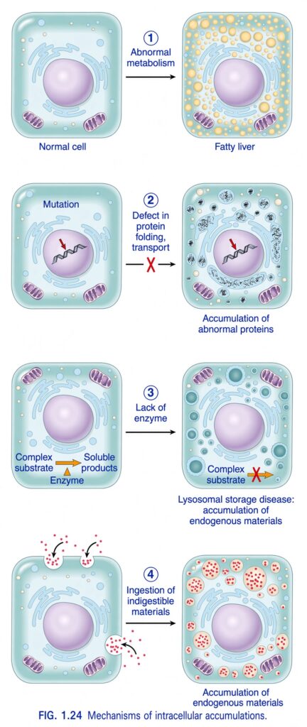
FIG. 1.24 — Mechanisms of Intracellular Accumulations
🧠 THE BIG IDEA
This figure explains why abnormal substances can accumulate inside cells.
Normally, a cell:
takes substances in → processes them → transports them → breaks them down → removes them
If any of these processes fails, the substance can build up inside the cell.
The figure shows 4 major mechanisms:
- Abnormal metabolism → Fatty liver
- Defective protein folding/transport → Abnormal proteins accumulate
- Lack of an enzyme → Lysosomal storage disease
- Ingestion of indigestible materials → Exogenous materials accumulate
1️⃣ ABNORMAL METABOLISM → FATTY LIVER
Look at the top row.
Left: Normal cell
The cell has a normal amount of substances and normal organelles.
↓
Abnormal metabolism
Something goes wrong with the cell’s normal metabolism.
↓
Right: Fatty liver
Many yellow fat droplets accumulate inside the cell.
🟡 What happened?
The cell is unable to handle fat normally.
Therefore:
Abnormal lipid metabolism
↓
Fat accumulates inside cells
↓
Fatty change / steatosis
In this figure, the example is:
Fatty liver
🧠 Simple analogy
Imagine a warehouse.
Normally:
Fat comes in → processed → transported out
But if processing or transport becomes abnormal:
Fat comes in
↓
Fat cannot be handled properly
↓
Fat piles up inside the warehouse
That’s what happens in fatty change.
Remember:
Abnormal metabolism → fat accumulation → fatty liver
2️⃣ MUTATION → DEFECTIVE PROTEIN FOLDING/TRANSPORT → ABNORMAL PROTEIN ACCUMULATION
Now look at the second row.
Left side:
The cell contains DNA.
A:
Mutation
occurs.
↓
The cell produces a protein with an abnormal structure or abnormal handling.
The figure specifically says:
Defect in protein folding, transport
The red ❌ means this process is defective.
↓
Right side:
Abnormal proteins accumulate inside the cell.
🧠 What is happening?
Think of making a protein like folding a complicated piece of paper.
Normally:
Protein made → correctly folded → transported to its proper location
But because of a mutation:
Protein made → folds incorrectly / transport fails
↓
Protein cannot be handled normally
↓
Protein accumulates
⭐ Key concept
Mutation can produce abnormal proteins that cannot fold properly or cannot be transported correctly, causing intracellular accumulation.
3️⃣ LACK OF ENZYME → LYSOSOMAL STORAGE DISEASE
Now look at the third row.
This one is extremely important.
Normal cell:
The figure shows:
Complex substrate
↓
Enzyme
↓
Soluble products
So the enzyme normally breaks down a complex substance into smaller soluble products.
🚫 What happens if the enzyme is missing?
The figure says:
Lack of enzyme
The red ❌ shows that the normal breakdown reaction cannot occur.
Therefore:
Complex substrate
↓
❌ Cannot be broken down
↓
Accumulates inside lysosomes
↓
Lysosomal storage disease
🧠 Simple analogy
Imagine a garbage disposal machine.
Normally:
Large waste → disposal machine → small waste
But if the machine is missing:
Large waste → cannot be broken down → piles up
The lysosome is like the cell’s recycling/digestion compartment.
If a necessary lysosomal enzyme is missing:
The material cannot be degraded and accumulates inside lysosomes.
⭐ Remember this sequence
Missing enzyme → substrate cannot be degraded → intracellular accumulation → lysosomal storage disease
4️⃣ INGESTION OF INDIGESTIBLE MATERIALS → EXOGENOUS ACCUMULATION
Now look at the bottom row.
Here, material comes from outside the cell.
The cell takes it in:
Ingestion of indigestible materials
↓
The cell cannot break down the material.
↓
The material remains inside the cell.
↓
Accumulation of exogenous materials
🧠 What does EXOGENOUS mean?
Exogenous = comes from outside the body/cell
So the material is not produced normally by the cell.
It enters from the external environment.
Think:
Outside → enters cell → cannot be digested → accumulates
🔴 Why can’t the cell remove it?
Because the material is:
Indigestible
The cell can take it up, but it cannot properly break it down.
Therefore:
Indigestible material
↓
Cannot be degraded
↓
Accumulates
⭐ NOW COMPARE ALL FOUR MECHANISMS
| Mechanism | What goes wrong? | What accumulates? | Example |
|---|---|---|---|
| 1. Abnormal metabolism | Metabolic processing is abnormal | Fat | Fatty liver |
| 2. Defective protein folding/transport | Protein is abnormal or cannot be handled properly | Abnormal proteins | Protein accumulation |
| 3. Lack of enzyme | Substance cannot be degraded | Complex endogenous substrates | Lysosomal storage disease |
| 4. Indigestible material | Cell cannot digest/remove material | Exogenous material | Exogenous material accumulation |
🧠 THE EASIEST WAY TO MEMORIZE THE 4
Think:
F → P → E → X
F — Fat
Abnormal metabolism → Fat accumulation
P — Protein
Mutation → defective protein → protein accumulation
E — Enzyme
Missing enzyme → substrate accumulation
X — eXternal
Indigestible external material → accumulation
🔍 NOW UNDERSTAND EVERY ARROW IN THE FIGURE
① Abnormal metabolism
Normal cell
    ↓
Abnormal metabolism
    ↓
Fat cannot be handled normally
    ↓
🟡 Fat droplets accumulate
    ↓
Fatty liver
② Mutation
Normal DNA
    ↓
Mutation
    ↓
Abnormal protein
    ↓
Defective folding / transport
    ↓
Protein accumulates
③ Lack of enzyme
Complex substrate
       ↓
     Enzyme
       ↓
Soluble products
Normally.
But:
Complex substrate
       ↓
❌ Enzyme missing
       ↓
Cannot be degraded
       ↓
Accumulates in lysosomes
       ↓
Lysosomal storage disease
④ Indigestible material
Material outside cell
       ↓
Cell ingests it
       ↓
Cannot digest it
       ↓
Remains inside cell
       ↓
Exogenous material accumulates
⭐ VERY IMPORTANT DIFFERENCE: ENDOGENOUS vs EXOGENOUS
Endogenous
Produced within the body/cell
Examples in the figure:
- Fat
- Abnormal proteins
- Complex substrates
Exogenous
Comes from outside
Example:
- Indigestible material taken up by the cell
So:
Endogenous = made inside
Exogenous = comes from outside
🧠 LOOK AT THE COLORS
The colors help you understand what is accumulating:
🟡 Yellow droplets
→ Fat
⚫/gray-blue deposits
→ Abnormal proteins
🔵 Large blue-green lysosomal deposits
→ Undegraded substrates
🔴 Red particles
→ Indigestible exogenous material
🎯 THE WHOLE FIGURE AS ONE STORY
A normal cell is constantly doing three major things:
1. Making and processing substances
2. Breaking substances down
3. Removing unwanted substances
If something goes wrong:
                    INTRACELLULAR ACCUMULATION
                              │
        ┌─────────────────────┼─────────────────────┐
        ↓                     ↓                     ↓
  METABOLISM              PROTEIN               DEGRADATION
    abnormal               defect                  defect
        ↓                     ↓                     ↓
      FAT                  PROTEIN               SUBSTRATE
   accumulates            accumulates            accumulates
        ↓                     ↓                     ↓
 Fatty liver          Abnormal protein      Lysosomal storage
                          accumulation            disease

                              +
                              ↓
                    INDIGESTIBLE MATERIAL
                              ↓
                    EXOGENOUS ACCUMULATION
🔥 FINAL EXAM CONCEPT
Intracellular substances accumulate when the cell cannot properly metabolize, fold/transport, degrade, or remove them. The four mechanisms shown are abnormal metabolism causing fatty change, defective protein folding or transport causing abnormal protein accumulation, enzyme deficiency causing lysosomal storage, and ingestion of indigestible exogenous materials causing intracellular accumulation.
🧠 One-line memory:
Metabolism problem → FAT
Protein handling problem → PROTEIN
Enzyme deficiency → SUBSTRATE
Indigestible outside material → EXOGENOUS MATERIAL
Extracellular Deposits: Pathologic Calcification
- Pathologic calcification means abnormal deposition of calcium salts.
- It occurs in many different disease conditions.
- Pathologic calcification can occur in two ways:
- Dystrophic calcification
- Metastatic calcification
- Dystrophic calcification occurs when calcium metabolism is normal, but calcium deposits in injured or dead tissue.
- It can occur in areas of necrosis of any type.
- It is almost always present in the arterial lesions of advanced atherosclerosis (Chapter 8).
- Sometimes dystrophic calcification is simply an incidental finding, showing that there was previous cell injury that may not be important.
- However, it can also cause organ dysfunction.
- For example, calcification can develop in old or damaged heart valves.
- This can severely interfere with normal valve movement (Chapter 9).
- Dystrophic calcification begins when crystalline calcium phosphate is deposited outside cells in membrane-bound vesicles.
- These vesicles may come from injured cells.
- Calcium can also accumulate inside the mitochondria of dying cells.
- Extracellular calcium is thought to become concentrated inside vesicles because calcium has an affinity for membrane phospholipids.
- Phosphates accumulate because of the action of membrane-bound phosphatases.
- The calcium phosphate crystals then grow and spread, forming larger calcium deposits.
- Metastatic calcification is associated with hypercalcemia, meaning an increased level of calcium in the blood.
- It can occur even in otherwise normal tissues.
- The major causes of hypercalcemia are:
- Increased parathyroid hormone (PTH) secretion
- Due to primary parathyroid tumors or hyperplasia
- Or due to production of PTH-related protein by malignant tumors
- Bone destruction
- Due to increased bone turnover, such as Paget disease
- Immobilization
- Tumors, including increased bone breakdown associated with multiple myeloma, leukemia, or widespread skeletal metastases
- Vitamin D–related disorders
- Vitamin D intoxication
- Sarcoidosis, in which macrophages activate a vitamin D precursor
- Renal failure
- Phosphate retention causes secondary hyperparathyroidism
- Increased parathyroid hormone (PTH) secretion
KEY CONCEPT
Pathologic calcification = abnormal calcium salt deposition.
- Dystrophic calcification → normal calcium metabolism + injured/dead tissue → calcium deposits
- Metastatic calcification → hypercalcemia + otherwise normal tissue → calcium deposits
Conceptual examples:
- Dead/necrotic tissue + normal blood calcium → dystrophic calcification
- Advanced atherosclerosis → arterial injury → dystrophic calcification
- Damaged aging heart valve → calcium deposition → impaired valve movement
- Hypercalcemia → normal tissue → metastatic calcification
- Hyperparathyroidism → ↑ calcium → metastatic calcification
- Bone destruction → ↑ calcium → metastatic calcification
- Vitamin D intoxication → ↑ calcium → metastatic calcification
- Renal failure → phosphate retention → secondary hyperparathyroidism → hypercalcemia → metastatic calcification
MORPHOLOGY
- Regardless of where calcium is deposited, calcium salts appear on gross examination as fine white granules or clumps.
- These deposits often feel gritty when touched.
- Dystrophic calcification is commonly seen in:
- Atherosclerotic plaques
- Areas of caseous necrosis in tuberculosis
- Sometimes, a tuberculous lymph node becomes almost completely converted into a radiopaque stone-like mass.
- On histologic examination, calcification appears as basophilic deposits.
- These calcium deposits may be located:
- Outside cells (extracellular)
- Inside cells (intracellular)
- Over time, heterotopic bone may develop within the area of calcification.
- Metastatic calcification can occur throughout the body.
- It mainly affects the interstitial tissues of:
- Blood vessels
- Kidneys
- Lungs
- Gastric mucosa
- The calcium deposits in metastatic calcification look morphologically similar to those seen in dystrophic calcification.
- Usually, these deposits do not cause clinical dysfunction.
- However, extensive calcification in the lungs may be visible on radiographs.
- Extensive pulmonary calcification may also cause respiratory deficits.
- Massive calcium deposits in the kidneys, called nephrocalcinosis, can cause renal damage.
KEY CONCEPT
Morphology of calcification = white, gritty calcium deposits grossly + basophilic deposits microscopically.
Conceptual examples:
- Atherosclerotic plaque → dystrophic calcification → white, gritty deposits
- Tuberculosis with caseous necrosis → dystrophic calcification → sometimes stone-like lymph node
- Calcification on microscopy → basophilic intracellular or extracellular deposits
- Long-standing calcification → possible heterotopic bone formation
- Metastatic calcification → mainly vessels, kidneys, lungs, gastric mucosa
- Massive kidney calcification → nephrocalcinosis → renal damage
