


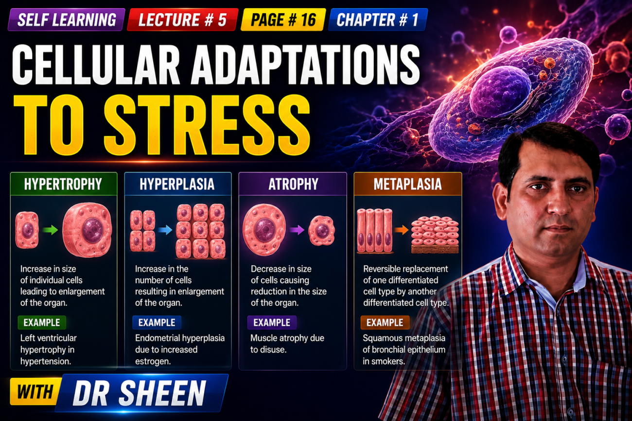
- Adaptations are reversible changes that occur in cells when their environment changes.
- These changes can affect:
- Number of cells
- Size of cells
- Phenotype (cell characteristics)
- Metabolic activity (how actively the cell works)
- Functions of cells
- Physiologic adaptations are normal changes that occur when cells receive normal stimulation from hormones or natural chemical mediators.
- Example: During pregnancy, hormones cause the breast and uterus to enlarge.
- Cells can also adapt normally to increased mechanical stress.
- This occurs especially in bones and muscles when they have increased physical demands.
- Pathologic adaptations occur when cells are exposed to stress or abnormal conditions.
- These adaptations allow cells to change their structure and function so they can avoid injury.
- However, this protection comes at a cost: the cell may not perform its normal function properly.
- Both physiologic and pathologic adaptations can occur in several different forms, which are described below.
KEY CONCEPT
Cellular adaptation = Cell changes itself to handle a change in its environment, and the change is reversible.
- Physiologic adaptation → normal demand
- Example: Pregnancy → breast and uterus enlarge
- Pathologic adaptation → abnormal stress
- Example concept: Stress → cell changes its structure/function to escape injury, but normal function may be reduced.
Hypertrophy
- Hypertrophy means cells become larger, which makes the whole organ larger.
- Hyperplasia, in contrast, means an increase in the number of cells.
- In pure hypertrophy, no new cells are formed; instead, the existing cells become larger because they contain more structural proteins and organelles.
- Pure hypertrophy mainly occurs in cell types that have a limited ability to divide.
- In other tissues, hypertrophy and hyperplasia can occur together, causing the organ to become enlarged.
- Hypertrophy can be physiologic (normal) or pathologic (abnormal).
- It occurs because of either:
- Increased functional demand, or
- Growth factor or hormonal stimulation.
- During pregnancy, the uterus becomes larger because estrogen stimulates both smooth muscle hypertrophy and hyperplasia (Fig. 1.20).
- In contrast, when workload increases, the striated muscle cells of the skeletal muscle and heart undergo only hypertrophy because these cells have a limited ability to divide.
- Pathologic hypertrophy of the heart occurs in hypertension and other conditions that increase pressure inside the heart, such as aortic stenosis (narrowing of the aortic valve) (Fig. 1.21).
- In these conditions, heart muscle cells experience a continuously increased workload.
- The cells adapt by becoming larger so they can produce the greater contractile force that is required.
- Hypertrophy can also be a prelude to cell injury.
- The mechanisms of hypertrophy have been studied most thoroughly in the heart.
- Cardiac hypertrophy can occur because of mechanical stimuli, such as stretch.
- Stretch causes the release of soluble mediators, such as growth factors and adrenergic hormones, which stimulate cell growth.
- These stimuli activate signal transduction pathways.
- These pathways increase the expression of genes that produce various cellular proteins.
- One important result is the production of more myofilaments in each cell.
- More myofilaments increase the force produced by each contraction.
- This allows the cell to meet the increased workload.
- Contractile proteins may also change from adult forms to fetal or neonatal forms.
- For example, during hypertrophy, α-myosin heavy chain is replaced by the β-myosin heavy chain.
- The β form produces slower but more energy-efficient contractions.
- An adaptation to stress, such as hypertrophy, can eventually progress to cell injury if the stress is not removed or becomes greater than the tissue’s adaptive capacity.
- In the heart, sustained hypertension can cause these harmful changes.
- Degenerative changes then appear in the myocardial fibers.
- The most important changes are fragmentation and loss of myofibrillar contractile elements.
- It is not fully understood why hypertrophy progresses to these regressive changes.
- One possibility is that the blood vessels may have a limited ability to supply enough blood to the enlarged muscle fibers.
- Another possibility is that the mitochondria may not be able to provide enough ATP.
- Another possibility is that the biosynthetic machinery may not be able to produce enough contractile proteins or other cytoskeletal elements.
- The final result of these degenerative changes is ventricular dilation.
- Ultimately, this can lead to cardiac failure.
KEY CONCEPT
Hypertrophy = bigger cells → bigger organ; the number of cells does not increase in pure hypertrophy.
Conceptual examples:
- Pregnancy → estrogen → uterine smooth muscle hypertrophy + hyperplasia → enlarged uterus
- Increased skeletal muscle workload → larger muscle cells → hypertrophy
- Hypertension/aortic stenosis → increased cardiac workload → enlarged heart muscle cells → cardiac hypertrophy
- Persistent excessive stress → hypertrophy → cell injury → ventricular dilation → cardiac failure
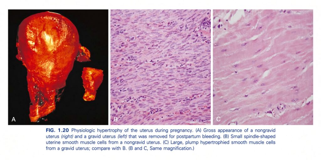
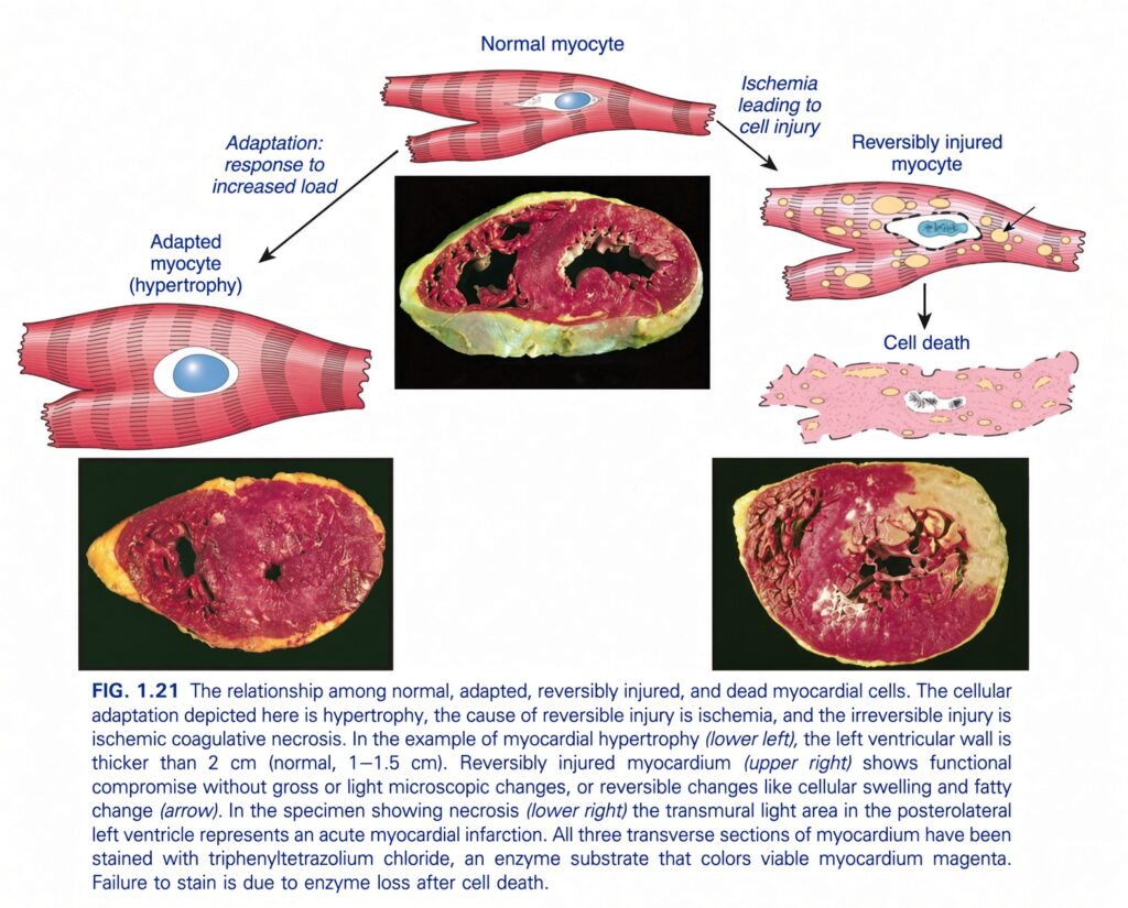
FIG. 1.21 — Relationship Among Normal, Adapted, Reversibly Injured, and Dead Myocardial Cells
🧠 THE BIG IDEA
This figure shows four possible states of heart muscle cells (myocytes):
- Normal myocyte → normal heart muscle cell
- Adapted myocyte (hypertrophy) → cell becomes larger because workload increases
- Reversibly injured myocyte → cell is injured but can recover if the cause is removed
- Dead myocyte → severe injury causes irreversible damage and cell death
The easiest way to remember the whole figure is:
Normal → Adaptation OR Injury → Recovery OR Death
1. ❤️ NORMAL MYOCYTE
Look at the top center.
This is the normal heart muscle cell.
It has:
- Normal size
- Normal structure
- Normal nucleus
- Normal contractile apparatus
Think:
Normal workload → normal-sized, normally functioning myocyte
This is the starting point of the figure.
2. 🔨 ADAPTATION TO INCREASED LOAD
The arrow from the normal myocyte points toward:
Adapted myocyte (hypertrophy)
The important word is:
Hypertrophy
Hypertrophy = cells become larger
The individual myocardial cells increase in size.
They do not primarily increase in number.
Why does the heart muscle hypertrophy?
The figure says:
Adaptation: response to increased load
Imagine the heart is being asked to pump against a greater resistance.
For example:
Increased workload
↓
Heart muscle must generate more force
↓
Myocardial cells adapt
↓
Cells become larger
↓
Hypertrophy
3. 🫀 WHAT DOES THE LOWER-LEFT HEART IMAGE SHOW?
The lower-left specimen demonstrates:
Myocardial hypertrophy
The caption specifically states that:
The left ventricular wall is thicker than 2 cm.
Normal left ventricular wall thickness is approximately:
1–1.5 cm
So:
Normal → 1–1.5 cm
Hypertrophy → >2 cm
Easiest concept:
Increased workload → heart muscle cells enlarge → ventricular wall becomes thicker.
4. ⚠️ ISCHEMIA → CELL INJURY
Now move to the upper-right side.
The normal myocyte has an arrow pointing toward:
Ischemia leading to cell injury
Ischemia = inadequate blood supply
The heart muscle is not receiving enough blood.
Therefore:
↓ Blood supply
↓
↓ Oxygen and nutrients
↓
Cell injury
5. 🔄 REVERSIBLY INJURED MYOCYTE
The next picture is labeled:
Reversibly injured myocyte
This means:
The cell is injured, but it is still alive.
If the cause of injury is removed soon enough:
Injury → recovery → normal cell
What changes can occur?
The figure shows examples such as:
💧 Cellular swelling
The injured cell takes up water and becomes swollen.
Think:
Injury → cell cannot maintain normal ion/water balance → water enters → swelling
🟡 Fatty change
Fat droplets accumulate inside the cell.
The figure shows these as small yellowish droplets.
So:
Reversible injury can produce cellular swelling and fatty change.
6. ⭐ WHY IS IT CALLED “REVERSIBLE”?
This is one of the most important pathology concepts.
Reversible injury:
The cell is:
Injured but alive
↓
If the cause is removed:
Cell can recover
Example:
Ischemia
   ↓
Cell injury
   ↓
REVERSIBLE
   ↓
Restore blood supply
   ↓
Cell recovers
But if the ischemia is severe or prolonged:
Ischemia
   ↓
Severe/prolonged injury
   ↓
IRREVERSIBLE injury
   ↓
Cell death
7. ☠️ CELL DEATH
Look at the arrow below the reversibly injured myocyte.
It leads to:
Cell death
This means the injury has crossed the point of no return.
The cell can no longer recover.
In this figure, the type of cell death is:
Coagulative necrosis
8. 🔥 ISCHEMIC COAGULATIVE NECROSIS
The caption tells us:
The cause of irreversible injury is ischemia.
In myocardial infarction:
Ischemia
↓
Severe/prolonged oxygen deprivation
↓
Irreversible cell injury
↓
Cell death
↓
Coagulative necrosis
So the important sequence is:
Ischemia → irreversible injury → coagulative necrosis
9. 🫀 LOWER-RIGHT IMAGE — MYOCARDIAL INFARCTION
The lower-right photograph shows an area of myocardial necrosis.
The caption says:
Acute myocardial infarction
There is a:
Transmural light area
in the posterolateral part of the left ventricle.
Transmural means:
Trans = through
Mural = wall
So:
Transmural infarction = infarction extending through the thickness of the ventricular wall.
10. 🧠 WHY DOES THE INFARCTED AREA LOOK DIFFERENT?
The figure uses a special stain:
Triphenyltetrazolium chloride (TTC)
This stain helps distinguish living myocardium from dead myocardium.
Living myocardium:
It contains functioning enzymes.
↓
These enzymes react with TTC.
↓
The viable myocardium becomes magenta/red.Dead myocardium:
After cell death:
Important enzymes are lost
↓
TTC cannot produce the normal color
↓
The dead area fails to stain
↓
It appears pale/light
⭐ VERY IMPORTANT TTC CONCEPT
Viable myocardium → stains magenta
Dead myocardium → fails to stain → pale
So when you see a pale region in the infarct specimen:
That pale region represents dead myocardium.
11. WHY DOES DEAD MYOCARDIUM FAIL TO STAIN?
This is specifically mentioned in the caption.
Cell death
↓
Loss of cellular enzymes
↓
TTC cannot be converted normally
↓
No magenta staining
Therefore:
Failure to stain is due to enzyme loss after cell death.
12. 🧩 UNDERSTAND THE THREE HEART SPECIMENS
The figure contains photographs that demonstrate different situations.
Central specimen
Shows the myocardium in the context of the heart and provides the overall myocardial appearance.
Lower-left specimen
Shows:
Myocardial hypertrophy
The left ventricular wall is >2 cm compared with the normal 1–1.5 cm.
Lower-right specimen
Shows:
Acute myocardial infarction with a pale/light infarcted region
The pale area represents myocardium that has died and therefore does not stain with TTC.
🔥 NOW FOLLOW THE ENTIRE FIGURE AS A STORY
              NORMAL MYOCYTE
                    │
          ┌─────────┴─────────┐
          ↓                   ↓
   INCREASED LOAD           ISCHEMIA
          ↓                   ↓
      ADAPTATION          CELL INJURY
          ↓                   ↓
     HYPERTROPHY       ┌──────┴──────┐
                       ↓             ↓
                  REVERSIBLE     IRREVERSIBLE
                    INJURY          INJURY
                       ↓             ↓
                  RECOVERY       CELL DEATH
                                     ↓
                           COAGULATIVE NECROSIS
                                     ↓
                           MYOCARDIAL INFARCTION
🧠 THE MOST IMPORTANT DIFFERENCE
🟢 Adaptation
The cell is not injured.
It changes to handle increased workload.
Increased load → hypertrophy
🟡 Reversible injury
The cell is injured but alive.
If the cause is removed:
Cell can recover.
Examples shown:
- Cellular swelling
- Fatty change
🔴 Irreversible injury
The cell has passed the point of no return.
Severe/prolonged ischemia → cell death
↓
Coagulative necrosis
📊 QUICK COMPARISON
| State | What happens? | Can it recover? |
|---|---|---|
| Normal myocyte | Normal structure/function | — |
| Hypertrophy | Cell becomes larger due to increased load | Adaptation |
| Reversible injury | Cell is injured but remains alive | Yes |
| Irreversible injury | Severe/prolonged injury | No |
| Cell death | Myocyte dies | No |
| Coagulative necrosis | Pattern of necrosis caused by ischemic injury in myocardium | No |
🎯 THE 3 THINGS YOU MUST REMEMBER
1. Increased workload
↑ Load → hypertrophy
Cell becomes bigger.
2. Mild/temporary ischemia
Ischemia → reversible injury
Cell swelling + fatty change
Remove the cause → cell can recover
3. Severe/prolonged ischemia
Ischemia → irreversible injury → cell death → coagulative necrosis
Dead myocardium fails to stain with TTC because of enzyme loss.
⭐ FINAL MEMORY LINE
The heart muscle can adapt to increased workload by hypertrophy; if ischemia causes injury, the cell may initially be reversibly injured, but severe or prolonged ischemia causes irreversible injury and coagulative necrosis, producing myocardial infarction.
Ultra-short memory:
↑ Load → HYPERTROPHY
Temporary ischemia → REVERSIBLE INJURY
Severe/prolonged ischemia → NECROSIS → CELL DEATH
Hyperplasia
- Hyperplasia means an increase in the number of cells in an organ.
- This increase happens because cells multiply (proliferate).
- The cells that multiply may be differentiated cells or, in some cases, progenitor cells.
- Hyperplasia can occur only when the tissue contains cells that are able to replicate.
- Hyperplasia may occur together with hypertrophy and often develops in response to the same stimuli.
- Hyperplasia can be physiologic (normal) or pathologic (abnormal).
- In both situations, cell multiplication is stimulated by hormones or growth factors.
- The two types of physiologic hyperplasia are:
- Hormonal hyperplasia
- Compensatory hyperplasia
- Hormonal hyperplasia occurs when hormones stimulate cells to multiply.
- Example: The glandular epithelium of the female breast proliferates at puberty and during pregnancy.
- Compensatory hyperplasia occurs when the remaining tissue grows after part of an organ is removed or lost.
- For example, when part of the liver is surgically removed, the remaining liver cells begin mitotic activity as early as 12 hours later.
- The continued cell multiplication eventually restores the liver to its normal weight.
- In this situation, polypeptide growth factors produced by uninjured hepatocytes and other nonparenchymal liver cells stimulate hyperplasia (Chapter 2).
- After the liver returns to its normal size, growth inhibitors stop further cell proliferation.
- Hormonal imbalances can cause pathologic hyperplasia.
- For example, after menstruation, there is normally a burst of uterine epithelial cell proliferation.
- Normally, this proliferation is carefully controlled by:
- Pituitary hormones and ovarian estrogen → stimulate proliferation
- Progesterone → inhibits proliferation
- If this balance is disturbed and estrogenic stimulation increases, endometrial hyperplasia can develop.
- Endometrial hyperplasia is a common cause of abnormal menstrual bleeding.
- Benign prostatic hyperplasia is another common example of pathologic hyperplasia.
- In this case, hyperplasia results from hormonal stimulation by androgens and estrogens.
- An important point is that hyperplasia remains controlled in all these situations.
- When the signals that started the hyperplasia decrease or disappear, the hyperplasia stops.
- This ability to respond to normal regulatory controls distinguishes pathologic hyperplasia from cancer.
- In cancer, the mechanisms controlling cell growth become permanently dysregulated or ineffective (Chapter 6).
- However, in many cases, pathologic hyperplasia can create a favorable setting in which cancer may eventually develop.
- For example, patients with endometrial hyperplasia have an increased risk of developing endometrial cancer (Chapter 17).
KEY CONCEPT
Hyperplasia = more cells → organ/tissue becomes larger because cells multiply.
Conceptual examples:
- Puberty/pregnancy → hormonal stimulation → breast glandular cells multiply → hormonal hyperplasia
- Part of liver removed → remaining hepatocytes multiply → liver returns to normal weight → compensatory hyperplasia
- Excess estrogen → excessive endometrial cell proliferation → endometrial hyperplasia → abnormal menstrual bleeding
- Androgens + estrogens → prostate cell proliferation → benign prostatic hyperplasia
- Hyperplasia signals stop → cell proliferation stops
- Cancer → growth control becomes permanently dysregulated or ineffective
Atrophy
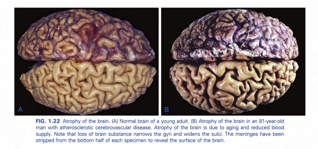
- Atrophy means a reduction in the size of an organ or tissue.
- This happens because the size and number of cells decrease (Fig. 1.22).
- Causes of atrophy include:
- Decreased workload — e.g., immobilization of a limb to allow a fracture to heal.
- Loss of innervation — loss of nerve supply.
- Reduced blood supply.
- Inadequate nutrition.
- Loss of endocrine stimulation.
- Aging — called senile atrophy.
- Some causes of atrophy are a normal part of life, such as loss of hormone stimulation during menopause.
- Other causes are pathologic, such as denervation.
- However, the basic cellular changes are similar in both situations.
- Atrophy can be understood as an adaptive retreat in which the cell becomes smaller so that it can still survive.
- However, if atrophy continues and becomes severe, the affected cells may cross a threshold and undergo apoptosis.
- Atrophy occurs because of a combination of:
- Decreased protein synthesis
- Increased protein degradation
- Protein synthesis decreases because the cell has reduced metabolic activity.
- Cellular proteins are mainly degraded through the ubiquitin-proteasome pathway.
- Nutrient deficiency and disuse may activate ubiquitin ligases.
- These enzymes attach multiple copies of the small peptide ubiquitin to cellular proteins.
- The ubiquitin-marked proteins are then targeted for degradation in the proteasome.
- In many situations, atrophy is also associated with increased autophagy.
- This causes an increase in the number of autophagic vacuoles.
- Autophagy is a process in which a starved cell breaks down and uses its own organelles in an attempt to survive.
KEY CONCEPT
Atrophy = smaller organ/tissue → cells become smaller and may also decrease in number.
Conceptual examples:
- Limb immobilization → decreased workload → decreased cell activity → muscle atrophy
- Loss of nerve supply → denervation → atrophy
- Reduced blood supply → reduced cell activity → atrophy
- Poor nutrition → protein degradation > protein synthesis → atrophy
- Aging/menopause → reduced hormonal stimulation → atrophy
- Severe/prolonged atrophy → cell crosses survival threshold → apoptosis
Metaplasia
- Metaplasia means that one adult cell type is replaced by another adult cell type.
- In this adaptation, a cell type that is sensitive to a particular stress is replaced by another cell type that can better tolerate the harmful environment.
- Metaplasia usually develops because stem cells are reprogrammed to develop into a different cell type, rather than because already-differentiated cells directly change into another type (transdifferentiation).
- Epithelial metaplasia is seen in the respiratory tract with prolonged cigarette smoking.
- Normally, the trachea and bronchi contain relatively delicate ciliated columnar epithelial cells.
- With prolonged cigarette smoking, these cells are replaced by tough stratified squamous epithelial cells (Fig. 1.23).
- The stratified squamous cells are better able to withstand the harmful chemicals in cigarette smoke.
- Although this metaplastic squamous epithelium has a survival advantage, important protective functions are lost.
- These lost protective functions include mucus secretion and ciliary clearance of particulate matter.
- Therefore, epithelial metaplasia is a double-edged sword because it improves cell survival but causes loss of important protective functions.
- In other situations, such as chronic gastric reflux, the normal stratified squamous epithelium of the lower esophagus may undergo metaplastic transformation.
- It may be replaced by gastric-type or intestinal-type columnar epithelium.
- Metaplasia can also occur in mesenchymal cells.
- In these situations, however, it is generally a response to a pathologic change rather than an adaptation to stress.
- For example, bone may sometimes form in soft tissues, particularly at sites of injury.
- If the influences causing metaplasia continue for a long time, they can increase the risk of malignant transformation of the epithelium.
- Many examples of this relationship exist.
- For example, squamous metaplasia of respiratory epithelium creates a favorable setting for the development of lung cancers made of malignant squamous cells.
- Similarly, intestinal metaplasia of the stomach is associated with the development of gastric cancer.
KEY CONCEPT
Metaplasia = one adult cell type → replaced by another adult cell type that is better able to tolerate the stress.
Conceptual examples:
- Cigarette smoke → respiratory ciliated columnar epithelium → stratified squamous epithelium → better stress resistance but loss of mucus/ciliary protection
- Chronic gastric reflux → lower esophageal squamous epithelium → gastric/intestinal-type columnar epithelium
- Injury → soft tissue → bone formation (mesenchymal metaplasia)
- Persistent metaplastic stimulus → increased risk of malignant transformation → cancer
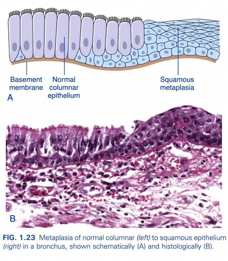
FIG. 1.23 — Metaplasia of Normal Columnar to Squamous Epithelium
🧠 THE BIG IDEA
This figure shows metaplasia in a bronchus.
Normally, the bronchus has:
Columnar epithelium
But under chronic irritation, the normal lining can change into:
Squamous epithelium
This change is called:
Squamous metaplasia
The easiest concept:
The cells change from one mature cell type to another mature cell type that is better able to tolerate the stress.
1. 🫁 NORMAL COLUMNAR EPITHELIUM — LEFT SIDE
Look at the left side of the diagram.
The cells are:
Tall and column-shaped
Therefore, they are called:
Columnar epithelial cells
They are arranged on the:
Basement membrane
What do the cells look like?
Think of them as:
🏢 Tall buildings standing next to each other
They are:
- Tall
- Narrow
- Closely packed
- Arranged along the basement membrane
At their top surface, the diagram shows many small hair-like structures.
These represent:
Cilia
2. 🚧 BASEMENT MEMBRANE
Look at the brown layer underneath the epithelial cells.
This is labeled:
Basement membrane
It forms the supporting boundary underneath the epithelium.
So the basic arrangement is:
          AIRWAY LUMEN
              ↓
      Tall columnar cells
      │ │ │ │ │ │ │ │
      │ │ │ │ │ │ │ │
      ────────────────
       Basement membrane
              ↓
       Connective tissue
3. 🔄 WHAT IS METAPLASIA?
Metaplasia = one mature cell type is replaced by another mature cell type.
It is an:
Adaptive response to chronic stress or irritation.
The important thing is:
The original cells are not simply transforming directly into the new cells.
Instead, the tissue’s stem/progenitor cells are reprogrammed so that they produce a different mature epithelial type.
4. ⚠️ WHY DOES THE BRONCHUS DEVELOP METAPLASIA?
The figure itself demonstrates the change, while the classic bronchial example is associated with chronic irritation, such as cigarette smoke.
The basic logic is:
Chronic irritation
       ↓
Normal epithelium is repeatedly stressed
       ↓
Tissue adapts
       ↓
Columnar epithelium
       ↓
Squamous epithelium
5. 🛡️ WHY SQUAMOUS EPITHELIUM?
Squamous epithelium is:
Better able to tolerate persistent mechanical/chemical irritation.
So the tissue is essentially making a trade-off:
Before:
Columnar epithelium
→ specialized for normal airway functions.
After:
Squamous epithelium
→ more resistant to chronic irritation.
Think:
The tissue sacrifices some specialized function to gain greater resistance.
6. 🔵 RIGHT SIDE — SQUAMOUS METAPLASIA
Now look at the right side of the diagram.
The cells are no longer tall.
Instead, they become:
Flatter and more squamous
This is:
Squamous metaplasia
Compare the two sides
LEFT — Normal
Tall → columnar
RIGHT — Metaplasia
Flat → squamous
So:
Columnar → Squamous
7. 🧠 Why is this an ADAPTATION?
Because the change helps the tissue survive a stressful environment.
The sequence is:
Chronic irritation
↓
Adaptive response
↓
Metaplasia
↓
More resistant epithelial lining
Therefore:
Metaplasia is initially an adaptive and potentially reversible change.
8. ⚠️ BUT THERE IS A COST
This is a very important pathology concept.
The new squamous epithelium may be more resistant, but it does not perform all the specialized functions of the original bronchial columnar epithelium.
For example, the normal respiratory epithelium has:
Cilia
which help move mucus and trapped particles.
When squamous metaplasia develops:
The normal specialized respiratory epithelial function is lost/reduced.
So:
Protection from irritation ↑
but
Specialized function ↓
9. 🔬 NOW LOOK AT IMAGE B — HISTOLOGY
The lower image is the actual microscopic appearance.
B = Histologic image
The left portion shows the:
Normal columnar epithelium
You can recognize it because the cells are:
- Tall
- Closely packed
- Column-shaped
- Lining the surface
10. 🔬 RIGHT SIDE OF IMAGE B
The right side shows:
Squamous metaplasia
The epithelial cells are:
- More numerous in layers
- More polygonal toward the deeper region
- Flatter toward the surface
- No longer arranged as the normal tall columnar lining
Therefore:
The microscopic appearance confirms the transition from columnar epithelium to squamous epithelium.
11. A vs B — WHAT IS THE DIFFERENCE?
The figure has two ways of showing the same process.
A = Schematic drawing
It gives you a simple cartoon showing:
Normal columnar epithelium → squamous metaplasia
B = Histology
It shows what the same change actually looks like under the microscope.
⭐ THE MOST IMPORTANT VISUAL COMPARISON
NORMAL BRONCHUS

   Cilia
 ↓↓↓↓↓↓↓↓↓
│ │ │ │ │ │ │
│ │ │ │ │ │ │   ← Tall columnar cells
│ │ │ │ │ │ │
──────────────   ← Basement membrane


          ↓
   CHRONIC IRRITATION
          ↓
       METAPLASIA
          ↓


SQUAMOUS METAPLASIA

──────────────
▱ ▱ ▱ ▱ ▱ ▱    ← Squamous cells
 ▱ ▱ ▱ ▱ ▱
  ▱ ▱ ▱ ▱
──────────────   ← Basement membrane
🧩 EACH LABEL IN THE FIGURE
Basement membrane
→ Supporting layer underneath the epithelium.
Normal columnar epithelium
→ Tall epithelial cells normally lining the bronchus.
Squamous metaplasia
→ Replacement of the normal columnar epithelial lining by squamous epithelium.
A
→ Schematic representation.
B
→ Histologic representation.
🔥 VERY IMPORTANT: METAPLASIA ≠ CANCER
Do not confuse them.
Metaplasia:
Adaptive cell-type change
↓
May be reversible if the stimulus is removed
Cancer:
Uncontrolled abnormal cellular growth
↓
Not simply an adaptive change.
⚠️ BUT METAPLASIA CAN INCREASE CANCER RISK
Persistent metaplastic tissue can provide a setting in which additional genetic abnormalities may develop.
Therefore:
Metaplasia itself is not cancer, but persistent metaplasia can be associated with increased risk of subsequent dysplasia and malignancy.
🎯 THE WHOLE FIGURE IN ONE STORY
NORMAL BRONCHUS
      ↓
Columnar epithelium
      ↓
Chronic irritation
      ↓
Adaptive response
      ↓
METAPLASIA
      ↓
Squamous epithelium
      ↓
Better resistance to irritation
      ↓
But loss of some specialized
respiratory epithelial functions
⭐ FINAL EXAM CONCEPT
Squamous metaplasia of the bronchus is an adaptive, potentially reversible replacement of normal ciliated columnar epithelium by stratified squamous epithelium in response to chronic irritation. It increases resistance to injury but causes loss of normal specialized respiratory epithelial functions.
🧠 One-line memory:
Chronic irritation → Columnar epithelium changes to Squamous epithelium = Squamous metaplasia.
Made by self learning Dr sheen
