


- During early life in multicellular animals, natural selection strongly favors genetic changes that improve reproduction.
- These beneficial genetic changes are passed to offspring and help maintain the population.
- In contrast, DNA repair mechanisms do not have to be perfect, as long as they are good enough to allow survival through the reproductive years.
- Because DNA repair is not perfect, mutations gradually accumulate with time.
- Some of these mutations are harmful and contribute to cellular aging.
- Aging is caused by a progressive decline in normal physiologic, cellular, and molecular mechanisms that maintain homeostasis after the reproductive years.
- Aging has important effects on health because increasing age is one of the strongest independent risk factors for many chronic diseases.
- These diseases include:
- Cancer
- Alzheimer disease
- Ischemic heart disease
- One important discovery about cellular aging is that aging is not simply because cells gradually “run out of steam” with time.
- Instead, aging results from changes in genes and signaling pathways that have been conserved through evolution from yeast to mammals.
- Experimental studies have shown that aging can be delayed.
- For example, in animals, some features of aging can be slowed by specific interventions such as calorie restriction and certain therapeutic drugs.
- Cellular aging results from a decrease in the ability of cells to replicate and a decrease in their functional activity.
- Several mechanisms contribute to cellular aging (Fig. 1.27).
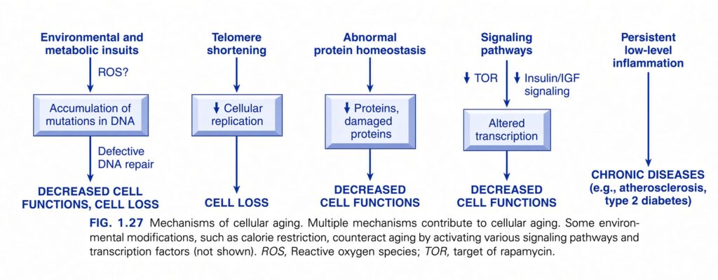
DNA damage
- Nuclear DNA and mitochondrial DNA frequently develop mutations.
- These mutations can include:
- Base substitutions
- Copy number variations
- Deletions
- Insertions
- Many mutations are produced by the spontaneous deamination of cytosine residues.
- This type of DNA damage occurs repeatedly over time.
- DNA damage becomes faster because of endogenous stresses, such as reactive oxygen species (ROS).
- It can also be increased by exogenous insults, such as UV radiation and chemotherapeutic agents.
- Most DNA changes are detected by the cell and corrected by DNA repair enzymes.
- However, some DNA changes are not repaired.
- These unrepaired changes accumulate as cells age.
- Several inherited syndromes that cause premature aging result from mutations in genes that produce DNA repair proteins needed to maintain genomic stability.
- Damage to nuclear and mitochondrial DNA may contribute to aging through several harmful effects:
- Telomere dysfunction
- Epigenetic alterations that change the expression of many genes
- Production of defective proteins that disturb protein homeostasis
- Mitochondrial dysfunction, which may trigger cell death
- Cellular senescence and loss of stem cells
- Changes in signaling pathways that regulate aging
Decreased cellular replication
- Normal cells, except stem cells, have a limited ability to replicate.
- After a fixed number of divisions, cells stop dividing and enter a permanently nondividing state called replicative senescence.
- Aging is associated with a progressive increase in replicative senescence.
- Cells from children can undergo more rounds of replication than cells from older people.
- In contrast, cells from patients with Werner syndrome, a rare disease that resembles aging, have a much lower ability to replicate.
- Replicative senescence occurs as cells age because their telomeres progressively become shorter.
- Eventually, telomere shortening causes cell cycle arrest.
- Telomeres are short, repeated DNA sequences located at the ends of chromosomes.
- They are important for:
- Ensuring complete replication of chromosome ends
- Protecting chromosome ends from fusion and degradation
- When somatic cells replicate, a small part of the telomere is not duplicated.
- Therefore, telomeres become progressively shorter with each replication.
- When telomeres become severely eroded, the chromosome ends are no longer properly protected.
- The cell recognizes these exposed chromosome ends as broken DNA.
- This signals the cell to stop the cell cycle.
- Telomere length is maintained by an enzyme called telomerase.
- Telomerase adds nucleotides to the ends of chromosomes.
- Telomerase is a specialized RNA-protein complex.
- It uses its own RNA as a template to add nucleotides to chromosome ends.
- Telomerase is active in germ cells.
- It is present at low levels in stem cells.
- It is absent in most somatic cells (Fig. 1.28).
- Therefore, as somatic cells age, their telomeres become shorter.
- The cells eventually leave the cell cycle.
- As a result, they cannot produce enough new cells to replace damaged cells.
- In contrast, immortalized cancer cells usually reactivate telomerase.
- This stabilizes their telomere length.
- As a result, cancer cells can continue proliferating indefinitely (Chapter 6).
- However, the exact relationship between telomerase activity, telomere length, and aging is still not completely established.
- Inherited deficiencies of telomerase activity have been associated with several diseases.
- These include:
- Aplastic anemia, thought to result from failure of hematopoietic stem cells
- Pulmonary fibrosis
- Liver fibrosis
- Premature graying of hair
- Characteristic skin pigment and nail abnormalities
- These disorders are sometimes called telomeropathies.
Altered protein homeostasis
- With time, cells become less able to maintain normal protein homeostasis.
- This happens because of:
- Increased protein turnover
- Decreased protein synthesis
- Defective activity of chaperones
- Defective activity of proteasomes
- Chaperones help proteins fold normally.
- Proteasomes break down misfolded proteins.
- Abnormal protein production can have many harmful effects on:
- Cell survival
- Cell replication
- Cell functions
- At the same time, misfolded proteins accumulate inside cells.
- This accumulation may trigger apoptosis.
Biochemical signaling pathways
- Biochemical signaling pathways may also help regulate the aging process.
- Certain environmental stresses, such as calorie restriction, can change signaling pathways that influence aging.
- The biochemical changes produced by calorie restriction may counteract aging and prolong lifespan.
- In experimental models, some agents that reduce aging include inhibitors of:
- Insulin-like growth factor (IGF-1)
- mTOR
- Both IGF-1 and mTOR influence signaling pathways that regulate cellular metabolism.
- Partially inhibiting these pathways may shift cells away from focusing mainly on growth and proliferation.
- Instead, cells may focus more on repairing damage.
- These strategies have increased the lifespan of model organisms.
- However, their relevance to humans remains uncertain.
Persistent inflammation
- As people age, damaged cells, lipids, and DNA accumulate.
- These accumulated materials may activate the inflammasome pathway (Chapter 5).
- This can produce low-level inflammation.
- Persistent inflammation can contribute to chronic diseases such as:
- Atherosclerosis
- Type 2 diabetes
- Cytokines produced during inflammation can themselves cause cellular changes that worsen aging.
- Chronic metabolic disorders may further accelerate aging.
- Clinical observations and epidemiologic studies show that physical activity and calorie restriction can slow aging.
- In contrast, many types of stress can accelerate aging.
- The exact mechanisms responsible for these effects are not yet completely understood.
- Therefore, we all remain vulnerable to the effects of aging.
- The different cellular changes and adaptations described in this chapter cover a wide spectrum.
- They range from:
- Reversible and irreversible acute cell injury
- Adaptations in cell size, growth, and function
- The largely unavoidable effects of aging
- These cellular changes are referred to throughout the book because organ injury and ultimately all clinical diseases arise from abnormalities in cell structure and function.
KEY CONCEPT
Cellular aging = progressive decrease in cell replication and function caused by accumulated cellular damage and changes in mechanisms that maintain cellular homeostasis.
Conceptual examples:
- DNA damage → unrepaired mutations accumulate → cellular aging
- Repeated cell division → telomeres shorten → cell cycle arrest → replicative senescence
- Low telomerase in somatic cells → telomeres progressively shorten → reduced ability to replace damaged cells
- Cancer cells → telomerase reactivated → telomeres stabilized → continued proliferation
- Aging → defective protein homeostasis → misfolded proteins accumulate → possible apoptosis
- Calorie restriction → altered signaling pathways → may slow aging
- Aging → damaged cells/lipids/DNA → inflammasome activation → persistent inflammation → chronic disease
- Physical activity → aging may be slowed; excessive stress → aging may be accelerated
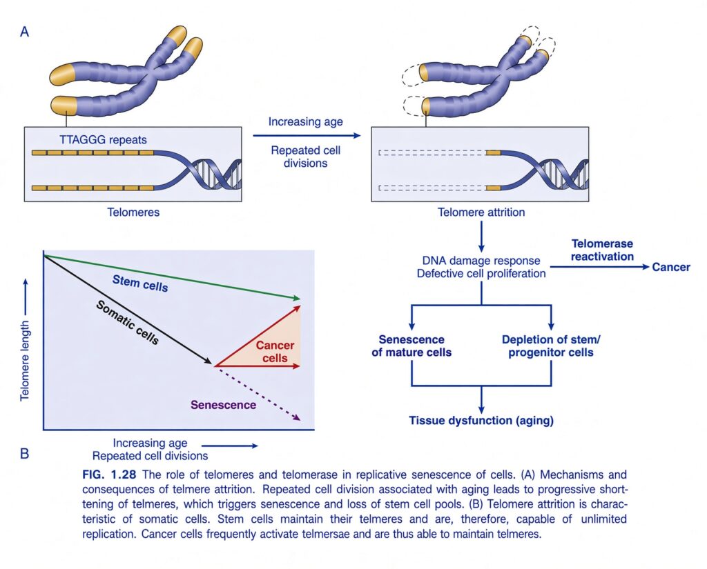
FIG. 1.28 — The Role of Telomeres and Telomerase in Replicative Senescence
🧠 THE BIG IDEA
This figure explains why many normal cells cannot divide forever and how telomeres, telomerase, aging, and cancer are connected.
The easiest central idea is:
Every time most normal cells divide, their telomeres become shorter. When telomeres become critically short, the cell stops dividing → senescence → tissue dysfunction and aging.
But:
Cancer cells can reactivate telomerase → maintain telomeres → continue dividing.
1. 🧬 What are telomeres?
Look at the ends of the chromosomes in the upper part of the figure.
At the ends are special DNA sequences called:
Telomeres
The figure shows:
TTAGGG repeats
These are repeated DNA sequences located at the ends of chromosomes.
Think of telomeres as the protective caps on shoelaces.
Without the plastic cap on a shoelace, the shoelace starts to unravel.
Similarly:
Telomeres protect chromosome ends from being damaged or confused with broken DNA.
2. 🧬 What happens when a cell divides repeatedly?
Look at the arrow:
Increasing age → repeated cell divisions
Every time a normal somatic cell divides:
Telomeres become progressively shorter.
This is called:
Telomere attrition
Attrition = gradual wearing away/shortening.
So:
Long telomeres
      ↓
Cell division
      ↓
Telomeres become shorter
      ↓
Cell division
      ↓
Even shorter telomeres
      ↓
Repeated divisions
      ↓
Critical telomere shortening
3. ✂️ Why do telomeres become shorter?
During DNA replication, the very ends of chromosomes cannot be copied completely by the normal DNA replication machinery.
Therefore, with repeated cell divisions:
A small amount of telomeric DNA is lost each time.
So the telomeres act somewhat like a countdown mechanism for many somatic cells.
4. 👴 What happens when telomeres become too short?
Look at the right side of the figure.
Telomere attrition
↓
DNA damage response
↓
Defective cell proliferation
The cell recognizes critically short/damaged telomeres as a serious problem.
Therefore:
The cell stops proliferating normally.
This leads to two major consequences shown in the figure.
5. 🛑 SENESCENCE OF MATURE CELLS
The first pathway is:
Senescence of mature cells
Senescence means:
The cell remains alive but permanently stops dividing.
This is different from cell death.
Senescent cell:
- Alive ✅
- Metabolically active ✅
- Normal proliferation ❌
Think:
The cell is alive, but it has permanently retired from cell division.
6. 🌱 DEPLETION OF STEM/PROGENITOR CELLS
The second pathway is:
Depletion of stem/progenitor cells
Stem and progenitor cells are important because they:
Replace and replenish cells in tissues.
If their ability to divide becomes limited:
↓ Stem/progenitor cells
↓
↓ Tissue repair and regeneration
↓
Tissue dysfunction
7. 👴 HOW DOES THIS CAUSE AGING?
The figure shows:
Senescence of mature cells
+
Depletion of stem/progenitor cells
↓
Tissue dysfunction (aging)
This is the main connection.
In simple terms:
Repeated cell divisions
        ↓
Telomeres shorten
        ↓
Critical telomere attrition
        ↓
Cells stop dividing
        ↓
Less cell replacement + repair
        ↓
Tissue dysfunction
        ↓
AGING
8. 🧬 NOW UNDERSTAND PANEL B — THE GRAPH
Panel B is very important.
Y-axis:
Telomere length
Going upward means:
Longer telomeres
Going downward means:
Shorter telomeres
X-axis:
Increasing age / repeated cell divisions
Moving right means:
More cell divisions and increasing age
9. ⚫ SOMATIC CELLS — BLACK LINE
Look at the black line labeled:
Somatic cells
The line goes downward.
That means:
As somatic cells undergo repeated divisions, their telomeres become shorter.
So:
More divisions → shorter telomeres
10. 🟢 STEM CELLS — GREEN LINE
Now look at the green line.
It decreases only slightly.
Why?
Because stem cells have mechanisms that help maintain their telomeres.
The important enzyme is:
Telomerase
Therefore:
Stem cells can maintain telomeres much better than ordinary somatic cells.
This allows them to undergo many more divisions.
11. 🟣 SENESCENCE — PURPLE DOTTED LINE
Eventually, somatic-cell telomeres become critically short.
At this point:
Senescence
occurs.
The purple dotted line continues downward from the point where the cell enters senescence.
This represents:
Progressive telomere shortening associated with cellular senescence.
12. 🔴 CANCER CELLS — RED LINE
Now look at the red line.
Cancer cells are different.
The figure shows:
Cancer cells
with telomere maintenance.
Cancer cells frequently:
Reactivate telomerase
↓
Telomeres are maintained
↓
Cancer cells can continue dividing.
13. ⭐ WHAT IS TELOMERASE?
Telomerase is an enzyme that:
Maintains and extends telomeres.
Think of telomeres as the ends of a candle that become shorter every time it is used.
Normal somatic cell:
🔥 Use → shorter
🔥 Use → shorter
🔥 Use → shorter
→ eventually stops dividing
Telomerase-active cell:
🔥 Use → telomere maintained
🔥 Use → telomere maintained
🔥 Use → telomere maintained
Therefore:
Telomerase gives cells the ability to maintain telomere length and continue proliferating.
14. 🚨 WHY IS TELOMERASE IMPORTANT IN CANCER?
This is one of the most important concepts in the figure.
Normally:
Telomeres shorten
↓
Cell stops dividing
↓
Limits the number of cell divisions.
But cancer cells can escape this limitation.
Cancer:
Telomerase reactivation
↓
Telomeres maintained
↓
Continued cell proliferation
↓
Cancer
So:
Telomerase helps cancer cells overcome the normal replicative limit.
15. 🧠 UNDERSTAND THE TOP PART OF PANEL A
Starting point:
Long telomeres
The chromosome ends have many:
TTAGGG repeats
Then:
Increasing age + repeated cell divisions
↓
Telomere attrition
The yellow telomere regions become progressively shorter.
16. 🚨 CRITICALLY SHORT TELOMERES
When telomeres become critically short:
DNA damage response is activated.
The cell essentially says:
“My chromosome ends are becoming unsafe.”
Therefore:
DNA damage response
↓
Defective cell proliferation
↓
Either:
Senescence of mature cells
or
Depletion of stem/progenitor cells
↓
Tissue dysfunction → aging
17. 🔥 BUT CANCER TAKES A DIFFERENT PATH
The figure shows another arrow:
Telomerase reactivation → Cancer
This means:
Cancer cells can turn telomerase back on.
↓
Telomeres are maintained.
↓
The cell avoids the normal telomere-shortening limit.
↓
It can continue proliferating.
Therefore:
The same mechanism that helps stem cells maintain their ability to divide can be misused by cancer cells to support essentially unlimited proliferation.
🧩 NOW UNDERSTAND THE ENTIRE FIGURE AS ONE STORY
              NORMAL CELL
                   ↓
          Repeated cell division
                   ↓
            Telomeres shorten
                   ↓
          Telomere attrition
                   ↓
           DNA damage response
                   ↓
       Defective cell proliferation
              ↙         ↘
             ↓           ↓
      Senescence      Stem/progenitor
     of mature cells      depletion
             ↘           ↙
                ↓
         Tissue dysfunction
                ↓
              AGING
But cancer can escape:
Cancer cell
    ↓
Telomerase reactivation
    ↓
Telomeres maintained
    ↓
Continued proliferation
    ↓
CANCER
⭐ THE MOST IMPORTANT COMPARISON
| Cell type | Telomere behavior | Result |
|---|---|---|
| Somatic cells | Telomeres progressively shorten | Limited replication → senescence |
| Stem cells | Telomeres maintained better | Continued ability to replicate |
| Cancer cells | Frequently activate telomerase | Telomeres maintained → continued proliferation |
🧠 SUPER-EASY ANALOGY
Imagine every cell division uses one piece of a protective rope at the end of each chromosome.
🧍 Somatic cell
Has a limited supply of rope.
Division → rope gets shorter
↓
Eventually:
Rope too short → cell stops dividing
= Senescence
🌱 Stem cell
Has a system that can replace the rope.
= Telomere maintenance
Therefore:
Stem cell can keep dividing for much longer.
🦠 Cancer cell
Turns that rope-repair system back on:
Telomerase reactivation
↓
Keeps the telomeres maintained.
↓
Allows continued proliferation.
🎯 FINAL EXAM CONCEPT
Repeated division of most somatic cells causes progressive telomere shortening. Critically short telomeres activate a DNA-damage response that causes cellular senescence and contributes to depletion of stem/progenitor cells, leading to tissue dysfunction and aging. Stem cells maintain their telomeres and can therefore replicate extensively. Cancer cells frequently reactivate telomerase, allowing telomere maintenance and continued proliferation.
🔑 ONE-LINE MEMORY
Somatic cells:
Division → telomere shortening → senescence → aging
Stem cells:
Telomere maintenance → continued replication
Cancer cells:
Telomerase reactivation → telomere maintenance → continued proliferation
Superfast RAPID REVIEW – Chapter # 1
Patterns of Cell Injury and Cell Death
- Causes of cell injury include:
- Ischemia
- Toxins
- Infections
- Immunologic reactions
- Genetic abnormalities
- Nutritional imbalances
- Physical agents, such as trauma and burns
- Aging
- Reversible cell injury is characterized by:
- Cell swelling
- Fatty change
- Plasma membrane blebbing
- Loss of microvilli
- Mitochondrial swelling
- Dilation of the ER
- Eosinophilia due to decreased cytoplasmic RNA
- Myelin figures
- Necrosis is characterized by:
- Eosinophilia
- Nuclear shrinkage
- Nuclear fragmentation
- Nuclear dissolution
- Breakdown of the plasma membrane and organellar membranes
- Leakage and enzymatic digestion of cellular contents
- Inflammation
- The morphologic types of tissue necrosis are:
- Coagulative
- Liquefactive
- Gangrenous
- Caseous
- Fat
- Fibrinoid
- Apoptosis is a regulated form of cell death that removes unnecessary or irreparably damaged cells.
- It occurs without an injurious reaction from the host.
- Apoptosis is characterized by:
- Enzymatic breakdown of proteins and DNA
- Activation by caspases
- Recognition and removal of dead cells by phagocytes
- There are two major pathways of apoptosis.
- The mitochondrial (intrinsic) pathway is triggered by:
- Loss of survival signals
- DNA damage
- Accumulation of misfolded proteins due to ER stress
- It involves leakage of proapoptotic proteins from mitochondria into the cytoplasm.
- These proteins then trigger caspase activation.
- The pathway is inhibited by antiapoptotic BCL family proteins.
- These antiapoptotic proteins are induced by survival signals, including growth factors.
- The death receptor (extrinsic) pathway helps eliminate:
- Self-reactive lymphocytes
- Cells targeted by cytotoxic T lymphocytes
- It begins when death receptors, which belong to the TNF receptor family, bind to ligands on adjacent cells.
- Autophagy is triggered by nutrient deprivation.
- It involves the degradation and recycling of cellular contents to provide energy during stress.
- If the stress is not relieved, autophagy can trigger apoptosis.
- Other unusual pathways of cell death include:
- Necroptosis — has features of both necrosis and apoptosis and is controlled by specific signaling pathways.
- Pyroptosis — cell death associated with the release of proinflammatory cytokines.
Mechanisms of Cell Injury
- Different initiating events can cause cell injury and death through different mechanisms.
- Mitochondrial damage and increased cell membrane permeability are often late events in cell injury and necrosis caused by different conditions.
- Oxidative stress means accumulation of reactive oxygen species (ROS).
- ROS can damage:
- Lipids
- Proteins
- DNA
- Oxidative stress is associated with many different causes of cell injury.
- ER stress occurs when proteins become misfolded.
- Protein misfolding can reduce essential proteins.
- If misfolded proteins accumulate inside cells, they can trigger apoptosis.
- DNA damage, such as damage caused by radiation, can also trigger apoptosis if the damage cannot be repaired.
- Hypoxia and ischemia cause ATP depletion.
- ATP depletion causes failure of many energy-dependent cellular functions.
- This initially produces reversible cell injury.
- If the condition is not corrected, it progresses to necrosis.
- In ischemia-reperfusion injury, restoring blood flow to ischemic tissue can actually increase the damage.
- This occurs because reperfusion increases:
- ROS production
- Inflammation
Cellular Adaptations to Stress
- Hypertrophy means increased cell size and organ size.
- It commonly occurs in response to increased workload.
- It is induced by growth factors produced in response to mechanical stress or other stimuli.
- It occurs in tissues whose cells are unable to divide.
- Hyperplasia means an increased number of cells.
- It occurs in response to hormones and other growth factors.
- It occurs in tissues whose cells can divide or contain many tissue stem cells.
- Atrophy means decreased cell size and organ size.
- It can result from decreased nutrient supply or disuse.
- It is associated with:
- Decreased synthesis of cellular building blocks
- Increased breakdown of cellular organelles
- Metaplasia means a change in the phenotype of differentiated cells.
- It often occurs in response to chronic irritation.
- The new cell type is better able to withstand the stress.
- It is usually caused by an altered differentiation pathway of tissue stem cells.
- It may cause reduced cellular functions or increase the tendency toward malignant transformation.
Abnormal Intracellular Depositions and Calcifications
- Abnormal substances can accumulate in cells and tissues because of:
- Excessive intake
- Defective transport
- Defective catabolism
- Lipids can accumulate abnormally.
- Fatty change means accumulation of free triglycerides inside cells.
- It results from excessive intake or defective transport, often because of defects in the synthesis of transport proteins.
- Fatty change is a manifestation of reversible cell injury.
- Cholesterol deposition results from:
- Defective catabolism
- Excessive intake
- It occurs in macrophages and smooth muscle cells of vessel walls in atherosclerosis.
- Proteins can accumulate as:
- Reabsorbed proteins in kidney tubules
- Immunoglobulins in plasma cells
- Glycogen can accumulate in macrophages of patients with defects in lysosomal enzymes that break down glycogen.
- These conditions are called glycogen storage diseases.
- Pigments are typically indigestible substances.
- Examples include:
- Carbon
- Lipofuscin, a breakdown product of lipid peroxidation
- Hemosiderin, usually associated with iron overload
- Pathologic calcifications are abnormal calcium deposits.
- Dystrophic calcification means deposition of calcium at sites of cell injury and necrosis.
- Metastatic calcification means deposition of calcium in normal tissues.
- It is caused by hypercalcemia.
- Hypercalcemia is usually a consequence of excess parathyroid hormone.
Cellular Aging
- Cellular aging results from a combination of multiple progressive cellular changes.
- These changes include:
- Accumulation of DNA damage and mutations
- Replicative senescence — reduced ability of cells to divide because of progressive shortening of telomeres at chromosome ends
- Defective protein homeostasis — loss of normal proteins and accumulation of misfolded proteins
- Cellular aging is accelerated by chronic diseases, especially diseases associated with prolonged inflammation.
- Stress can also accelerate aging.
- Calorie restriction and exercise can slow down aging.
KEY CONCEPT
Rapid Review = Cell response can be understood as:
- Injury → reversible injury → if severe/persistent → cell death
- Cell death → necrosis or apoptosis
- Stress → adaptation → hypertrophy, hyperplasia, atrophy, or metaplasia
- Abnormal accumulation → lipids, proteins, glycogen, pigments, or calcium
- Aging → DNA damage + telomere shortening + protein problems → reduced cell replication and function
Made by self Learning Dr sheen
