


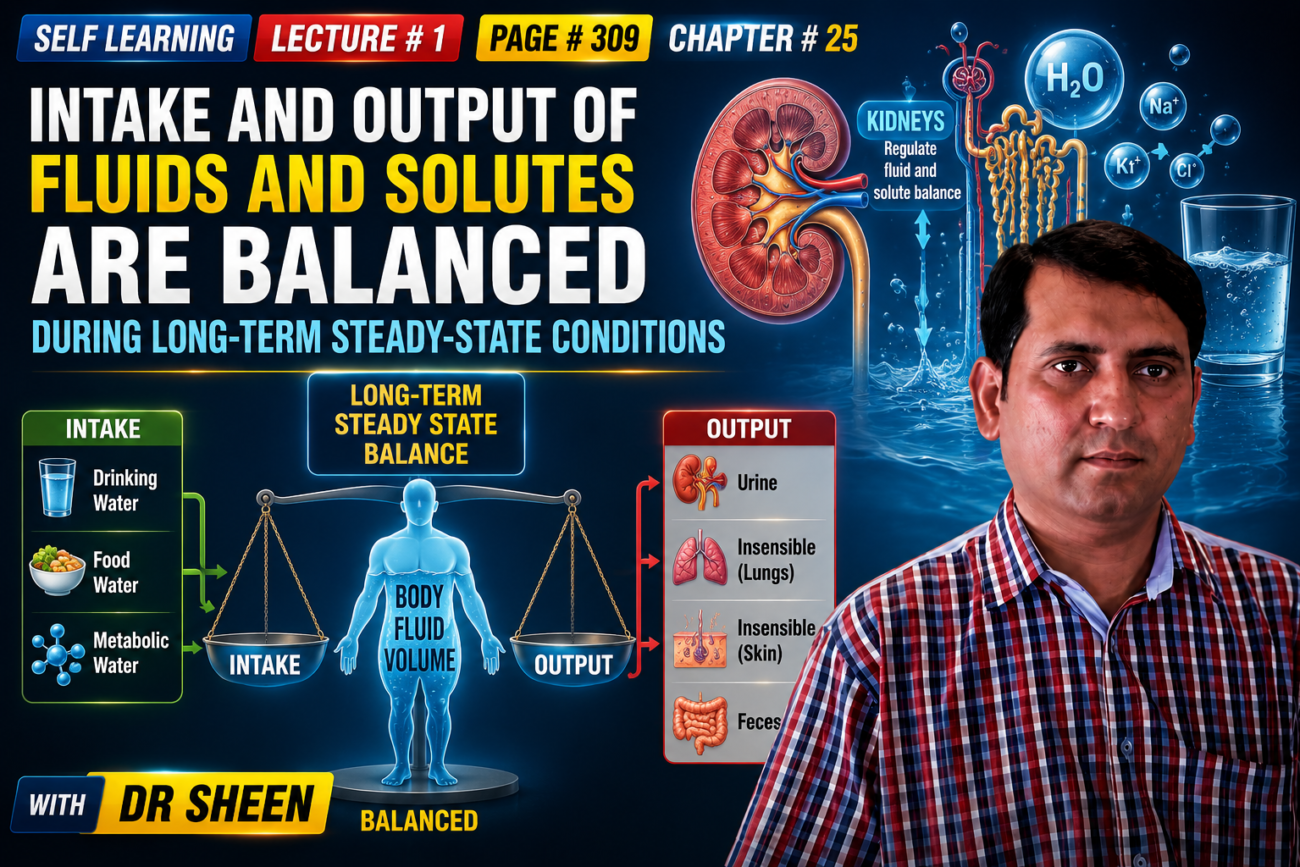
- Body fluids remain relatively constant even though fluid and solutes are continuously exchanged:
- Between the body and the outside environment.
- Between different body compartments.
- Fluid and electrolyte intake can vary greatly.
- Therefore, the body must match fluid and electrolyte output with intake to prevent body fluid volumes and electrolyte levels from becoming too high or too low.
DAILY INTAKE OF WATER
- Water enters the body mainly from two sources:
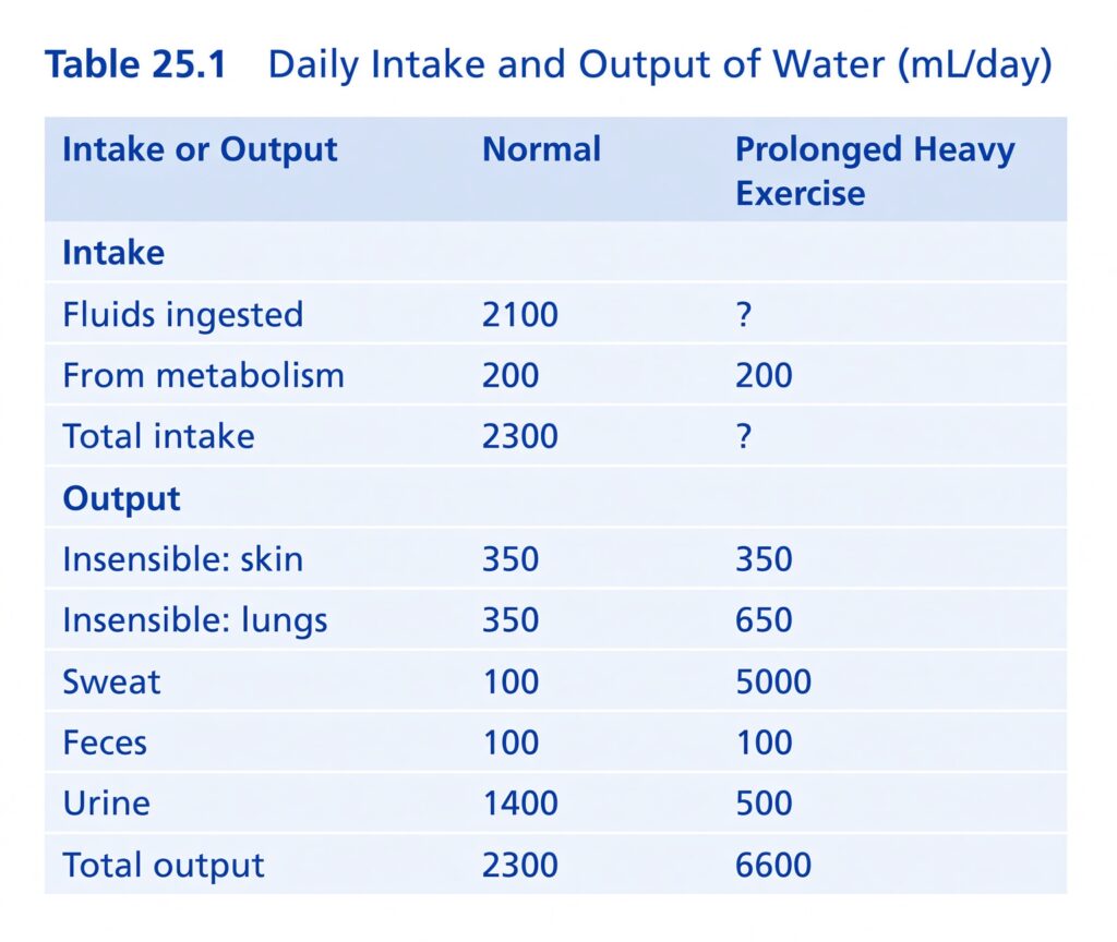
- Drinking water and water present in food: about 2100 mL/day.
- Water produced inside the body from carbohydrate oxidation: about 200 mL/day.
- Therefore:
- Total daily water intake = 2100 + 200 = 2300 mL/day.
- Water intake varies greatly between people and even in the same person from day to day.
- It depends on:
- Climate
- Habits
- Physical activity
Daily Loss of Body Water
Insensible Water Loss
- Some water losses cannot be precisely controlled.
- Water is continuously lost by:
- Evaporation from the respiratory tract.
- Diffusion through the skin.
- Together, these normally cause about 700 mL/day of water loss.
- This is called insensible water loss because we do not consciously notice it.
- Water loss through the skin by diffusion is about 300–400 mL/day.
- This occurs even without sweating and even in people born without sweat glands.
- The skin’s cholesterol-filled, cornified layer limits this water loss.
- When this protective layer is lost, such as in extensive burns, evaporation can increase up to 10 times, reaching about 3–5 L/day.
- Therefore, burn patients need large amounts of fluid, usually intravenously, to replace the fluid they lose.
- Water loss through the respiratory tract is normally about 300–400 mL/day.
- As air enters the respiratory tract, it becomes saturated with water vapor at a vapor pressure of about 47 mm Hg before being exhaled.
- Inspired air usually has a vapor pressure below 47 mm Hg.
- Therefore, water continuously leaves the body through the lungs during breathing.
- In cold weather, atmospheric vapor pressure becomes very low, nearly zero.
- This increases water loss from the lungs as temperature decreases.
- This explains why the respiratory passages feel dry in cold weather.
Fluid Loss in Sweat
- Water loss through sweating varies greatly with:
- Physical activity
- Environmental temperature
- Normal sweat loss is about 100 mL/day.
- In hot weather or during heavy exercise, sweat loss can sometimes increase to 1–2 L/hour.
- Such a large loss could rapidly decrease body fluids if water intake did not also increase through activation of the thirst mechanism.
Water Loss in Feces
- Normally, only about 100 mL/day of water is lost in feces.
- With severe diarrhea, water loss can increase to several liters per day.
- Therefore, severe diarrhea can become life-threatening within a few days if the fluid loss is not corrected.
Water Loss By the Kidneys
- The remaining major water loss occurs through urine produced by the kidneys.
- Several mechanisms control how much urine is excreted.
- The most important way the body maintains:
- Water intake = water output
- Electrolyte intake = electrolyte output
- is by controlling how much water and electrolytes the kidneys excrete.
- For example:
- In dehydration, urine output can fall to about 0.5 L/day.
- After drinking extremely large amounts of water, urine output can increase to about 20 L/day.
- Intake also varies greatly for electrolytes such as:
- Sodium
- Chloride
- Potassium
- Sodium intake may be as low as 20 mEq/day in some people.
- In others, sodium intake may reach 300–500 mEq/day.
- The kidneys adjust the excretion of water and electrolytes to closely match their intake.
- They also compensate for excessive fluid and electrolyte losses that occur during certain diseases.
- The mechanisms that allow the kidneys to perform these functions are discussed in Chapters 26–32.
KEY CONCEPT
- Main principle:
Long-term steady state = Intake ≈ Output - The body keeps fluid volume and electrolytes relatively constant by adjusting mainly kidney excretion.
- Approximate daily water intake:
- Food + drinks = 2100 mL
- Metabolic production = 200 mL
- Total = 2300 mL/day
- Major water losses:
- Insensible loss = ~700 mL/day
- Sweat = ~100 mL/day normally, but can reach 1–2 L/hour
- Feces = ~100 mL/day normally
- Kidneys = highly variable, about 0.5–20 L/day
- Conceptual example:
If water intake increases greatly → kidneys increase urine output → body water remains relatively stable. - Conceptual example:
Severe burns → skin barrier is lost → water loss can rise to 3–5 L/day → large fluid replacement is required. - Conceptual example:
Dehydration → kidneys conserve water → urine output can fall to ~0.5 L/day.
BODY FLUID COMPARTMENTS
- Total body fluid is mainly divided into two major compartments:
- Extracellular fluid (ECF)
- Intracellular fluid (ICF) (Fig. 25.1)
- The extracellular fluid is further divided into:
- Interstitial fluid
- Blood plasma
- There is also a small fluid compartment called transcellular fluid.
- It includes fluid found in:
- Synovial spaces
- Peritoneal spaces
- Pericardial spaces
- Intraocular spaces
- Cerebrospinal fluid
- Transcellular fluid is usually considered a special type of extracellular fluid.
- However, in some situations, its composition can be very different from plasma or interstitial fluid.
- All transcellular fluids together contain about 1–2 liters.
- In a 70-kg adult man:
- Total body water is about 60% of body weight.
- Therefore, total body water is about 42 liters.
- The percentage of body water depends on:
- Age
- Muscle mass
- Percentage of body weight that is fat
- As a person gets older, the percentage of body weight that is water gradually decreases.
- This happens partly because aging is usually associated with:
- Decreased muscle mass
- Increased body fat
- Because fat contains less water than muscle, increased body fat lowers the percentage of water in the body.
- Women normally have a higher percentage of body fat than men.
- Therefore, total body water in women averages about 50% of body weight.
- This percentage also varies considerably among women according to:
- Age
- Muscle mass
- Body fat
- In premature and newborn babies, total body water is about 70–75% of body weight.
- Therefore, average body-fluid compartments vary according to:
- Age
- Sex
- Muscle mass
- Body fat
- In many countries, average body weight and fat mass have increased rapidly during the past 30 years.
- In the United States, the estimated average body weight is:
- Adult men older than 20 years: about 90 kg (198 lb)
- Adult women: about 77.5 kg (171 lb)
- Therefore, when using data based on an average 70-kg man, the values should be adjusted when considering body-fluid compartments in many people.
KEY CONCEPT
- Total body water → mainly two compartments:
- ICF = fluid inside cells
- ECF = fluid outside cells
- ECF → interstitial fluid + plasma
- Transcellular fluid → small specialized part of ECF, about 1–2 L
- Conceptual example:
A 70-kg man → 60% body water → 42 L total body water. - Conceptual example:
More body fat → lower percentage of total body water. - Conceptual example:
Newborn → 70–75% body weight is water, whereas an adult woman averages about 50%. - Key relationship:
Age ↑ / body fat ↑ / muscle mass ↓ → percentage of body water ↓.
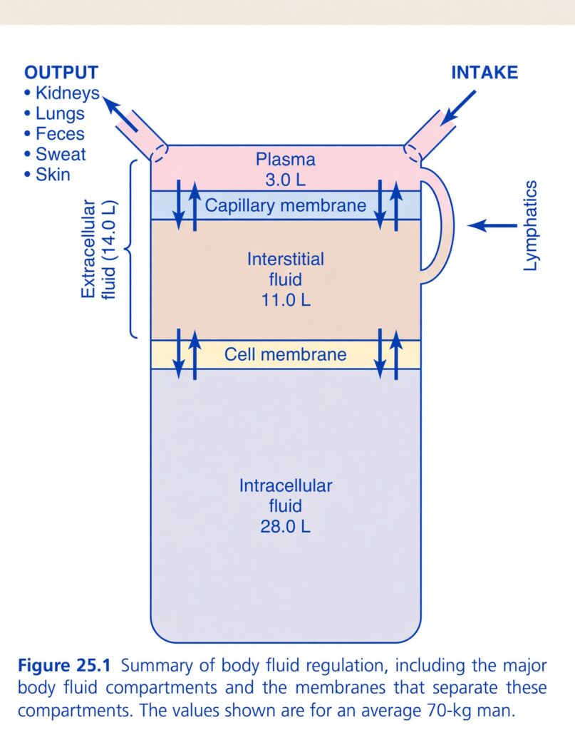
Figure 25.1 — Body Fluid Compartments and Fluid Regulation
🧠 The BIG IDEA
This figure shows where the water in an average 70-kg person’s body is located and how water moves between the different compartments.
Think of the body as having three main water tanks:
                 TOTAL BODY WATER ≈ 42 L
                         ↓
          ┌──────────────┴──────────────┐
          ↓                             ↓
   EXTRACELLULAR FLUID            INTRACELLULAR FLUID
          14 L                         28 L
          ↓
     ┌────┴────┐
     ↓         ↓
 Plasma     Interstitial
  3 L          11 L
⭐ The most important numbers:
- Intracellular fluid (ICF) = 28 L
- Extracellular fluid (ECF) = 14 L
- Plasma = 3 L
- Interstitial fluid = 11 L
- Total body water = 42 L
So:
28 L inside cells + 14 L outside cells = 42 L total body water
1. 💧 INTRACELLULAR FLUID — 28 L
What does intracellular mean?
Intra = inside
So intracellular fluid means:
Fluid inside the body’s cells.
It is the largest fluid compartment.
Amount:
≈ 28 L
That is about:
2/3 of total body water
2. 🌊 EXTRACELLULAR FLUID — 14 L
Extra = outside
Extracellular fluid means:
Fluid outside the cells.
Total:
ECF ≈ 14 L
It is divided mainly into:
- Plasma = 3 L
- Interstitial fluid = 11 L
Therefore:
3 L + 11 L = 14 L ECF
3. 🩸 PLASMA — 3 L
Look at the top pink compartment.
This represents:
Plasma
Amount:
≈ 3 L
Plasma is the fluid portion of blood.
It is located inside the blood vessels.
Think:
Blood vessel → plasma
4. 🌿 INTERSTITIAL FLUID — 11 L
The large beige compartment underneath plasma is:
Interstitial fluid
Amount:
≈ 11 L
This fluid surrounds and bathes the cells.
Think of it like this:
Blood vessel
   ↓
Plasma
   ↓
Interstitial fluid
   ↓
Cells
So interstitial fluid acts like the middle fluid environment surrounding cells.
5. 🧫 INTRACELLULAR FLUID — 28 L
The large lower compartment represents:
Fluid inside cells
Amount:
≈ 28 L
Notice that it is much larger than either plasma or interstitial fluid.
That’s because most body water is inside cells.
6. 🚧 CAPILLARY MEMBRANE
Look at the horizontal boundary between:
Plasma ↔ Interstitial fluid
This is labeled:
Capillary membrane
It separates:
Blood plasma from interstitial fluid
Water and many small substances can move across this barrier.
Simple picture:
PLASMA
  3 L
   ↕
CAPILLARY MEMBRANE
   ↕
INTERSTITIAL FLUID
  11 L
The arrows in the figure indicate that fluid can move in both directions between these compartments.
7. 🧱 CELL MEMBRANE
Now look at the boundary between:
Interstitial fluid ↔ Intracellular fluid
This is the:
Cell membrane
It separates:
Fluid outside the cells from fluid inside the cells.
Water can move across the cell membrane, allowing the cells and extracellular fluid to maintain appropriate fluid balance.
Simple picture:
INTERSTITIAL FLUID
       11 L
        ↕
   CELL MEMBRANE
        ↕
INTRACELLULAR FLUID
       28 L
8. 🔄 WHAT DO THE DOUBLE ARROWS MEAN?
You can see arrows going up and down at both membranes.
These indicate:
Fluid can move between the compartments in both directions.
Across the capillary membrane:
Plasma ↔ Interstitial fluid
Across the cell membrane:
Interstitial fluid ↔ Intracellular fluid
The direction and amount of movement depend on the body’s physiological conditions.
9. 🟡 LYMPHATICS
On the right side, you can see:
Lymphatics
This is extremely important.
Some fluid leaves the blood vessels and enters the interstitial space.
The lymphatic system helps:
Collect excess interstitial fluid and return it to the circulation.
Think of lymphatics as a:
🚰 Drainage and return system
Blood plasma
     ↓
Interstitial fluid
     ↓
Excess fluid
     ↓
Lymphatic vessels
     ↓
Returned to circulation
⭐ Key concept:
Lymphatics help prevent excessive accumulation of fluid in the interstitial space.
If lymphatic drainage becomes severely impaired, edema can develop.
10. 🍽️ INTAKE — WHERE DOES BODY WATER COME FROM?
At the upper right, the figure shows:
INTAKE
This represents water entering the body.
Water enters mainly through:
- Drinks
- Food
- Metabolic water produced inside the body
The figure simplifies this as intake entering the body-fluid system.
11. 🚽 OUTPUT — WHERE DOES WATER LEAVE?
On the upper left, the figure lists:
OUTPUT
Water leaves the body through several routes:
🫘 Kidneys
The kidneys remove water in urine.
🫁 Lungs
Water leaves in exhaled air as water vapor.
💩 Feces
Some water leaves with stool.
💦 Sweat
Water is lost through sweating.
🧴 Skin
Water can also be lost through the skin as insensible water loss, apart from obvious sweating.
12. ⚖️ INTAKE = OUTPUT
This is the central idea behind body fluid regulation.
Under normal steady-state conditions:
Water entering the body ≈ water leaving the body
If:
Intake > Output
Body water increases.
Output > Intake
Body water decreases.
The body therefore regulates water balance by adjusting intake and especially renal output.
13. 🧠 FOLLOW THE WATER THROUGH THE WHOLE FIGURE
Imagine drinking a glass of water.
Step 1
Water enters the body:
INTAKE
↓
Step 2
It enters the body’s fluid system.
↓
Step 3
It distributes between:
ECF ↔ ICF
↓
Step 4
Within ECF, fluid is distributed between:
Plasma ↔ Interstitial fluid
↓
Step 5
Interstitial fluid surrounds the cells.
↓
Step 6
Excess interstitial fluid can enter:
Lymphatics
↓
Step 7
Water eventually leaves the body through:
Kidneys + lungs + feces + sweat + skin
🔥 THE MOST IMPORTANT NUMBERS
For an average 70-kg man:
Total body water
≈ 42 L
Divided into:
🟦 Intracellular fluid
28 L
🟨 Extracellular fluid
14 L
ECF further divides into:
Plasma = 3 L
Interstitial fluid = 11 L
Therefore:
28 + 3 + 11 = 42 L
⭐ ECF vs ICF — VERY EASY
| Compartment | Where? | Volume |
|---|---|---|
| ICF | Inside cells | 28 L |
| ECF | Outside cells | 14 L |
| Plasma | Inside blood vessels | 3 L |
| Interstitial fluid | Around cells | 11 L |
Remember:
ICF = 28 L
ECF = 14 L
ECF = Plasma 3 L + Interstitial 11 L
🧩 UNDERSTAND THE TWO MEMBRANES
🩸 Capillary membrane
Separates:
Plasma ↔ Interstitial fluid
🧫 Cell membrane
Separates:
Interstitial fluid ↔ Intracellular fluid
So the sequence is:
       BLOOD VESSEL
            ↓
       🩸 PLASMA
          3 L
            ↕
   CAPILLARY MEMBRANE
            ↕
   INTERSTITIAL FLUID
          11 L
            ↕
      CELL MEMBRANE
            ↕
   INTRACELLULAR FLUID
          28 L
🎯 ONE-MINUTE REVISION
Total body water = 42 L
Inside cells:
→ ICF = 28 L
Outside cells:
→ ECF = 14 L
ECF consists of:
→ Plasma = 3 L
→ Interstitial fluid = 11 L
Barriers:
Capillary membrane
→ separates plasma from interstitial fluid.
Cell membrane
→ separates interstitial fluid from intracellular fluid.
Lymphatics:
→ return excess interstitial fluid to the circulation.
Intake:
→ adds water to the body.
Output:
→ kidneys, lungs, feces, sweat, and skin remove water.
🧠 FINAL CONCEPT
The body’s water is divided mainly into two big compartments: 28 L inside cells and 14 L outside cells. The 14 L of extracellular fluid consists of 3 L of plasma and 11 L of interstitial fluid. Fluid continuously exchanges across the capillary and cell membranes, while the lymphatic system returns excess interstitial fluid to the circulation. Overall water balance is maintained by matching intake with output.
INTRACELLULAR FLUID COMPARTMENT
- About two-thirds of the total body water is inside the trillions of cells.
- In a 70-kg person, this is about 28 L out of 42 L of total body water.
- This fluid inside cells is called intracellular fluid (ICF).
- Therefore, ICF makes up about 40% of total body weight in an average person.
- Each cell has its own mixture of different substances.
- However, the concentrations of these substances are generally similar from one cell to another.
- In fact, cell-fluid composition is remarkably similar even among different animals, from very simple microorganisms to humans.
- Therefore, the intracellular fluid of all cells together is considered one large fluid compartment.
EXTRACELLULAR FLUID COMPARTMENT
- All fluid outside the cells is collectively called extracellular fluid (ECF).
- ECF makes up about 20% of body weight, or about 14 L in a 70-kg lean man.
- The two largest parts of ECF are:
- Interstitial fluid → more than three-fourths of ECF, about 11 L
- Plasma → almost one-fourth of ECF, about 3 L
- Plasma is the noncellular part of blood.
- Plasma continuously exchanges substances with interstitial fluid through pores in the capillary membranes.
- These pores allow almost all ECF solutes to pass through, except proteins.
- Therefore, plasma and interstitial fluid are continuously mixing.
- Because of this, plasma and interstitial fluid have almost the same composition, except that proteins are present in higher concentration in plasma.
BLOOD VOLUME
- Blood contains:
- Extracellular fluid → plasma
- Intracellular fluid → fluid inside red blood cells
- However, blood is considered a separate fluid compartment because it is contained within its own chamber: the circulatory system.
- Blood volume is especially important for controlling cardiovascular dynamics.
- Average adult blood volume is about 7% of body weight.
- In an average adult, this is about 5 L.
- About:
- 60% = plasma
- 40% = red blood cells
- These percentages can vary considerably between people depending on:
- Sex
- Weight
- Other factors
Hematocrit (Packed Red Blood Cell Volume)
- Hematocrit is the fraction of blood made up of red blood cells.
- It is measured by centrifuging blood in a hematocrit tube until the red cells become tightly packed at the bottom.
- The centrifuge does not completely pack the red cells together.
- Therefore, about 3–4% of plasma remains trapped between the red cells.
- Because of this, the true hematocrit is about 96% of the measured hematocrit.
- In men, the normal measured hematocrit is about 0.42 (42%).
- In women, it is about 0.38 (38%).
- In severe anemia, hematocrit may fall as low as 0.10 (10%).
- A hematocrit of 0.10 is barely enough to sustain life.
- In polycythemia, excessive production of red blood cells can increase hematocrit to about 0.65 (65%).
KEY CONCEPT
- Total body water ≈ 42 L in a 70-kg person
- ICF ≈ 28 L → inside cells
- ECF ≈ 14 L → outside cells
- Interstitial fluid ≈ 11 L
- Plasma ≈ 3 L
- Conceptual example: If total body water is 42 L, about 2/3 (28 L) is inside cells and about 1/3 (14 L) is outside cells.
- Conceptual example: ECF is mainly interstitial fluid + plasma, and they continuously exchange substances through capillary pores.
- Conceptual example: Proteins are the major exception: plasma has a higher protein concentration than interstitial fluid.
- Blood ≈ 5 L in an average adult
- 60% plasma
- 40% red blood cells
- Hematocrit = fraction of blood occupied by RBCs
- Men ≈ 42%
- Women ≈ 38%
- Severe anemia can be ≈ 10%
- Polycythemia can reach ≈ 65%
CONSTITUENTS OF EXTRACELLULAR
AND INTRACELLULAR FLUIDS
Comparisons of the composition of the extracellular
fluid, including the plasma and interstitial fluid, and the
intracellular fluid are shown in Figs. 25.2 and 25.3 and
in Table 25.2.
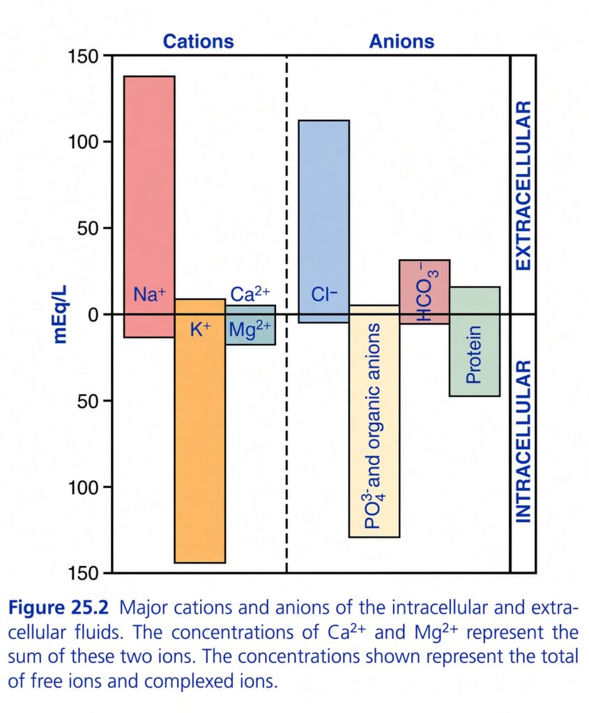
Figure 25.2 — Major Cations and Anions of Intracellular and Extracellular Fluids
🧠 The BIG IDEA
This graph compares the main positively charged ions (cations) and negatively charged ions (anions) found inside cells versus outside cells.
The easiest way to understand it is:
Outside the cell → Na⁺ and Cl⁻ dominate.
Inside the cell → K⁺ and phosphate/organic anions dominate.
This is one of the most important concepts in body-fluid physiology.
1. First understand the graph
The graph is divided into two sides:
Left side → Cations (+)
These are positively charged ions:
- Na⁺
- K⁺
- Ca²⁺
- Mg²⁺
Right side → Anions (−)
These are negatively charged substances:
- Cl⁻
- HCO₃⁻
- PO₄³⁻ and organic anions
- Proteins
2. What does the vertical scale mean?
The vertical axis is:
mEq/L
This means milliequivalents per liter.
For this figure, you can simply think:
The taller the bar, the greater the concentration of that ion in that fluid.
3. 🚧 The most important dividing line
Look at the horizontal line at 0.
The figure uses:
Above the horizontal line
→ Extracellular fluid (ECF)
Below the horizontal line
→ Intracellular fluid (ICF)
The dashed vertical line in the middle separates:
Cations | Anions
So mentally divide the figure into four areas:
                 CATIONS       ANIONS
                    |             |
ECF  ───────────────┼─────────────┼────
                    |             |
ICF  ───────────────┼─────────────┼────
                    |             |
4. 🩸 EXTRACELLULAR FLUID — MAIN CATION = Na⁺
Look at the large pink Na⁺ bar.
It extends strongly above the zero line.
This means:
Sodium (Na⁺) is the major cation of extracellular fluid.
ECF’s most important positive ion:
Na⁺
Think:
Na⁺ = Outside
5. 🧫 INTRACELLULAR FLUID — MAIN CATION = K⁺
Now look at the large orange K⁺ bar.
It extends strongly below the zero line.
This represents intracellular fluid.
Therefore:
Potassium (K⁺) is the major cation inside cells.
ICF’s most important positive ion:
K⁺
Think:
K⁺ = Inside
⭐ The most important memory rule
Na⁺ = ECF
K⁺ = ICF
This is extremely important in physiology.
6. What about Ca²⁺ and Mg²⁺?
Look at the smaller bars labeled:
- Ca²⁺
- Mg²⁺
The figure notes that the concentrations of Ca²⁺ and Mg²⁺ represent the sum of these two ions.
The important point is:
Their concentrations are much smaller than the major Na⁺ and K⁺ concentrations shown here.
Especially remember:
Extracellular Ca²⁺ is much higher than intracellular free Ca²⁺.
This very large Ca²⁺ gradient is important for:
- Muscle contraction
- Neurotransmitter release
- Cell signaling
But for this particular figure, the main exam concept is simply:
Na⁺ and K⁺ are the dominant cations in ECF and ICF, respectively.
7. 🔵 CHLORIDE — MAIN ECF ANION
Now move to the anion side.
Look at the large blue Cl⁻ bar.
It extends upward above the zero line.
Therefore:
Chloride (Cl⁻) is a major extracellular anion.
Think:
Na⁺ + Cl⁻ → major ECF ions
8. 🟡 Phosphate and Organic Anions — MAIN ICF ANIONS
Look at the large yellow bar extending downward.
It is labeled:
PO₄³⁻ and organic anions
These are major negatively charged substances inside cells.
Therefore:
Phosphate and organic anions are major intracellular anions.
Think:
K⁺ + phosphate/organic anions → major ICF ions
9. 🩷 Bicarbonate — ECF ANION
Look at the pink bar labeled:
HCO₃⁻
It is above the zero line.
Therefore:
Bicarbonate is an extracellular anion.
It is particularly important for:
Acid-base balance
10. 🟢 PROTEINS — IMPORTANT ICF ANIONS
Look at the green Protein bar.
It extends below the zero line.
Therefore:
Proteins are important negatively charged substances inside cells.
Intracellular proteins contribute substantially to the negative charge inside cells.
🔥 Now compare ECF vs ICF
🩸 ECF
Major cation:
Na⁺
Major anions:
Cl⁻ and HCO₃⁻
So:
ECF = Na⁺ + Cl⁻/HCO₃⁻
🧫 ICF
Major cation:
K⁺
Major anions:
Phosphate + organic anions + proteins
So:
ICF = K⁺ + phosphate/organic anions/proteins
🧠 THE WHOLE FIGURE IN ONE TABLE
| Fluid compartment | Major cation (+) | Major anions (−) |
|---|---|---|
| 🩸 ECF | Na⁺ | Cl⁻, HCO₃⁻ |
| 🧫 ICF | K⁺ | Phosphate, organic anions, proteins |
11. Why are the bars drawn ABOVE and BELOW zero?
This can initially look confusing.
The graph is not saying that intracellular ions have negative concentrations.
❌ K⁺ concentration is NOT −150 mEq/L.
Instead:
- Above zero = extracellular concentration
- Below zero = intracellular concentration
The downward direction is simply a visual way to separate ICF from ECF.
Very important:
The minus sign-like direction of the K⁺ bar does NOT mean K⁺ has a negative charge.
K⁺ is still positively charged.
Similarly, the downward phosphate/protein bars represent their intracellular concentrations, not negative numerical concentrations.
12. Why are cations and anions separated?
The dashed vertical line separates:
Cations (+)
from
Anions (−)
This helps you compare the major positive and negative ions in each compartment.
And notice something very important:
Each fluid compartment remains electrically neutral overall.
That means:
Total positive charge ≈ Total negative charge
So even though individual ions are distributed very differently, the fluid as a whole does not have a huge net electrical charge.
13. ⭐ Why is Na⁺ high outside and K⁺ high inside?
The main reason is the activity of the:
Na⁺/K⁺ pump
It moves:
3 Na⁺ OUT
and
2 K⁺ IN
using ATP.
Therefore it helps maintain:
Outside:
High Na⁺
Inside:
High K⁺
14. Why is this difference so important?
The unequal distribution of ions across the cell membrane creates the conditions necessary for:
- Resting membrane potential
- Nerve impulses
- Muscle contraction
- Cellular signaling
So this simple graph is actually the foundation for understanding electrical activity in nerves and muscles.
🧠 SUPER-EASY VISUAL MEMORY
Imagine a cell as a house:
🏠 OUTSIDE THE HOUSE = ECF
The main positive ion outside is:
Na⁺
The important negative ions outside are:
Cl⁻ + HCO₃⁻
🏠 INSIDE THE HOUSE = ICF
The main positive ion inside is:
K⁺
The important negative substances inside are:
Phosphate + organic anions + proteins
🎯 ONE-LINE MEMORY TRICK
“Na outside, K inside; Cl outside, phosphate/protein inside.”
Or:
ECF = Na⁺ + Cl⁻/HCO₃⁻
ICF = K⁺ + phosphate/organic anions/proteins
🔑 What the figure’s caption adds
The caption specifically tells us two additional things:
1. Ca²⁺ and Mg²⁺
The concentrations shown for Ca²⁺ and Mg²⁺ represent the sum of these two ions.
2. Free + complexed ions
The concentrations shown represent the total of free ions and complexed ions.
So the bars are showing the total concentration represented in the figure, not only the freely dissolved fraction.
⭐ FINAL CONCEPT
                 OUTSIDE CELL
                     ECF
                      ↓
              Na⁺ = MAIN CATION
              Cl⁻ = MAIN ANION
              HCO₃⁻ = IMPORTANT ANION
                      │
                CELL MEMBRANE
                      │
                     ICF
                      ↓
              K⁺ = MAIN CATION
       Phosphate/organic anions
          + proteins = MAIN ANIONS
                 INSIDE CELL
🎯 The single most important takeaway:
Extracellular fluid is mainly a Na⁺-rich compartment, whereas intracellular fluid is mainly a K⁺-rich compartment; Cl⁻ and HCO₃⁻ are important ECF anions, while phosphate, organic anions, and proteins are important ICF anions.
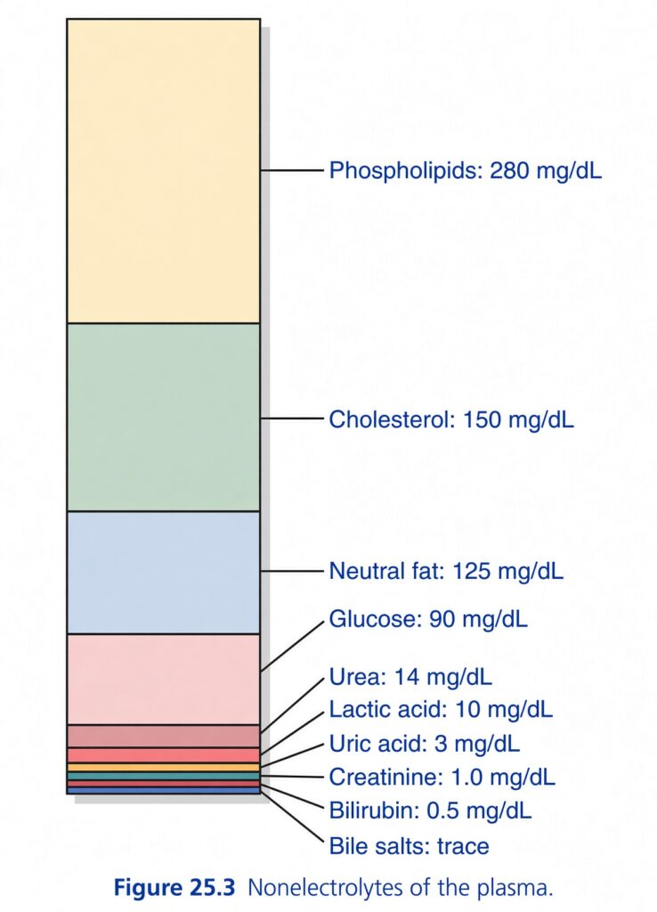
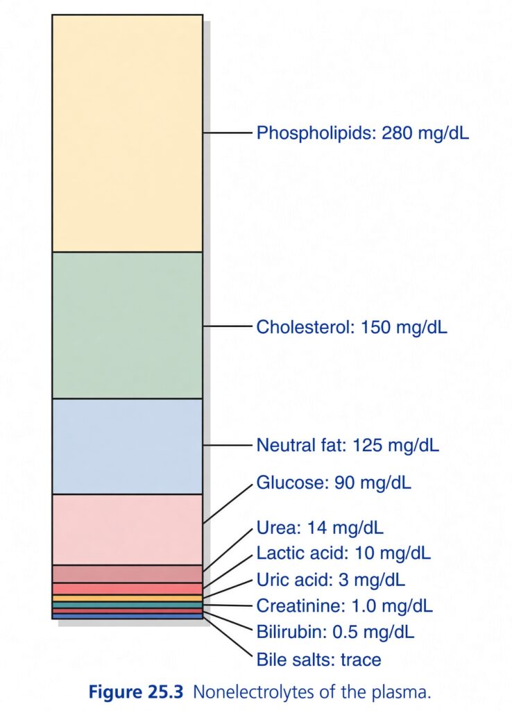
Figure 25.3 — Nonelectrolytes of the Plasma
🧠 The BIG IDEA
This figure shows the main substances in plasma that are NOT electrolytes.
What is a nonelectrolyte?
An electrolyte is a substance that forms ions in solution, such as:
- Na⁺
- K⁺
- Cl⁻
A nonelectrolyte does not significantly dissociate into ions.
The figure shows that plasma contains several important nonelectrolytes, including:
- Phospholipids
- Cholesterol
- Neutral fat
- Glucose
- Urea
- Lactic acid
- Uric acid
- Creatinine
- Bilirubin
- Bile salts
The values are given in mg/dL.
1. 🟡 Phospholipids — 280 mg/dL
This is the largest section of the figure.
Amount:
280 mg/dL
Phospholipids are important components of:
- Cell membranes
- Lipoproteins
Simple idea:
Phospholipids are major lipid substances present in plasma.
2. 🟢 Cholesterol — 150 mg/dL
Amount:
150 mg/dL
Cholesterol is another important plasma lipid.
It is needed for things such as:
- Cell membranes
- Steroid hormone synthesis
- Bile acid production
For this figure, remember:
Cholesterol = 150 mg/dL
3. 🔵 Neutral fat — 125 mg/dL
Amount:
125 mg/dL
“Neutral fat” mainly refers to:
Triglycerides
These are important energy-storage lipids.
Remember:
Neutral fat = 125 mg/dL
4. 🩷 Glucose — 90 mg/dL
Amount:
90 mg/dL
Glucose is the body’s major circulating carbohydrate.
It is an important fuel for cells.
Simple idea:
Glucose in plasma = readily available energy source.
The figure gives:
90 mg/dL
5. 🟣 Urea — 14 mg/dL
Amount:
14 mg/dL
Urea is a major waste product produced during:
Protein/amino acid metabolism
It is transported in the blood to the:
Kidneys
where it is excreted in urine.
Remember:
Urea = nitrogenous waste
14 mg/dL
6. 🟠 Lactic acid — 10 mg/dL
Amount:
10 mg/dL
Lactic acid is produced during metabolism, especially when tissues produce energy with increased reliance on anaerobic glycolysis.
It is transported in the blood and can be metabolized, particularly by the:
- Liver
- Other tissues
Remember:
Lactic acid = metabolic product
10 mg/dL
7. 🟤 Uric acid — 3 mg/dL
Amount:
3 mg/dL
Uric acid is produced from:
Purine metabolism
It is also eliminated mainly through the kidneys.
Remember:
Uric acid = purine breakdown product
3 mg/dL
8. ⚫ Creatinine — 1.0 mg/dL
Amount:
1.0 mg/dL
Creatinine is produced from:
Muscle creatine metabolism
It is filtered by the kidneys and is commonly used clinically as an indicator of kidney function.
Remember:
Creatinine = muscle metabolism + kidney function marker
1.0 mg/dL
9. 🟡 Bilirubin — 0.5 mg/dL
Amount:
0.5 mg/dL
Bilirubin is produced mainly from the breakdown of:
Hemoglobin/heme
It is processed by the liver and eventually excreted through bile.
Remember:
Bilirubin = heme breakdown product
0.5 mg/dL
10. 🟢 Bile salts — TRACE
At the bottom of the figure:
Bile salts: trace
This means:
Only a very small amount is normally present in plasma.
Bile salts are produced from cholesterol and are important for:
Digestion and absorption of fats
📊 NOW LOOK AT THE WHOLE FIGURE
The bars are arranged roughly from larger concentrations at the top to very small concentrations at the bottom.
The major values shown are:
| Substance | Plasma concentration |
|---|---|
| Phospholipids | 280 mg/dL |
| Cholesterol | 150 mg/dL |
| Neutral fat | 125 mg/dL |
| Glucose | 90 mg/dL |
| Urea | 14 mg/dL |
| Lactic acid | 10 mg/dL |
| Uric acid | 3 mg/dL |
| Creatinine | 1.0 mg/dL |
| Bilirubin | 0.5 mg/dL |
| Bile salts | Trace |
🧠 Why does the figure look like one big stacked bar?
Don’t interpret it as one combined concentration.
Each colored section represents a different substance and its approximate concentration.
Think of it like a tower:
🟡 Phospholipids      280 mg/dL
🟢 Cholesterol        150 mg/dL
🔵 Neutral fat        125 mg/dL
🩷 Glucose             90 mg/dL
🟣 Urea                14 mg/dL
🟠 Lactic acid         10 mg/dL
🟤 Uric acid            3 mg/dL
⚫ Creatinine           1.0 mg/dL
🟡 Bilirubin            0.5 mg/dL
🟢 Bile salts           trace
The figure is primarily giving you a visual comparison of their concentrations.
🔥 VERY IMPORTANT: WHY ARE THESE CALLED NONELECTROLYTES?
Because these substances are not being represented as the major freely dissociated ions of plasma.
Compare:
Electrolytes ⚡
Examples:
Na⁺, K⁺, Cl⁻, HCO₃⁻
These exist as ions in solution and contribute to plasma electrolyte composition.
Nonelectrolytes
Examples in this figure:
Glucose, urea, lipids, creatinine, bilirubin
These are not major sources of ionic charge in plasma.
🧩 ORGANIZE THEM INTO GROUPS
Instead of memorizing ten separate names randomly, organize them.
🥑 Lipids
- Phospholipids — 280
- Cholesterol — 150
- Neutral fat — 125
- Bile salts — trace
Think:
Plasma contains several lipid-related substances.
🍬 Energy / metabolic substances
- Glucose — 90
- Lactic acid — 10
🚽 Waste / metabolic products
- Urea — 14
- Uric acid — 3
- Creatinine — 1.0
🩸 Heme breakdown
🎯 EASIEST MEMORY ORDER
From the largest concentration to the smallest:
280 → 150 → 125 → 90 → 14 → 10 → 3 → 1 → 0.5 → trace
Corresponding to:
Phospholipids → Cholesterol → Neutral fat → Glucose → Urea → Lactic acid → Uric acid → Creatinine → Bilirubin → Bile salts
⭐ ONE-LINE CONCEPT
Figure 25.3 shows the major nonelectrolytes present in plasma, with phospholipids, cholesterol, and neutral fat being the largest quantities shown, while substances such as urea, uric acid, creatinine, and bilirubin occur in much smaller concentrations.
🧠 The most important numbers to remember from the figure:
Phospholipids 280
Cholesterol 150
Neutral fat 125
Glucose 90
Urea 14
Lactic acid 10
Uric acid 3
Creatinine 1.0
Bilirubin 0.5
Bile salts = trace
Plasma and Interstitial Fluid Have Similar Ionic Composition
- Plasma and interstitial fluid are separated only by highly permeable capillary membranes.
- Therefore, their ionic composition is similar.
- The most important difference is that plasma contains more protein.
- Capillaries have low permeability to plasma proteins.
- Therefore, only small amounts of protein normally leak into the interstitial spaces in most tissues.
- Because of the Donnan effect, positively charged ions (cations) are slightly more concentrated in plasma than in interstitial fluid.
- This difference is about 2%.
- Plasma proteins have a net negative charge.
- Therefore, they attract positively charged ions such as sodium (Na⁺) and potassium (K⁺).
- These cations are therefore held in slightly greater amounts in the plasma along with the plasma proteins.
- In contrast, negatively charged ions (anions) tend to be slightly more concentrated in interstitial fluid than in plasma.
- This happens because the negative charges of plasma proteins repel negatively charged anions.
- For practical purposes, however, the concentrations of ions in plasma and interstitial fluid are considered approximately equal.
- Referring again to Fig. 25.2, extracellular fluid, including plasma and interstitial fluid, contains:
- Large amounts of sodium and chloride ions
- Reasonably large amounts of bicarbonate ions
- Small amounts of potassium, calcium, magnesium, phosphate, and organic acid ions
- The composition of extracellular fluid is carefully controlled by various mechanisms, especially by the kidneys.
- This regulation keeps the cells continuously surrounded by fluid containing the correct concentrations of electrolytes and nutrients.
- This allows the cells to function optimally.
Intracellular Fluid Constituents
- Intracellular fluid is separated from extracellular fluid by the cell membrane.
- The cell membrane is highly permeable to water.
- However, it is not permeable to most of the body’s electrolytes.
- Compared with extracellular fluid, intracellular fluid contains:
- Only small amounts of sodium ions
- Only small amounts of chloride ions
- Almost no calcium ions
- Instead, intracellular fluid contains large amounts of:
- Potassium ions
- Phosphate ions
- It also contains moderate amounts of:
- Magnesium ions
- Sulfate ions
- These substances have relatively low concentrations in extracellular fluid.
- Cells also contain large amounts of protein.
- The amount of protein inside cells is almost four times greater than in plasma.
KEY CONCEPT
- Plasma ≈ Interstitial fluid
- Both have similar ionic composition.
- Main difference → plasma has more protein.
- Donnan effect:
- Plasma proteins are negative → attract cations → slightly more cations in plasma.
- Plasma proteins repel anions → slightly more anions in interstitial fluid.
- In practice → ionic concentrations are considered approximately equal.
- ECF → mainly Na⁺, Cl⁻, and HCO₃⁻
- ICF → mainly K⁺ and phosphate
- ICF has very little Ca²⁺ and much more protein than plasma.
- Conceptual example: Think of plasma and interstitial fluid as two neighboring rooms separated by a membrane that allows most small ions to move between them, so their ionic contents remain similar; plasma simply keeps more protein.
- Conceptual example: Inside the cell, the pattern is different: K⁺ is high and Na⁺ is low, while extracellular fluid has Na⁺ high and K⁺ low.
