


Learning Objectives
After studying this topic, students will be able to:
- Explain the basic function of the organ of Corti.
- Describe how hair cells convert sound vibrations into nerve signals.
- Explain the role of the endocochlear potential in hearing.
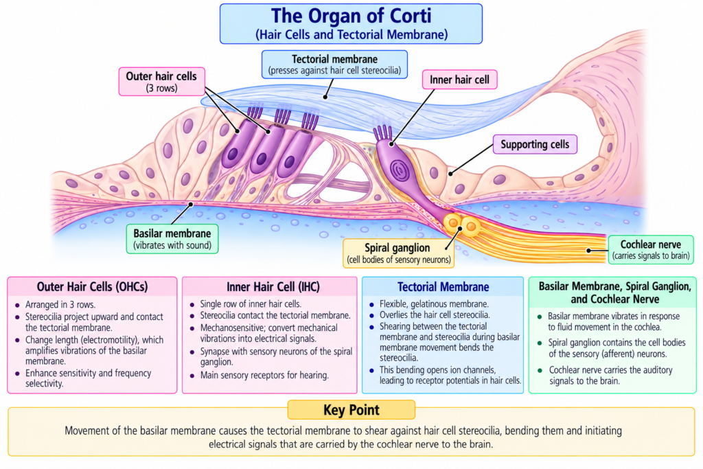
The organ of Corti is the receptor organ for hearing.
It lies on the basilar fibers and basilar membrane and generates nerve impulses in response to vibration of the basilar membrane.
Its sensory receptors are specialized hair cells.
Sound-induced movement of the basilar membrane causes movement of these hair cells and their stereocilia.
This mechanical movement is converted into electrical changes within the hair cells, which then stimulate cochlear nerve endings.
The special ionic composition and electrical potential of the scala media also help make the hair cells highly sensitive to very small sound vibrations.
MAIN TOPIC / CORE CONCEPT
Organ of Corti: The receptor organ that generates nerve impulses in response to vibration of the basilar membrane.
The organ of Corti is shown in Figs. 53.2 and 53.6.
Inner Hair Cells
Inner hair cells are arranged in:
- A single row.
Their approximate number is:
3500
Their diameter is about:
12 micrometers
Outer Hair Cells
Outer hair cells are arranged in:
- Three rows.
- Sometimes four rows.
Their approximate number is:
12,000
Their diameter is about:
8 micrometers
Comparison of Inner and Outer Hair Cells
| Feature | Inner Hair Cells | Outer Hair Cells |
|---|---|---|
| Arrangement | One row | Three to four rows |
| Approximate number | 3500 | 12,000 |
| Approximate diameter | 12 μm | 8 μm |
| Cochlear nerve endings | Receive about 90–95% | Receive a much smaller proportion |
| Main significance described | Especially important for sound detection | Proposed role in tuning and controlling sensitivity |
Cochlear Nerve Endings
- The bases and sides of the hair cells synapse with a network of cochlear nerve endings.
- Approximately 90–95% of these endings terminate on the inner hair cells.
This emphasizes the special importance of inner hair cells in detecting sound.
Spiral Ganglion and Cochlear Nerve
Hair-cell stimulation activates nerve fibers that pass to the:
Spiral ganglion of Corti
Spiral ganglion of Corti: A collection of auditory neuronal cell bodies located in the modiolus of the cochlea.
Modiolus: The central bony portion of the cochlea.
The neurons of the spiral ganglion send approximately:
30,000 axons
into the cochlear nerve.
Pathway
Hair cells
→ cochlear nerve endings
→ spiral ganglion of Corti
→ approximately 30,000 axons
→ cochlear nerve
→ central nervous system at the upper medulla
Excitation of the Hair Cells
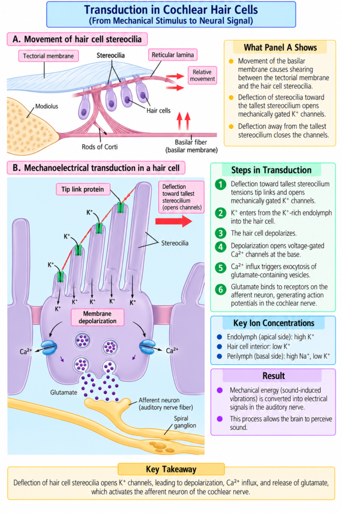
Hair-cell excitation is demonstrated in Fig. 53.7, including the mechanical movement of the organ of Corti and the ionic mechanism of hair-cell excitation.
Stereocilia
Stereocilia: Small, stiff hair-like projections extending upward from the hair cells.
- They project from the apical ends of hair cells.
- They either:
- Touch the tectorial membrane.
- Or are embedded in its surface gel coating.
Tectorial Membrane
Tectorial membrane: The membrane lying above the stereocilia in the scala media against which stereocilia move during vibration of the organ of Corti.
When stereocilia bend:
- Bending in one direction → depolarization
- Bending in the opposite direction → hyperpolarization
These changes influence the auditory nerve endings that synapse with the hair cells.
Mechanical Movement of the Organ of Corti
The outer ends of the hair cells are held in a rigid structure called the:
Reticular lamina
Reticular lamina: A flat plate supporting the outer ends of the hair cells.
The reticular lamina is supported by:
Rods of Corti
Rods of Corti: Triangular supporting structures attached tightly to the basilar fibers.
Together:
Basilar fibers
- rods of Corti
- reticular lamina
move as a relatively rigid unit.
Mechanism / Pathway / Step-by-Step Explanation
Upward Movement of the Basilar Fibers
Basilar fibers move upward
→ reticular lamina rocks upward and inward toward the modiolus
→ stereocilia shear against the tectorial membrane
→ hair cells are excited
Downward Movement
Basilar membrane moves downward
→ reticular lamina rocks downward and outward
→ stereocilia bend in the opposite direction
→ opposite electrical response occurs
Overall Mechanical Mechanism
Basilar membrane vibrates
→ basilar fibers move
→ rods of Corti and reticular lamina move with them
→ stereocilia shear back and forth against the tectorial membrane
→ hair cells alternate between depolarization and hyperpolarization
→ cochlear nerve endings are stimulated
Auditory Signals Are Transmitted Mainly by Inner Hair Cells
There are approximately:
- Three to four times as many outer hair cells as inner hair cells.
However:
- About 90% of auditory nerve fibers are stimulated by the inner hair cells rather than the outer hair cells.
Therefore, inner hair cells have the major role in transmitting auditory information.
Functional Importance of Outer Hair Cells
Outer hair cells are also important.
If:
Outer hair cells are damaged
while
inner hair cells remain fully functional
→ a large amount of hearing loss can still occur.
Therefore, it has been proposed that outer hair cells help control the sensitivity of inner hair cells to different sound pitches.
This process is called:
Tuning
Tuning: Proposed adjustment of the receptor system’s sensitivity to different sound pitches through the function of outer hair cells.
Retrograde Nerve Fibers
Many nerve fibers travel backward from the brain stem toward the region of the outer hair cells.
These are:
Retrograde nerve fibers
Stimulation of these fibers can:
- Shorten outer hair cells.
- Possibly alter their stiffness.
Concept
Brain stem
→ retrograde nerve fibers
→ outer hair cells
→ change in length and possibly stiffness
→ may modify sensitivity to different sound pitches
These observations suggest a retrograde nervous mechanism for controlling auditory sensitivity through the outer hair cells.
Hair-Cell Receptor Potentials
Hair-cell receptor potential: The electrical change generated within a hair cell when its stereocilia are bent.
Each hair cell has approximately:
100 stereocilia
on its apical border.
Arrangement of Stereocilia
- Stereocilia become progressively longer on the side of the hair cell away from the modiolus.
- The shorter stereocilia are connected by thin filaments to adjacent longer stereocilia.
When stereocilia bend toward the longer stereocilia:
→ the tips of the shorter stereocilia are pulled outward
This produces:
→ mechanical transduction
Opening of Cation Channels
Bending toward the longer stereocilia causes approximately:
200–300 cation-conducting channels
to open.
This allows positively charged:
Potassium ions (K⁺)
to move rapidly from the surrounding scala media fluid into the stereocilia.
Result
Stereocilia bend toward longer stereocilia
→ cation channels open
→ K⁺ enters
→ hair-cell membrane depolarizes
Calcium Entry
Depolarization then opens:
Voltage-sensitive calcium channels
Therefore:
Hair-cell depolarization
→ Ca²⁺ channels open
→ Ca²⁺ enters the cell
→ depolarization is further increased
Repolarization
Hair-cell repolarization occurs mainly because:
→ potassium ions leave the cell
through:
Calcium-sensitive potassium channels
Flow
Depolarization
→ Ca²⁺ enters
→ Ca²⁺-sensitive K⁺ channels participate
→ K⁺ leaves
→ repolarization occurs
Direction of Basilar Fiber Movement
When the basilar fibers bend toward the:
Scala vestibuli
→ hair cells depolarize
When the fibers bend in the opposite direction:
→ hair cells hyperpolarize
Therefore:
Basilar membrane vibrates back and forth
→ alternating depolarization and hyperpolarization
→ alternating hair-cell receptor potential
Neurotransmitter Release
The alternating receptor potential stimulates the cochlear nerve endings that synapse with the bases of the hair cells.
Current evidence described in the provided text indicates that the rapidly acting neurotransmitter released during hair-cell depolarization is:
Glutamate
Flow
Hair-cell depolarization
→ glutamate release
→ cochlear nerve endings stimulated
Complete Hair-Cell Transduction Mechanism
Sound vibration
→ basilar membrane vibrates
→ reticular lamina moves
→ stereocilia shear against tectorial membrane
→ stereocilia bend toward longer stereocilia
→ 200–300 cation channels open
→ K⁺ enters from scala media
→ hair cell depolarizes
→ voltage-sensitive Ca²⁺ channels open
→ Ca²⁺ enters
→ glutamate is released
→ cochlear nerve endings are stimulated
Endocochlear Potential
Endocochlear potential: The approximately +80 mV electrical potential of the endolymph in the scala media relative to the surrounding perilymph.
Understanding this potential requires understanding the two major cochlear fluids:
- Endolymph.
- Perilymph.
Endolymph
Endolymph: The fluid filling the scala media.
It is secreted by the:
Stria vascularis
Stria vascularis: A highly vascular area located on the outer wall of the scala media that secretes endolymph and continually secretes potassium ions into the scala media.
Endolymph contains:
- High concentration of K⁺.
- Low concentration of Na⁺.
Perilymph
Perilymph: The fluid filling the scala vestibuli and scala tympani.
The scala vestibuli and scala tympani communicate directly with the:
- Subarachnoid space around the brain.
Therefore:
- Perilymph is almost identical to cerebrospinal fluid.
Its ionic composition is opposite to that of endolymph.Endolymph vs Perilymph
| Feature | Endolymph | Perilymph |
|---|---|---|
| Location | Scala media | Scala vestibuli and scala tympani |
| Potassium | High | Opposite pattern to endolymph |
| Sodium | Low | Opposite pattern to endolymph |
| Source/relationship | Secreted by stria vascularis | Almost identical to cerebrospinal fluid |
Generation of Endocochlear Potential
An electrical potential of approximately:
+80 mV
exists between endolymph and perilymph.
The scala media is:
→ positive
relative to the perilymph outside it.
Mechanism
Stria vascularis
→ continuously secretes positively charged K⁺ into scala media
→ endolymph becomes electrically positive
→ endocochlear potential ≈ +80 mV
Relationship of Hair Cells to Endolymph and Perilymph
Different parts of the hair cells are exposed to different fluids.
Upper Surface
The tops of the hair cells:
- Project through the reticular lamina.
- Are bathed by endolymph in the scala media.
Lower Surface
The lower bodies of the hair cells are:
- Bathed by perilymph.
Hair-Cell Electrical Potential
The intracellular potential of the hair cell is approximately:
−70 mV relative to perilymph
Because endolymph is approximately:
+80 mV relative to perilymph
the hair-cell interior is approximately:
−150 mV relative to the endolymph at its upper surface
Easy Calculation
Hair-cell intracellular potential:
−70 mV
Endolymph potential:
+80 mV
Difference:
−70 − (+80)
= −150 mV
Therefore, the inside of the hair cell is approximately 150 mV negative relative to the endolymph at the stereocilia.
Importance of the Endocochlear Potential
This large electrical potential exists where the stereocilia project into the endolymph.
It likely increases the sensitivity of the hair cells, helping them respond to very small sound vibrations.
Concept
Endocochlear potential
- negative hair-cell interior
→ very large electrical difference at stereocilia
→ likely increases hair-cell sensitivity
→ very weak sounds can be detected more effectively
Original Educational Figure / Diagram
Mechanical-to-Electrical Conversion of Sound
Sound vibration
→ basilar membrane movement
→ reticular lamina moves
→ stereocilia shear against tectorial membrane
→ stereocilia bend
→ K⁺ channels open
→ K⁺ enters
→ depolarization
→ Ca²⁺ channels open
→ Ca²⁺ enters
→ glutamate released
→ cochlear nerve stimulated
Endocochlear Electrical Mechanism
Stria vascularis
→ K⁺ secretion into scala media
→ endolymph becomes K⁺ rich
→ endocochlear potential ≈ +80 mV
Hair-cell interior
→ approximately −70 mV relative to perilymph
Therefore:
Hair-cell interior relative to endolymph
→ approximately −150 mV
→ likely increases sensitivity of stereocilia to sound
Key Concept
The organ of Corti converts mechanical vibration into neural activity.
Main Sequence
Basilar membrane vibration
→ stereocilia bending
→ K⁺ entry
→ depolarization
→ Ca²⁺ entry
→ glutamate release
→ cochlear nerve stimulation
Functional Roles
Inner hair cells
→ main transmission of auditory signals
Outer hair cells
→ proposed role in tuning and controlling sensitivity to different pitches
Clinical / Functional Importance
- Inner hair cells are especially important for sound detection because approximately 90–95% of cochlear nerve endings terminate on them.
- About 90% of auditory nerve fibers are stimulated mainly by inner hair cells.
- Outer hair cells are also functionally important because damage to them can cause substantial hearing loss even when inner hair cells remain functional.
- Outer hair cells are proposed to contribute to tuning and sensitivity to different sound pitches.
- The endocochlear potential likely increases the sensitivity of hair cells to very small sound vibrations.
High-Yield / Exam Points
- Organ of Corti lies on:
- Basilar fibers.
- Basilar membrane.
- Inner hair cells:
- One row.
- About 3500.
- About 12 μm diameter.
- Outer hair cells:
- Three to four rows.
- About 12,000.
- About 8 μm diameter.
- About 90–95% of cochlear nerve endings terminate on inner hair cells.
- About 90% of auditory nerve fibers are stimulated mainly by inner hair cells.
- Spiral ganglion:
- Located in the modiolus.
- Spiral ganglion neurons contribute approximately:
- 30,000 axons to the cochlear nerve.
- Each hair cell has about:
- 100 stereocilia.
- Bending toward longer stereocilia:
- Opens about 200–300 cation channels.
- Initial depolarizing ion:
- K⁺
- K⁺ enters from:
- Endolymph of scala media.
- Depolarization opens:
- Voltage-sensitive Ca²⁺ channels.
- Repolarization occurs mainly through:
- K⁺ exit through Ca²⁺-sensitive K⁺ channels.
- Neurotransmitter indicated by current evidence:
- Glutamate
- Endolymph:
- High K⁺.
- Low Na⁺.
- Perilymph:
- Scala vestibuli and scala tympani.
- Almost identical to CSF.
- Endocochlear potential:
- About +80 mV.
- Hair-cell intracellular potential:
- About −70 mV relative to perilymph.
- Hair-cell interior relative to endolymph:
- About −150 mV.
- Stria vascularis:
- Maintains endolymph and endocochlear potential through K⁺ secretion.
Common Student Mistakes
- Do not think outer hair cells transmit most auditory nerve activity.
- Most auditory nerve endings and fibers are associated functionally with inner hair cells.
- Do not think outer hair cells are unimportant.
- Their damage can still cause major hearing loss.
- Do not state that the tuning role of outer hair cells is completely proven from this text.
- The text describes it as a proposed and suggested mechanism.
- Do not confuse endolymph with perilymph.
- Endolymph → scala media.
- Perilymph → scala vestibuli and scala tympani.
- Do not think Na⁺ is the main ion entering during hair-cell depolarization in this mechanism.
- The provided text describes K⁺ entering from endolymph.
- Do not think K⁺ leaves during the initial depolarizing event.
- K⁺ initially enters the stereocilia.
- Do not confuse the direction of movement.
- Basilar fibers toward scala vestibuli → depolarization.
- Opposite direction → hyperpolarization.
- Do not confuse the +80 mV and −150 mV values.
- +80 mV → endocochlear potential.
- −150 mV → hair-cell interior relative to endolymph at the upper surface.
Quick Revision
Organ of Corti
Basilar membrane
→ organ of Corti
→ hair cells
→ cochlear nerve
Inner Hair Cells
One row
→ about 3500
→ 90–95% of cochlear nerve endings
→ major auditory signal transmission
Outer Hair Cells
3–4 rows
→ about 12,000
→ proposed tuning and sensitivity control
Hair-Cell Excitation
Basilar membrane vibrates
→ reticular lamina moves
→ stereocilia bend
→ K⁺ channels open
→ K⁺ enters
→ depolarization
→ Ca²⁺ enters
→ glutamate released
→ cochlear nerve stimulated
Opposite Movement
Stereocilia bend opposite way
→ hyperpolarization
Endolymph
Scala media
→ high K⁺
→ low Na⁺
Perilymph
Scala vestibuli + scala tympani
→ almost identical to CSF
Endocochlear Potential
Stria vascularis
→ K⁺ secretion
→ scala media ≈ +80 mV
Hair-cell interior
→ −70 mV relative to perilymph
→ −150 mV relative to endolymph
→ likely increases hair-cell sensitivity
Final Take-Home Concept / Summary
The organ of Corti is the receptor organ that converts basilar membrane vibration into auditory nerve activity.
Its sensory receptors are:
- Inner hair cells.
- Outer hair cells.
Although outer hair cells are more numerous, most cochlear nerve endings terminate on inner hair cells.
Therefore:
Inner hair cells are especially important for transmitting auditory signals.
Outer hair cells remain essential for normal hearing and are proposed to help control receptor sensitivity and tuning for different sound pitches.
The basic transduction mechanism is:
Basilar membrane vibration
→ reticular lamina movement
→ stereocilia bending
→ cation channels open
→ K⁺ enters from endolymph
→ hair cell depolarizes
→ Ca²⁺ enters
→ glutamate is released
→ cochlear nerve ending is stimulated
Movement in the opposite direction causes:
→ hyperpolarization
The scala media contains endolymph, which has:
- High K⁺.
- Low Na⁺.
The scala vestibuli and scala tympani contain perilymph, which is almost identical to cerebrospinal fluid.
The stria vascularis continually secretes K⁺ into the scala media and produces an:
Endocochlear potential of approximately +80 mV
The hair-cell interior is approximately:
−70 mV relative to perilymph
and therefore approximately:
−150 mV relative to the endolymph at the stereocilia
This large electrical difference likely increases the sensitivity of hair cells to very slight sounds.
Final Take-Home Concept
Basilar membrane vibration → stereocilia bend → K⁺ enters → depolarization → Ca²⁺ enters → glutamate release → cochlear nerve stimulation.
Inner hair cells = main auditory signal transmission.
Outer hair cells = proposed tuning and sensitivity control.
Endocochlear potential = approximately +80 mV and helps make hair cells highly sensitive to sound.
References / Sources
- Guyton and Hall Textbook of Medical Physiology, 15th Edition, Chapter 53.
