


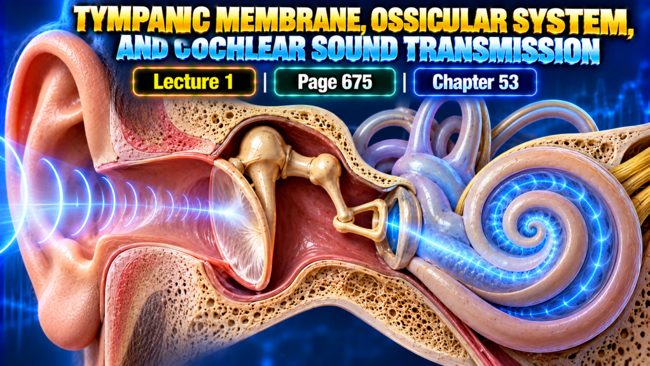
Learning Objectives
After completing this lecture, students will be able to:
- Explain how sound travels from the tympanic membrane to the cochlea.
- Describe impedance matching by the ossicular system.
- Explain attenuation of loud sounds and bone conduction.
- Describe the functional anatomy of the cochlea and basilar membrane.
- Explain traveling waves and frequency discrimination in the cochlea.
Introduction
Hearing requires efficient transfer of sound energy from air in the external and middle ear into the fluid of the cochlea.
The tympanic membrane first responds to incoming sound vibrations. These vibrations are then transmitted through the middle-ear ossicles:
- Malleus
- Incus
- Stapes
The ossicular system increases the force applied to cochlear fluid and therefore helps overcome the much greater inertia of fluid compared with air.
Inside the cochlea, sound produces fluid movement and generates a traveling wave along the basilar membrane.
Different sound frequencies produce their greatest vibration at different locations on the basilar membrane, allowing the auditory system to distinguish one frequency from another.
Main Topic / Core Concept
Conduction of Sound From the Tympanic Membrane to the Cochlea
The tympanic membrane and ossicular system are shown in Fig. 53.1.
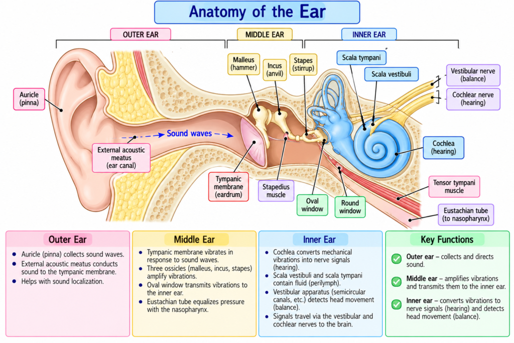
Tympanic membrane: The eardrum that receives sound vibrations and transfers them to the middle-ear ossicles.
Ossicles: The malleus, incus, and stapes that conduct sound vibrations through the middle ear toward the cochlea.
Basic Sound Pathway
Sound waves
→ tympanic membrane
→ malleus
→ incus
→ stapes
→ oval window
→ cochlear fluid
Malleus
- The handle of the malleus is attached to the tympanic membrane.
- The tip of the handle is attached near the center of the membrane.
- When the tympanic membrane moves:
- The malleus moves with it.
Incus
- The malleus is attached to the incus by small ligaments.
- Therefore:
Malleus moves
→ incus moves with it
- The opposite end of the incus articulates with the stapes.
Stapes
- The stapes is connected to the incus.
- Its faceplate lies at the oval window.
- The oval window communicates mechanically with the cochlear fluid.
Flow
Tympanic membrane vibrates
→ malleus moves
→ incus moves
→ stapes moves
→ oval window moves
→ cochlear fluid moves
Role of the Tensor Tympani Muscle
Tensor tympani muscle: A muscle that continuously pulls on the handle of the malleus and helps keep the tympanic membrane tense.
- The center of the tympanic membrane is constantly pulled inward by the tensor tympani muscle.
- This maintains tension in the membrane.
Importance
Tympanic membrane remains tense
→ vibration occurring at any part of membrane can be transmitted effectively
→ ossicles receive the vibration
If the tympanic membrane were lax:
→ sound transmission to the ossicles would be less effective.
Ossicles Act as a Lever System
The middle-ear ossicles are suspended by ligaments.
The:
- Malleus
- Incus
act together as a single lever.
Their approximate fulcrum lies near the border of the tympanic membrane.
Movement of the Stapes
When the tympanic membrane moves inward:
Tympanic membrane inward
→ malleus moves
→ incus moves
→ stapes pushes inward on oval window
→ cochlear fluid moves forward
When the malleus moves outward:
→ stapes moves backward
→ cochlear fluid is pulled backward
Key Concept
Tympanic membrane movement
→ ossicular lever movement
→ stapes movement at oval window
→ alternating movement of cochlear fluid
Impedance Matching by the Ossicular System
Impedance matching: The mechanism by which the tympanic membrane and ossicles increase the force of sound transmission so that vibrations in air can efficiently produce vibrations in cochlear fluid.
Fluid has much greater inertia than air.
Therefore:
Sound traveling in air
→ must generate greater force
→ to produce effective vibration of cochlear fluid
The tympanic membrane and ossicles provide this increased force.
Ossicular Lever Effect
The movement amplitude of the stapes faceplate is only about:
3/4 of the movement amplitude of the malleus handle
Therefore, the ossicular system does not increase the movement distance of the stapes.
Instead:
Movement distance decreases
→ force increases approximately 1.3 times
Key Concept
Ossicular lever:
Smaller movement distance
→ greater movement force
Surface Area Effect
Surface area of tympanic membrane:
≈ 55 mm²
Surface area of stapes:
≈ 3.2 mm²
Area Ratio
55 ÷ 3.2
≈ 17
Therefore:
The effective tympanic membrane area is approximately 17 times the stapes area.
Total Force Increase
Area effect:
17-fold
Lever effect:
1.3-fold
Therefore:
17 × 1.3
≈ 22
Result
Force exerted on cochlear fluid
≈ 22 times the force produced by the sound wave on the tympanic membrane
Conceptual Flow
Large tympanic membrane area
- ossicular lever effect
→ force concentrated onto small stapes footplate
→ approximately 22-fold force advantage
→ better movement of cochlear fluid
Why Impedance Matching Is Necessary
Air
→ relatively low inertia
Cochlear fluid
→ much greater inertia
Therefore:
Airborne sound alone
→ insufficiently efficient fluid vibration
Tympanic membrane + ossicular system
→ increase force
→ improve transfer of sound energy from air to fluid
Efficiency of Impedance Matching
The impedance-matching system is approximately:
50%–75% of perfect
for sound frequencies between:
300 and 3000 cycles/sec
This allows most of the incoming sound-wave energy to be used.
Sound Transmission Without the Ossicular System
Even without the tympanic membrane and ossicles:
- Sound can travel through the air in the middle ear.
- It can reach the cochlea through the oval window.
However:
Hearing sensitivity becomes approximately:
15–20 decibels lower
than with normal ossicular transmission.
The text compares this reduction approximately to:
Medium voice
→ barely perceptible voice
Key Concept
Direct air transmission can still produce hearing
but
normal ossicular transmission greatly improves hearing sensitivity.
Attenuation of Sound by Tensor Tympani and Stapedius Muscles
Attenuation reflex: A reflex contraction of middle-ear muscles that increases ossicular rigidity and reduces transmission of mainly low-frequency sound.
When loud sound reaches the auditory system:
Loud sound
→ central nervous system
→ reflex response after 40–80 milliseconds
→ stapedius contracts
- tensor tympani contracts to a lesser degree
Opposing Muscle Actions
Tensor tympani:
→ pulls malleus inward
Stapedius:
→ pulls stapes outward
These forces oppose each other.
Result
Opposing forces
→ ossicular chain becomes more rigid
→ sound conduction decreases
The reduction mainly affects frequencies:
Below 1000 cycles/sec
Amount of Attenuation
The attenuation reflex can reduce low-frequency sound transmission by approximately:
30–40 decibels
The text compares this difference to approximately:
Loud voice
versus
whisper
Functions of the Attenuation Reflex
The mechanism has two major functions.
Protection of the Cochlea
Very loud sound
→ attenuation reflex
→ ossicular transmission reduced
→ damaging cochlear vibrations reduced
Reduction of Background Noise
Low-frequency background sound
→ attenuated
Higher-frequency sound above approximately 1000 cycles/sec
→ becomes relatively easier to concentrate on
The text notes that much of the important information in voice communication is transmitted above this frequency.
Reduction of Hearing One’s Own Voice
Another function of the tensor tympani and stapedius muscles is:
→ hearing sensitivity to one’s own voice decreases
This occurs because:
Brain activates voice mechanism
→ collateral nerve signals simultaneously reach middle-ear muscles
→ hearing sensitivity to one’s own voice decreases transmission of Sound Through Bone
Bone conduction: Transmission of sound to the cochlea through vibration of the bones of the skull.
The cochlea is embedded within a bony cavity of the temporal bone called the:
Bony labyrinth
Therefore:
Skull vibration
→ bony labyrinth vibration
→ cochlear fluid vibration
→ sound can be heard
Demonstration of Bone Conduction
A:
- Tuning fork
- Electronic vibrator
placed on a bony prominence can generate hearing.
An especially effective site is:
- The mastoid process near the ear.
Flow
Vibrator on skull
→ skull vibrates
→ cochlear fluid vibrates
→ auditory sensation
Limitation of Normal Airborne Sound
Even loud airborne sound normally does not provide enough skull vibration to produce significant hearing through bone conduction.
For effective bone-conduction hearing:
→ a special electromechanical sound-amplifying device must be applied to the bone.
Cochlea
Functional Anatomy of the Cochlea
Cochlea: A system of coiled tubes in the inner ear involved in converting sound vibrations into neural signals.
Its cross-sectional organization is shown in Fig. 53.2.
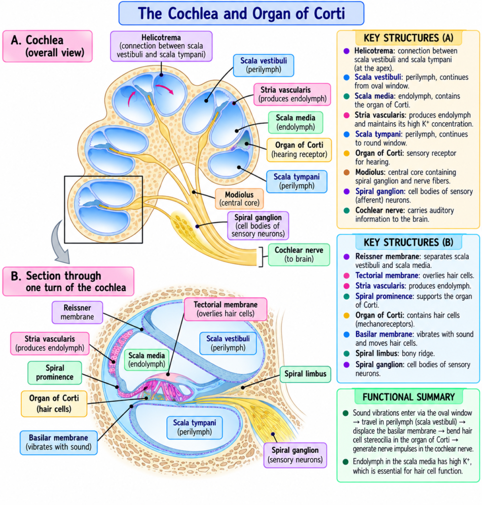
The cochlea contains three tubes arranged side by side:
- Scala vestibuli.
- Scala media.
- Scala tympani.
Scala Vestibuli
The scala vestibuli lies adjacent to the scala media.
It is separated from the scala media by:
Reissner membrane
also called:
Vestibular membrane
Scala Media
The scala media lies between:
- Scala vestibuli.
- Scala tympani.
Scala Tympani
The scala tympani is separated from the scala media by:
Basilar membrane
Major Cochlear Membranes
| Structure | Separates |
|---|---|
| Reissner membrane | Scala vestibuli from scala media |
| Basilar membrane | Scala media from scala tympani |
Organ of Corti
The organ of Corti lies on the surface of the basilar membrane.
Organ of Corti: The sound-receptive structure containing electromechanically sensitive hair cells.
The hair cells are:
- Receptive end organs for sound.
- Activated by sound vibrations.
- Responsible for generating nerve impulses in response to those vibrations.
Concept
Sound vibration
→ basilar membrane movement
→ organ of Corti hair cells stimulated
→ nerve impulses generated
Functional Relationship of Scala Vestibuli and Scala Media
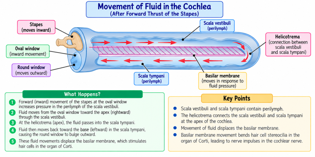
The functional pathway of cochlear vibration is illustrated in Fig. 53.3.
Reissner membrane is extremely:
- Thin.
- Easily moved.
Therefore:
- It does not significantly block sound vibrations passing from the scala vestibuli into the scala media.
For sound-fluid conduction:
Scala vestibuli + scala media
→ can function essentially as one chamber
However, the Reissner membrane remains important because it maintains the special fluid composition within the scala media needed for normal hair-cell function.
Sound Entry Into the Cochlea
Sound vibrations enter the cochlea through:
Oval window
The stapes faceplate covers this opening.
It is attached to the edges of the oval window by:
- A loose annular ligament.
This allows the stapes to move:
- Inward.
- Outward.
Inward Stapes Movement
Stapes moves inward
→ cochlear fluid moves forward through scala vestibuli and scala media
Outward Stapes Movement
Stapes moves outward
→ cochlear fluid moves backward
Basilar Membrane and Resonance in the Cochlea
Basilar membrane: Fibrous membrane separating the scala media from the scala tympani.
It contains approximately:
20,000–30,000 basilar fibers
These fibers extend from:
- Modiolus
toward - Outer wall of the cochlea.
Modiolus
Modiolus: The central bony structure of the cochlea from which the basilar fibers project.
Characteristics of Basilar Fibers
The fibers are:
- Stiff.
- Elastic.
- Reedlike.
They are fixed at:
- Their basal ends in the modiolus.
Their distal ends are relatively free except for being embedded in the loose basilar membrane.
Therefore:
→ they can vibrate somewhat like the reeds of a harmonica.
Changes in Basilar Fiber Length
Moving from the base toward the apex:
Basilar fibers become progressively longer.
Near the oval and round windows:
≈ 0.04 mm
Near the helicotrema:
≈ 0.5 mm
Length Increase
0.5 ÷ 0.04
= 12.5
This is approximately the stated:
12-fold increase
Changes in Basilar Fiber Stiffness
Moving from:
Oval window
→ helicotrema
fiber diameter decreases.
Therefore:
Overall stiffness decreases by more than:
100-fold
Relationship Between Fiber Properties and Frequency
Base of Cochlea
Fibers are:
- Short.
- Stiff.
Therefore:
→ resonate best with high-frequency sounds
Apex of Cochlea
Fibers are:
- Longer.
- More flexible.
Therefore:
→ resonate best with low-frequency sounds
Comparison of Cochlear Base and Apex
| Feature | Base of Cochlea | Apex / Near Helicotrema |
|---|---|---|
| Basilar fibers | Short | Long |
| Fiber stiffness | High | Low |
| Best resonance | High frequency | Low frequency |
| Relation to oval window | Near | Far |
Key Concept
Cochlear base
→ short + stiff fibers
→ high-frequency resonance
Cochlear apex
→ long + flexible fibers
→ low-frequency resonance
Low-Frequency Resonance Near the Helicotrema
Low-frequency resonance near the cochlear tip occurs mainly because:
- Fibers are less stiff.
It is also helped by:
- Greater fluid mass that must vibrate along the cochlear tubes.
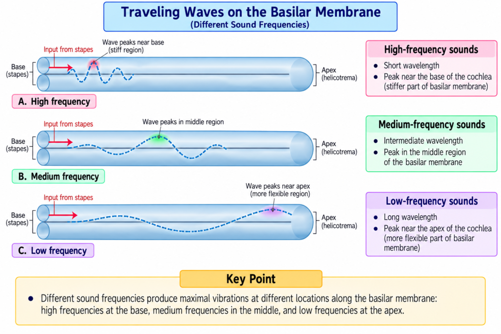
Transmission of Sound Waves in the Cochlea — Traveling Wave
Traveling wave: A fluid-induced wave that begins near the cochlear base and travels along the basilar membrane toward the helicotrema.
The traveling-wave patterns for different frequencies are shown in Fig. 53.4.
Beginning of the Traveling Wave
When the stapes moves inward:
Stapes pushes oval window inward
→ cochlear fluid is displaced
Because the cochlea is surrounded by rigid bony walls:
→ the round window must bulge outward
Initial Basilar Membrane Movement
Sound enters through oval window
→ basilar membrane near cochlear base bends toward round window
This bending produces:
→ elastic tension in basilar fibers
The elastic tension then initiates:
→ a wave traveling along the basilar membrane
→ toward the helicotrema traveling Wave for Different Frequencies
High-Frequency Sound
High-frequency wave
→ travels only a short distance
→ reaches resonant region near base
→ vibration becomes maximal
→ energy dissipates
→ wave dies
Medium-Frequency Sound
Medium-frequency wave
→ travels farther
→ reaches resonant region around intermediate cochlear area
→ becomes maximal
→ dies
Very Low-Frequency Sound
Very low-frequency wave
→ travels almost the entire basilar membrane
→ reaches region near helicotrema
→ maximum vibration occurs there
Mechanism / Pathway / Step-by-Step Explanation
Traveling Wave
Stapes moves inward
→ oval window moves inward
→ cochlear fluid displaced
→ round window bulges outward
→ basal basilar membrane bends
→ elastic tension develops
→ wave travels toward helicotrema
→ wave reaches region matching its frequency
→ maximum vibration occurs
→ wave energy is dissipated
Resonant Point of a Sound Wave
Each sound wave begins relatively weak.
As it travels:
Wave reaches a basilar membrane region whose natural frequency matches sound frequency
→ membrane vibrates very easily
→ vibration amplitude increases greatly
→ wave energy is dissipated
→ wave stops traveling farther
This location is the sound’s:
resonant region
Frequency-Dependent Traveling Distance
High frequency
→ short travel distance
Medium frequency
→ intermediate travel distance
Very low frequency
→ long travel distance
Key Concept
Frequency determines where the traveling wave reaches maximum vibration.
Speed of the Traveling Wave
The traveling wave:
- Moves rapidly in the initial portion of the basilar membrane.
- Becomes progressively slower as it moves toward the cochlear apex.
Why?
Near oval window:
→ basilar fibers have high elasticity
Farther toward apex:
→ elasticity progressively decreases
Importance of Rapid Initial Transmission
Rapid initial travel allows high-frequency sounds to:
- Move far enough into the cochlea.
- Spread apart from one another.
- Produce separate peak locations.
Without this rapid initial transmission:
High-frequency waves
→ would cluster within first millimeter of basilar membrane
→ individual frequencies could not be adequately distinguished
_ vibration Amplitude Pattern of the Basilar Membrane
The vibration pattern of a single sound frequency during different phases of the stapes cycle is demonstrated in Fig. 53.5.
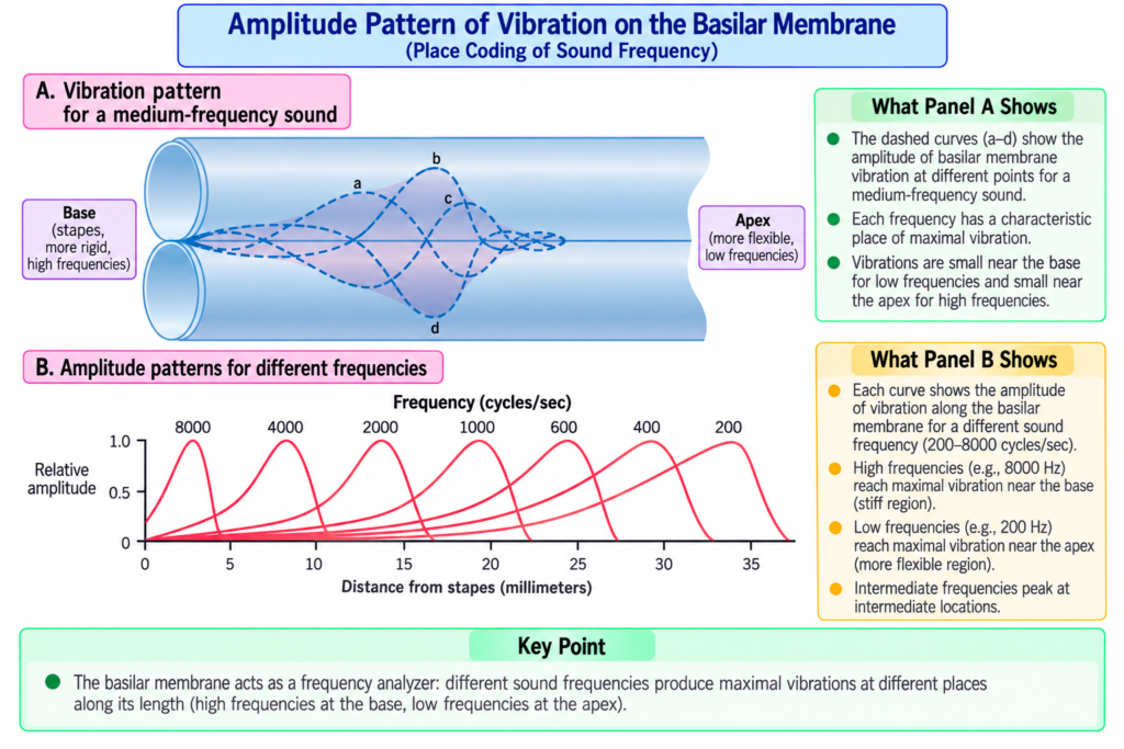
During one complete vibration:
- Stapes moves inward.
- Returns to neutral.
- Moves outward.
- Returns toward neutral while moving inward again.
The total region through which the basilar membrane moves during the cycle represents the:
Amplitude pattern of vibration
Location of Maximum Amplitude
For different frequencies, maximum vibration occurs at different points.
8000 cycles/sec
Maximum amplitude occurs:
→ near the base of the cochlea
Less Than 200 cycles/sec
Maximum amplitude occurs:
→ near the tip of the basilar membrane
→ close to the helicotrema
Helicotrema
Helicotrema: The small opening near the cochlear apex where the scala vestibuli and scala tympani communicate.
Frequency Discrimination
The major method for distinguishing sound frequencies is based on:
The location of maximum stimulation along the basilar membrane
Flow
Different sound frequency
→ different resonant location on basilar membrane
→ different organ of Corti nerve fibers stimulated maximally
→ sound frequency can be distinguished
Key Concept
High pitch
→ maximal stimulation near cochlear base
Low pitch
→ maximal stimulation near cochlear apex
Therefore:
Sound frequency is coded mainly by place of maximum basilar membrane vibration.
Original Educational Figure / Diagram
Sound Transmission From Air to Cochlea
Sound in air
→ tympanic membrane vibrates
→ malleus
→ incus
→ stapes
→ oval window
→ cochlear fluid movement
→ basilar membrane traveling wave
→ organ of Corti hair-cell stimulation
→ nerve impulses
Impedance Matching
Large tympanic membrane area
→ ossicular lever
→ small stapes footplate
→ approximately 22-fold force increase
→ efficient transfer of sound from air to cochlear fluid
Frequency Mapping
High frequency
Oval window
→ short, stiff basilar fibers near base
→ maximum vibration
Low frequency
Wave travels farther
→ long, flexible fibers near apex
→ maximum vibration
Key Concept
The auditory mechanical system performs two essential tasks.
Middle Ear
Airborne sound
→ tympanic membrane
→ ossicular system
→ force increased
→ cochlear fluid efficiently vibrated
Cochlea
Fluid wave
→ travels along basilar membrane
→ frequency-specific region resonates
→ organ of Corti stimulated
Therefore:
Middle ear improves energy transfer, while the basilar membrane separates sounds according to frequency.
Clinical / Functional Importance
- The ossicular system improves hearing sensitivity by efficiently transferring sound energy from air to cochlear fluid.
- Without normal tympanic membrane and ossicular transmission, hearing sensitivity decreases by approximately 15–20 dB.
- The attenuation reflex reduces transmission of intense low-frequency sounds by approximately 30–40 dB.
- This reflex helps:
- Protect the cochlea from excessively strong vibrations.
- Reduce low-frequency background noise.
- Reduce sensitivity to one’s own voice.
- Bone vibration can stimulate the cochlea directly because the cochlea is enclosed within the temporal bone.
- Different basilar membrane locations respond maximally to different frequencies, allowing sound-frequency discrimination.
High-Yield / Exam Points
- Ossicles:
- Malleus.
- Incus.
- Stapes.
- Stapes transmits vibrations through the oval window.
- Tensor tympani keeps the tympanic membrane tense.
- Malleus and incus act together as a lever.
- Stapes movement amplitude:
- About 3/4 that of the malleus handle.
- Lever force increase:
- About 1.3-fold.
- Tympanic membrane area:
- About 55 mm².
- Stapes area:
- About 3.2 mm².
- Area ratio:
- About 17-fold.
- Total force increase:
- About 22-fold.
- Best impedance matching:
- Approximately 300–3000 cycles/sec.
- Efficiency:
- About 50%–75% of perfect.
- Loss of ossicular transmission:
- Hearing decreases approximately 15–20 dB.
- Attenuation reflex latency:
- 40–80 ms.
- Mainly attenuates:
- Frequencies below 1000 cycles/sec.
- Attenuation:
- About 30–40 dB.
- Cochlear chambers:
- Scala vestibuli.
- Scala media.
- Scala tympani.
- Reissner membrane:
- Between scala vestibuli and scala media.
- Basilar membrane:
- Between scala media and scala tympani.
- Organ of Corti:
- Lies on basilar membrane.
- Basilar fibers:
- About 20,000–30,000.
- Fiber length:
- About 0.04 mm at base.
- About 0.5 mm near apex.
- Fiber stiffness decreases:
- More than 100-fold toward apex.
- Cochlear base:
- High-frequency sounds.
- Cochlear apex:
- Low-frequency sounds.
- 8000 cycles/sec:
- Maximum amplitude near base.
- <200 cycles/sec:
- Maximum amplitude near helicotrema.
- Frequency discrimination depends mainly on:
- Place of maximum basilar membrane stimulation.
Common Student Mistakes
- Do not think the ossicular lever increases the movement distance of the stapes.
- It actually reduces movement distance while increasing force.
- Do not confuse force amplification with amplitude amplification.
- Ossicles increase force, not stapes movement amplitude.
- Do not think all frequencies vibrate maximally at the same part of the basilar membrane.
- High frequencies → base.
- Low frequencies → apex.
- Do not confuse Reissner membrane with basilar membrane.
- Reissner membrane → scala vestibuli vs scala media.
- Basilar membrane → scala media vs scala tympani.
- Do not think the attenuation reflex equally reduces all sound frequencies.
- Its major effect is on low frequencies, particularly below 1000 cycles/sec.
Quick Revision
Sound conduction
Sound
→ tympanic membrane
→ malleus
→ incus
→ stapes
→ oval window
→ cochlear fluid
Impedance matching
Tympanic membrane area ≈ 55 mm²
→ stapes area ≈ 3.2 mm²
→ approximately 17-fold area effect
Lever effect ≈ 1.3-fold
17 × 1.3
→ approximately 22-fold force increase
Attenuation reflex
Loud sound
→ stapedius + tensor tympani contraction
→ ossicular rigidity increases
→ low-frequency transmission decreases
Bone conduction
Skull vibration
→ cochlear fluid vibration
→ hearing
Cochlear chambers
Scala vestibuli
→ Reissner membrane
→ scala media
→ basilar membrane
→ scala tympani
Basilar membrane
Base
→ short + stiff
→ high frequency
Apex
→ long + flexible
→ low frequency
Traveling wave
Oval window movement
→ fluid wave
→ basilar membrane traveling wave
→ resonant point reached
→ maximum vibration
→ wave dies
Frequency discrimination
Different frequency
→ different place of maximum basilar membrane vibration
→ different nerve fibers maximally stimulated
Final Take-Home Concept / Summary
Sound first reaches the tympanic membrane and is mechanically transferred through:
Malleus
→ incus
→ stapes
→ oval window
The middle-ear system does not mainly increase movement amplitude.
Instead:
Large tympanic membrane
- ossicular lever
- small stapes footplate
→ approximately 22-fold increase in force
→ efficient transfer from air to cochlear fluid
This is called:
Impedance matching
Loud sounds activate the attenuation reflex:
Stapedius + tensor tympani contraction
→ ossicular chain becomes more rigid
→ low-frequency sound transmission decreases
The cochlea contains:
Scala vestibuli
- scala media
- scala tympani
The organ of Corti lies on the basilar membrane and contains the sound-sensitive hair cells.
When the stapes moves:
Oval window movement
→ cochlear fluid movement
→ basilar membrane bends
→ traveling wave develops
Different frequencies travel different distances before reaching their resonant region.
High-frequency sound
→ short, stiff fibers near cochlear base
Low-frequency sound
→ long, flexible fibers near cochlear apex
At the resonant region:
Wave amplitude becomes maximal
→ energy is dissipated
→ wave dies
Therefore, the cochlea distinguishes sound frequency mainly according to:
Where maximum vibration occurs on the basilar membrane.
Final Take-Home Concept
Tympanic membrane receives sound.
Ossicles increase force and match air vibrations to cochlear fluid.
Stapes moves the oval window.
Cochlear fluid creates a traveling wave.
Basilar membrane separates frequencies by place.
Base = high frequency.
Apex = low frequency.
- Guyton and Hall Textbook of Medical Physiology, 15th Edition, Chapter 53.
