


Learning Objectives
After studying this topic, students will be able to:
- Explain how sound frequency and loudness are determined.
- Trace the major auditory pathway from the cochlea to the auditory cortex.
- Explain cortical processing and localization of sound.
- Differentiate nerve deafness from conduction deafness.
Introduction
The auditory system must determine several features of sound, including:
- Frequency or pitch.
- Loudness.
- Pattern of sound.
- Direction from which sound comes.
Different parts of the auditory system participate in these functions.
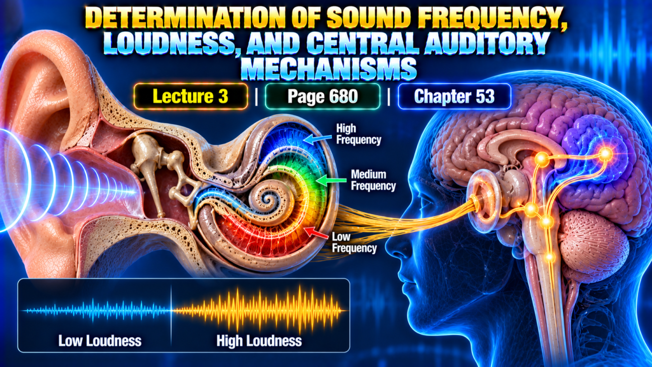
The cochlea begins the analysis by separating different sound frequencies along the basilar membrane. Auditory information then travels through organized pathways in the brain stem and thalamus before reaching the auditory cortex.
The auditory cortex further analyzes:
- Sound frequency.
- Sound patterns.
- Direction.
- Meaning of sounds.
Main Topic / Core Concept
Determination of Sound Frequency — The “Place” Principle
Different sound frequencies produce maximum stimulation at different locations along the basilar membrane.
Low-Frequency Sounds
Low-frequency sounds
→ maximum basilar membrane activation near the apex of the cochlea
High-Frequency Sounds
High-frequency sounds
→ maximum activation near the base of the cochlea
Intermediate-Frequency Sounds
Intermediate frequencies
→ maximum activation at intermediate positions
Place Principle
Place principle: The nervous system identifies sound frequency mainly by determining which location along the basilar membrane is stimulated most strongly.
Flow
Sound of specific frequency
→ maximum vibration at specific basilar membrane location
→ corresponding cochlear nerve fibers activated
→ specific neurons in auditory pathway activated
→ sound frequency recognized
This spatial organization continues:
Cochlea
→ brain stem auditory pathways
→ auditory cortex
Specific brain neurons therefore respond preferentially to specific sound frequencies.
Low-Frequency Sounds and the Volley Principle
The distal basilar membrane near the helicotrema is stimulated by all frequencies below approximately:
200 cycles/sec
Therefore, the place principle alone has difficulty explaining discrimination of very low frequencies between approximately:
20 and 200 cycles/sec
Another mechanism has been proposed:
Volley or frequency principle
Volley Principle
Volley principle: Low-frequency sounds produce groups of auditory nerve impulses that are synchronized with the frequency of the sound.
The text describes this mechanism for frequencies from approximately:
20 to 1500–2000 cycles/sec
Flow
Low-frequency sound
→ synchronized volleys of nerve impulses
→ cochlear nerve
→ cochlear nuclei
→ frequency of impulse volleys analyzed
The cochlear nuclei are suggested to distinguish different volley frequencies.
Support for this idea comes from the observation that destruction of the apical half of the cochlea does not completely eliminate discrimination of lower-frequency sounds.
Place Principle vs Volley Principle
| Feature | Place Principle | Volley / Frequency Principle |
|---|---|---|
| Main basis | Location of maximum basilar membrane stimulation | Frequency of synchronized nerve volleys |
| Especially important for | General frequency discrimination | Very low-frequency discrimination |
| Neural code | Place | Timing/frequency of impulses |
| Low-frequency range described | Limited below 200 cycles/sec | Approximately 20–1500/2000 cycles/sec |
Determination of Loudness
The auditory system determines loudness in at least three ways.
Increased Rate of Nerve Firing
As sound becomes louder:
Sound intensity increases
→ basilar membrane vibration increases
→ hair-cell vibration increases
→ auditory nerve endings fire more rapidlySpatial Summation
As vibration amplitude becomes greater:
- More hair cells around the edges of the resonating region become stimulated.
- More nerve fibers participate.
This is called:
Spatial summation
Flow
Greater vibration amplitude
→ larger area of hair cells activated
→ more nerve fibers stimulated
→ stronger sensation of loudness
Stimulation of Outer Hair Cells
Outer hair cells are not significantly stimulated until basilar membrane vibration becomes relatively intense.
Therefore:
High-intensity vibration
→ outer hair cells stimulated
→ nervous system receives an additional indication that the sound is loud
Three Mechanisms of Loudness Detection
| Mechanism | Effect of Louder Sound |
|---|---|
| Increased firing rate | Hair cells stimulate nerve endings more rapidly |
| Spatial summation | More hair cells and nerve fibers become active |
| Outer hair-cell activation | Indicates high-intensity sound |
Detection of Changes in Loudness — The Power Law
The perceived change in sound intensity does not increase directly in proportion to the actual physical sound intensity.
For sound:
Perceived sensation
→ changes approximately according to the cube root of actual sound intensity
This greatly compresses the enormous physical range of sound intensity into a range that the nervous system can interpret.
Large Range of Hearing
The ear can discriminate sounds ranging from:
- The softest whisper.
- To the loudest possible noise.
This represents approximately:
1 trillion-fold increase in sound energy
and approximately:
1 million-fold increase in basilar membrane movement amplitude
Yet the auditory system interprets this range as approximately:
10,000-fold change in perceived sound level
Key Concept
Very large physical intensity range
→ auditory processing compresses the range
→ nervous system can interpret both very weak and very strong sounds
Decibel Unit
Because sound intensities vary over an enormous range, they are expressed logarithmically.
Bel
A 10-fold increase in sound energy is:
1 bel
Decibel
0.1 bel = 1 decibel
One decibel corresponds to an actual increase in sound energy of approximately:
1.26 times
Why Decibels Are Useful
Within the usual intensity range of communication:
- The ear can barely distinguish approximately a 1-decibel change in sound intensity.
Threshold for Hearing at Different Frequencies
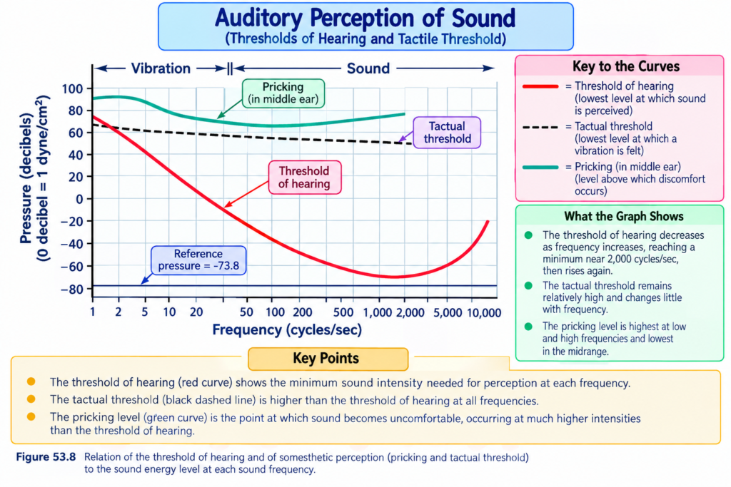
The hearing threshold is different for different sound frequencies, as demonstrated in Fig. 53.8.
3000 cycles/sec
A sound at approximately 3000 cycles/sec can be heard at a very low intensity.
100 cycles/sec
A 100 cycles/sec sound must have approximately:
10,000 times greater intensity
to be detected compared with the very sensitive range described for 3000 cycles/sec.
Key Concept
The ear is not equally sensitive to all frequencies.
Frequency Range of Hearing
A young person can usually hear frequencies from approximately:
20 to 20,000 cycles/sec
However, the range depends strongly on sound intensity.At Lower Sound Intensity
At a loudness approximately:
60 decibels below 1 dyne/cm² sound pressure level
the detectable range is approximately:
500–5000 cycles/sec
With Intense Sound
Only with stronger sounds can the complete approximate range of:
20–20,000 cycles/sec
be achieved.
Effect of Aging
With aging, gradual hearing loss may occur.
This is called:
Presbycusis
The hearing range may decrease to approximately:
50–8000 cycles/sec or less
Central Auditory Mechanisms
Auditory Nervous Pathways
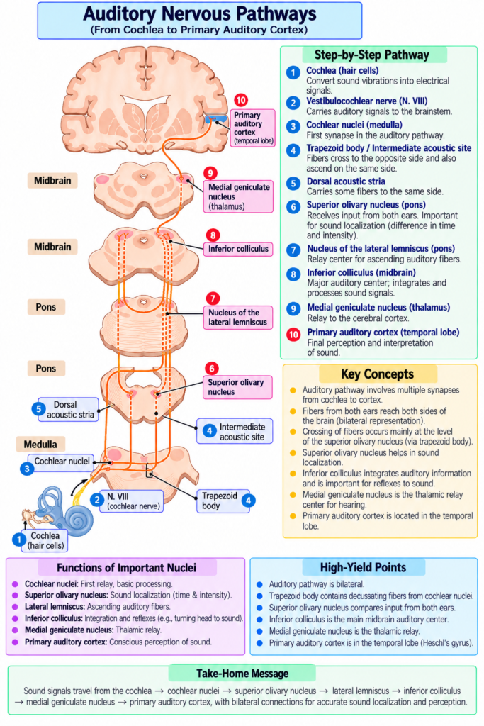
The major auditory pathway is shown in Fig. 53.9.
Cochlear Nuclei
Nerve fibers from the spiral ganglion of Corti enter:
- Dorsal cochlear nucleus.
- Ventral cochlear nucleus.
These nuclei are located in the:
- Upper medulla.
All auditory fibers synapse here.
Superior Olivary Nucleus
Second-order neurons then travel mainly:
→ to the opposite side of the brain stem
and terminate in:
→ superior olivary nucleus
A smaller number travel to the:
→ superior olivary nucleus on the same side
Lateral Lemniscus
From the superior olivary nucleus:
→ auditory signals travel upward through the lateral lemniscus
Some fibers terminate in:
→ nucleus of the lateral lemniscus
Many continue onward to:
→ inferior colliculus
Inferior Colliculus
At the inferior colliculus:
- All or almost all auditory fibers synapse.
From here:
→ signals pass to the medial geniculate nucleus
Medial Geniculate Nucleus
At the medial geniculate nucleus:
- Auditory fibers synapse.
Then:
→ auditory radiation
→ auditory cortex
Auditory Cortex
The auditory cortex is located mainly in:
Superior gyrus of the temporal lobe
Complete Auditory Pathway
Spiral ganglion
→ dorsal and ventral cochlear nuclei
→ mainly opposite superior olivary nucleus
→ lateral lemniscus
→ inferior colliculus
→ medial geniculate nucleus
→ auditory radiation
→ auditory cortex
Bilateral Transmission of Auditory Signals
Signals from each ear travel through auditory pathways on both sides of the brain.
However:
- More transmission occurs through the contralateral pathway.
Major Crossing Points
Crossing between the two auditory pathways occurs at least at:
- Trapezoid body.
- Commissure between the two nuclei of the lateral lemnisci.
- Commissure connecting the two inferior colliculi.
Key Concept
One ear
→ sends auditory information to both sides of the brain
→ contralateral transmission is greater
Collateral Auditory Pathways
Some auditory fibers send collateral branches to other parts of the nervous system.
Reticular Activating System
Auditory collateral fibers
→ reticular activating system
A loud sound can therefore:
→ activate large portions of the nervous system
Cerebellar Vermis
Other auditory collaterals travel to:
→ vermis of the cerebellum
A sudden noise can activate this area almost immediately.
Spatial Organization of Sound Frequencies
A high degree of spatial organization is preserved from the cochlea to the auditory cortex.
The text describes:
- Three frequency patterns in the cochlear nuclei.
- Two patterns in the inferior colliculi.
- One precise pattern in the auditory cortex.
- At least five additional less precise patterns in auditory cortex and association areas.
Firing Rates in the Auditory Pathway
Individual auditory nerve fibers entering the cochlear nuclei can fire at rates up to approximately:
1000 impulses/sec
The firing rate depends mainly on:
→ sound loudness
Synchronization With Sound Frequency
At sound frequencies up to approximately:
2000–4000 cycles/sec
auditory nerve impulses may be synchronized with the sound waves.
However:
- An impulse does not necessarily occur with every sound wave.
Brain Stem Processing
In auditory tracts of the brain stem:
- Synchronization with sound frequency is usually lost.
It may persist mainly for frequencies below:
200 cycles/sec
Above the inferior colliculi:
→ even this synchronization is mainly lost
Important Concept
Sound information is not simply passed unchanged from the ear to the brain.
Instead:
Sound enters cochlea
→ cochlear nuclei begin analysis
→ auditory information is progressively dissected and processed
→ higher centers receive analyzed information
Function of the Cerebral Cortex in Hearing
The auditory cortex is demonstrated in Fig. 53.10.
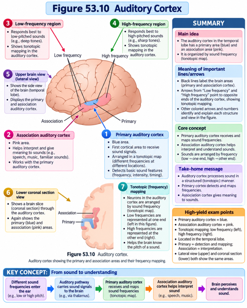
It lies mainly on:
- Supratemporal plane of the superior temporal gyrus.
It also extends onto:
- Lateral temporal lobe.
- Much of the insular cortex.
- Lateral part of the parietal operculum.
Primary and Association Auditory Cortex
Two major cortical divisions are described:
- Primary auditory cortex.
- Auditory association cortex.
Primary Auditory Cortex
The primary auditory cortex receives direct projections mainly from:
→ medial geniculate body
Auditory Association Cortex
The auditory association cortex receives:
- Signals from the primary auditory cortex.
- Some projections from nearby thalamic association areas.
Sound Frequency Perception in the Auditory Cortex
At least six tonotopic maps have been identified in:
- Primary auditory cortex.
- Auditory association areas.
Tonotopic map: An organized cortical representation in which different sound frequencies activate different locations.
Organization
Within each map:
High frequencies
→ one end
Low frequencies
→ opposite end
In most maps:
Low frequencies
→ anterior
High frequencies
→ posterior
This arrangement is not identical in every map.
Why Are There Multiple Tonotopic Maps?
Different maps may analyze different characteristics of sound.
Possible functions described include:
Sharpening of Frequency Response
Individual neurons in the auditory cortex respond to a much narrower frequency range than neurons at earlier levels.
Therefore:
Broad frequency response in cochlea/brain stem
→ progressively processed
→ narrow cortical frequency response
This sharpening is believed to depend mainly on:
Lateral inhibition
Lateral Inhibition
A pathway activated by one sound frequency:
→ inhibits neighboring frequency pathways
Therefore:
Main frequency response strengthened relatively
- adjacent frequencies suppressed
→ frequency discrimination becomes sharper
Auditory Association Neurons
Many neurons in the auditory association cortex do not respond only to one sound frequency.
They may:
- Associate different frequencies with each other.
- Associate auditory information with information from other sensory areas.
The parietal part of the auditory association cortex partly overlaps:
→ somatosensory area II
This may allow association between:
- Auditory information.
- Somatosensory information.
Discrimination of Sound Patterns
The auditory cortex is especially important for discriminating:
- Sound pitches.
- Tonal patterns.
- Sequential sound patterns.
Complete bilateral removal of the auditory cortex in experimental animals does not completely prevent detection of sound.
However:
→ discrimination of pitch and especially sound patterns becomes greatly impaired.
Effect of Auditory Cortex Damage in Humans
Bilateral Primary Auditory Cortex Damage
Destruction of both primary auditory cortices:
→ greatly reduces hearing sensitivity
Unilateral Damage
Destruction of one auditory cortex:
→ only slightly decreases hearing in the opposite ear
It does not cause complete deafness because auditory pathways have extensive crossover connections.
However:
→ sound localization becomes impaired
because localization requires comparison of signals in both cortices.
Auditory Association Cortex Damage
Damage to auditory association areas while the primary auditory cortex remains intact may leave the person able to:
- Hear sounds.
- Distinguish tones.
- Interpret simple sound patterns.
However:
→ understanding the meaning of sound can be impaired.
Wernicke Area
The posterior part of the superior temporal gyrus is called:
Wernicke area
It forms part of the auditory association cortex.
Damage here may cause a person to:
- Hear words normally.
- Repeat words.
- Yet fail to understand their meaning.
Determination of the Direction From Which Sound Comes
Horizontal sound direction is determined mainly by two mechanisms:
- Difference in arrival time between the two ears.
- Difference in sound intensity between the two ears.
Time-Lag Mechanism
This mechanism works best for sound frequencies below approximately:
3000 cycles/sec
Example
Sound directly in front
→ reaches both ears at the same time
Sound closer to right ear
→ reaches right ear first
→ reaches left ear later
The brain uses this time difference to determine direction.
Intensity-Difference Mechanism
This mechanism works best at:
→ higher sound frequencies
At high frequencies, the head acts as a greater barrier to sound.
Therefore:
Sound coming from one side
→ nearer ear receives stronger sound
→ farther ear receives weaker sound
→ intensity difference helps identify direction
Comparison of Direction Mechanisms
| Mechanism | Works Best |
|---|---|
| Time difference between ears | Below about 3000 cycles/sec |
| Intensity difference between ears | Higher frequencies |
The time-lag mechanism provides more precise directional information according to the provided text.ole of the Pinnae
The two basic binaural mechanisms cannot determine well whether a sound comes from:
- Front.
- Behind.
- Above.
- Below.
This discrimination depends mainly on:
Pinnae
Pinna: The visible external part of the ear that helps funnel sound into the ear.
Its shape changes the quality of incoming sound depending on direction.
Different directions
→ different frequencies emphasized
→ brain receives directional clues
Neural Mechanism for Detecting Sound Direction
Analysis of sound direction begins mainly in the:
Superior olivary nuclei
Although the entire pathway to the auditory cortex is required for conscious interpretation.
Lateral Superior Olivary Nucleus
The lateral superior olivary nucleus is concerned mainly with:
→ comparing sound intensities reaching the two ears
Flow
Sound intensity in right ear
vs
sound intensity in left ear
→ lateral superior olivary nucleus
→ auditory cortex
→ direction estimated
Medial Superior Olivary Nucleus
The medial superior olivary nucleus detects:
→ time differences between sound arriving at the two ears
Its neurons have two major dendrites:
- One projecting toward the right.
- One projecting toward the left.
Signals from:
Right ear
→ right dendrite
Left ear
→ left dendrite
Time-Lag Coding
Different neurons respond maximally to different time delays.
One region
→ short time lag
Opposite region
→ long time lag
Intermediate neurons
→ intermediate time lags
Therefore:
Different direction
→ different arrival-time difference
→ different set of medial olivary neurons activated
Spatial Pattern for Sound Direction
Sound directly ahead
→ one set of superior olivary neurons activated maximally
Sound from different side angles
→ other sets activated
This spatial pattern is transmitted to:
→ auditory cortex
The brain then determines sound direction according to:
→ location of the maximally stimulated neurons
Key Concept
The auditory system separates different qualities of sound at different neural levels.
At the superior olivary nuclei:
Sound direction
→ separated from
sound tonal information
Centrifugal Signals to Lower Auditory Centers
Centrifugal pathways: Retrograde pathways carrying signals from higher auditory centers back toward lower auditory structures.
Retrograde pathways are present:
Auditory cortex
→ brain stem centers
→ cochlea
The final pathway described travels mainly:
Superior olivary nucleus
→ hair cells in organ of Corti
Function of Retrograde Fibers
These fibers are mainly:
Inhibitory
Direct stimulation of specific points in the superior olivary nucleus can reduce sensitivity of specific parts of the organ of Corti by approximately:
15–20 decibels
Functional Importance
This mechanism may help a person:
Focus attention on a particular sound
→ suppress sensitivity to other sound qualities
Example from the text:
→ listening specifically to one instrument in a symphony orchestra
Clinical Importance / Clinical Correlation
Types of Deafness
Deafness is broadly divided into:
- Nerve deafness.
- Conduction deafness.
Nerve Deafness
Nerve deafness: Hearing loss caused by impairment of:
- Cochlea.
- Auditory nerve.
- Central auditory nervous system pathways.
If the cochlea or auditory nerve is destroyed:
→ permanent deafness occurs.
Conduction Deafness
Conduction deafness: Hearing loss caused by impairment of structures that physically conduct sound toward the cochlea.
Examples include damage to:
- Tympanic membrane–ossicular system.
- Middle-ear conducting structures.
If the cochlea and auditory nerve remain intact:
→ sound can still reach the cochlea through bone conduction
even if the tympanum-ossicular system is damaged or fixed in place.
Nerve vs Conduction Deafness
| Feature | Nerve Deafness | Conduction Deafness |
|---|---|---|
| Main site | Cochlea, auditory nerve, or central pathways | Sound-conducting structures |
| Air conduction | Reduced | Reduced |
| Bone conduction | Reduced | Relatively preserved |
| If cochlea/nerve destroyed | Permanent deafness | Not the primary problem |
Audiometer
Audiometer: An instrument used to assess hearing loss at different sound frequencies.
It consists of:
- Earphone.
- Electronic oscillator.
- Calibrated volume control.
It can generate:
→ pure tones from low to high frequencies
Zero-Intensity Level
For each frequency:
0 intensity level
means:
→ loudness that can just barely be heard by a normal ear
If a person requires:
30 dB above normal
to hear a particular frequency:
→ hearing loss at that frequency is 30 dB
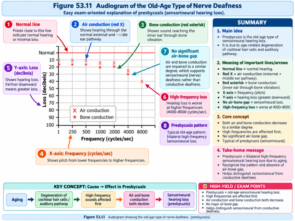
Audiogram
During audiometry:
- Approximately 8–10 frequencies across the auditory spectrum are tested.
- Hearing loss is measured for each frequency.
- Results are plotted as an:
Audiogram
Audiograms are illustrated in Figs. 53.11 and 53.12.
Air-Conduction Testing
Audiometer:
→ earphones applied to ears
→ tones delivered through normal air-conduction pathwayone-Conduction Testing
Audiometer also uses:
→ mechanical vibrator
→ mastoid process
→ skull vibration
→ cochlea
Audiogram in Nerve Deafness
In nerve deafness:
- Air conduction is impaired.
- Bone conduction is also impaired.
The example described in the text shows mainly:
→ high-frequency hearing loss
This may result from damage near:
→ base of cochlea
This type of high-frequency loss occurs to some extent in many older people.
Other Patterns of Nerve Deafness
The provided text also describes:
Low-Frequency Hearing Loss
Prolonged exposure to very loud sounds
→ may damage organ of Corti
→ low-frequency hearing loss
Loss at All Frequencies
Drug sensitivity of the organ of Corti may cause hearing loss at all frequencies.
Examples listed in the provided text include:
- Streptomycin.
- Gentamicin.
- Kanamycin.
- Chloramphenicol.
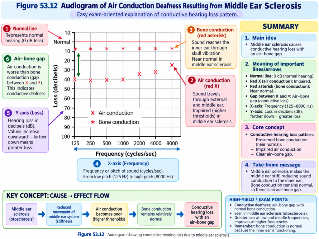
Middle-Ear Conduction Deafness
Conduction deafness may result from:
- Fibrosis after repeated middle-ear infections.
- Fibrosis in otosclerosis.
In these conditions:
Sound waves
→ not transmitted efficiently through ossicles
→ reduced movement at oval window
Audiogram Pattern
In middle-ear conduction deafness:
Bone conduction
→ essentially normal
Air conduction
→ greatly reduced
The impairment may be greater at:
→ lower frequencies
Stapes Ankylosis
In some conduction deafness:
- Stapes faceplate becomes fixed by bone overgrowth to the edges of the oval window.
This produces:
→ severe loss of ossicular conduction
The text describes restoration of hearing by:
Removal of stapes
→ replacement with small Teflon or metal prosthesis
→ sound transmitted from incus to oval window
Key Concept
Auditory analysis occurs at multiple levels.
Frequency
Basilar membrane location
→ place principle
Very low frequencies
→ volley/frequency mechanism also contributes
Loudness
Greater sound intensity
→ faster nerve firing
- more hair cells activated
- outer hair cells recruited
Direction
Time difference between ears
- intensity difference between ears
- pinna-related sound changes
→ sound location determined
Hearing Loss
Air + bone conduction reduced
→ nerve deafness
Air conduction reduced but bone conduction relatively preserved
→ conduction deafness
High-Yield / Exam Points
- High-frequency sound → cochlear base.
- Low-frequency sound → cochlear apex.
- Place principle:
- Frequency determined by position of maximum basilar membrane stimulation.
- Very low frequencies may also use:
- Volley/frequency principle.
- Loudness increases through:
- Faster nerve firing.
- Spatial summation.
- Outer hair-cell activation at high intensity.
- 1 bel = 10-fold increase in sound energy.
- 1 decibel = 0.1 bel.
- One decibel ≈ 1.26-fold increase in sound energy.
- Young hearing range:
- Approximately 20–20,000 cycles/sec.
- Presbycusis:
- Age-related reduction in hearing range.
- Auditory pathway:
- Spiral ganglion
- Cochlear nuclei
- Superior olive
- Lateral lemniscus
- Inferior colliculus
- Medial geniculate nucleus
- Auditory radiation
- Auditory cortex
- Auditory signals from one ear reach:
- Both sides of brain.
- Contralateral transmission is greater.
- Auditory cortex contains multiple:
- Tonotopic maps.
- Lateral inhibition sharpens frequency discrimination.
- Time-lag localization:
- Best below about 3000 cycles/sec.
- Intensity localization:
- More important at higher frequencies.
- Lateral superior olive:
- Intensity comparison.
- Medial superior olive:
- Time-lag comparison.
- Centrifugal inhibition can reduce cochlear sensitivity by:
- 15–20 dB.
- Nerve deafness:
- Air and bone conduction impaired.
- Conduction deafness:
- Air conduction impaired.
- Bone conduction relatively preserved.
Common Student Mistakes
- Do not think pitch is determined mainly by sound intensity.
- Pitch depends mainly on sound frequency and the location of maximal basilar membrane activation.
- Do not confuse the place principle with the volley principle.
- Place principle → location.
- Volley principle → timing/frequency of nerve impulses.
- Do not think louder sound activates only the same hair cells more strongly.
- It also recruits additional hair cells and nerve fibers.
- Do not think auditory information from one ear travels only to the opposite cerebral cortex.
- Each ear projects to both sides.
- Do not confuse medial and lateral superior olivary nuclei.
- Medial → time difference.
- Lateral → intensity difference.
- Do not confuse nerve deafness with conduction deafness.
- Nerve deafness affects both air and bone conduction.
- Conduction deafness mainly impairs air conduction.
Quick Revision
Frequency
High frequency
→ base
Low frequency
→ apex
Specific basilar membrane place
→ specific frequency Low Frequency
Low-frequency sound
→ synchronized nerve volleys
→ cochlear nuclei
→ frequency discrimination
Loudness
Louder sound
→ faster nerve firing
- more hair cells activated
- outer hair cells activated
Auditory Pathway
Spiral ganglion
→ cochlear nuclei
→ superior olive
→ lateral lemniscus
→ inferior colliculus
→ medial geniculate
→ auditory cortex
Auditory Cortex
Tonotopic organization
→ different frequencies represented at different locations
Lateral inhibition
→ sharpens frequency response
Sound Direction
Below ~3000 cycles/sec
→ time difference important
Higher frequencies
→ intensity difference important
Pinna
→ helps determine front/back/above/below
Superior Olive
Medial
→ time lag
Lateral
→ intensity difference
Deafness
Nerve deafness
→ air ↓ + bone ↓
Conduction deafness
→ air ↓ + bone relatively normal
Final Take-Home Concept / Summary
Sound frequency is determined mainly by the place principle:
High-frequency sound
→ cochlear base
Low-frequency sound
→ cochlear apex
For very low frequencies, synchronized nerve volleys may provide additional frequency information.
Loudness depends on:
Sound intensity increases
→ firing rate increases
→ more hair cells are recruited
→ more nerve fibers participate
→ outer hair cells become active at high intensity
Auditory signals then travel through:
Spiral ganglion
→ cochlear nuclei
→ superior olivary nuclei
→ lateral lemniscus
→ inferior colliculus
→ medial geniculate nucleus
→ auditory cortex
The auditory cortex contains organized frequency maps and is particularly important for:
- Pitch discrimination.
- Sound-pattern discrimination.
- Sound localization.
- Interpretation of auditory information.
Horizontal sound direction depends mainly on:
Time difference between ears
- intensity difference between ears
The superior olivary nuclei begin this analysis:
Medial superior olive → time difference
Lateral superior olive → intensity difference
Finally, hearing loss can be broadly divided into:
Nerve deafness
→ cochlea / auditory nerve / central pathway problem
→ air and bone conduction impaired
Conduction deafness
→ sound-conducting structures impaired
→ air conduction reduced while bone conduction is relatively preserved
References / Sources
- Guyton and Hall Textbook of Medical Physiology, 15th Edition, Chapter 53.
