


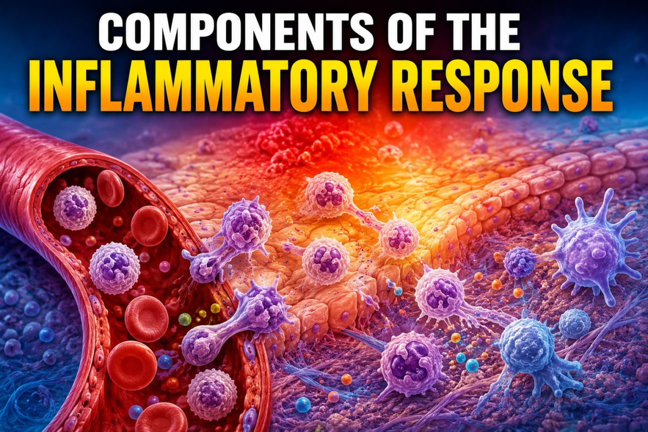
- The inflammatory response starts when:
- Foreign microbes enter the body and damage tissues.
- Or the body faces severe stress such as hypothermia or hypotension.
- Many cellular pathways work at the same time to:
- Limit further tissue damage.
- Start the healing process.
- A localized inflammatory response is usually helpful.
- A major injury can cause an uncontrolled (dysregulated) inflammatory response.
- This uncontrolled response can become very dangerous.
- A patient’s outcome depends not only on the original injury but also on how the body responds to that injury.
- Surgeons commonly deal with both acute and chronic inflammatory responses.
- Understanding how inflammation:
- Starts,
- Is controlled,
- Or becomes stronger,
is important for understanding patients with: - Local reaction after surgery.
- Systemic inflammatory response syndrome (SIRS).
- Multisystem organ failure.
- Chronic critical illness.
Cells of the Immune System
Neutrophils
- Neutrophils are polymorphonuclear (PMN) leukocytes.
- They are one of the most important cells in acute inflammation.
- They are usually the first immune cells to reach the site of injury or infection.
- Neutrophils circulate in the blood.
- Their normal life span is about 8 hours.
- During inflammation, their survival becomes longer, although the exact duration is still uncertain.
- Neutrophils are continuously produced in the bone marrow.
- Their production is stimulated by granulocyte colony-stimulating factor (G-CSF).
- Their production is also regulated by:
- IL-17 from T cells.
- IL-23 from macrophages.
- To move from blood into tissues, neutrophils pass through these steps (Fig. 3.1):
- Tethering.
- Rolling.
- Adhesion.
- Crawling.
- Transmigration.
- Neutrophils contain three types of inflammatory granules:
- Azurophilic (primary) granules.
- Specific (secondary) granules.
- Gelatinase (tertiary) granules.
- These granules contain proteolytic enzymes.
- The enzymes are released:
- Outside the cell.
- Or inside the phagosome.
- These enzymes help destroy invading microbes.
- Neutrophils also release a fiber-like mesh called the neutrophil extracellular trap (NET).
- Histones, proteins, and enzymes attach to this mesh.
- The NET traps microbes outside the cell.
- This prevents the microbes from spreading.
- It also helps other immune cells remove the microbes by phagocytosis.
- Neutrophils do more than just start inflammation.
- Their granules contain proteases that help:
- Tissue remodeling.
- Wound healing.
- They also directly promote angiogenesis by releasing vascular endothelial growth factors (VEGFs).
- Neutrophils can change their function depending on the situation (plasticity).
- Although they are usually proinflammatory, some neutrophils become anti-inflammatory in certain disease conditions.
Macrophages
- Macrophages are named because they eat and digest extracellular debris.
- They are one of the main cells of innate immunity.
- Monocytes develop into macrophages after infection or tissue injury.
- Unlike immature monocytes, macrophages have many pattern recognition receptors (PRRs).
- PRRs detect different danger signals inside and outside cells.
- When PRRs are activated, macrophages:
- Destroy microbes by phagocytosis.
- Digest them inside lysosomes.
- Macrophages also release inflammatory cytokines such as:
- IL-1β
- TNF-α
- These cytokines attract more immune cells to the injured tissue.
- Macrophages also process antigens.
- They display these antigens on their surface.
- This helps activate helper T cells.
- Therefore, macrophages are professional antigen-presenting cells (APCs).
- Like neutrophils, macrophages also show plasticity.
- Their function changes according to their surrounding environment.
- M1 macrophages:
- Produce proinflammatory cytokines.
- Release proteolytic substances.
- Are mainly active during bacterial and viral infections.
- Stimulate proinflammatory helper T cells.
- The inflammatory products of M1 macrophages help fight microbes.
- However, excessive M1 activity can produce a harmful inflammatory response.
- High levels of M1 cytokines are associated with increased death in sepsis.
- M2 macrophages:
- Help tissue remodeling.
- Promote wound healing.
- Produce anti-inflammatory substances such as IL-10.
- Macrophages are found throughout the body.
- Their function changes according to the tissue where they live.
- Examples:
- Kupffer cells are macrophages of the liver.
- Microglia are macrophages of the central nervous system.
KEY CONCEPT
- Inflammation protects the body after injury or infection, but excessive inflammation can cause serious disease.
- Neutrophils are the first responders that kill microbes, form NETs, and later help wound healing and new blood vessel formation.
- Macrophages remove microbes and dead tissue, activate T cells, and change their function depending on the body’s needs.
- M1 macrophages = Fight infection (proinflammatory).
- M2 macrophages = Repair tissues (anti-inflammatory).
Conceptual Examples
- Cut finger with bacteria → Neutrophils arrive first to destroy bacteria.
- After infection is controlled → M2 macrophages repair the damaged tissue and help wound healing.
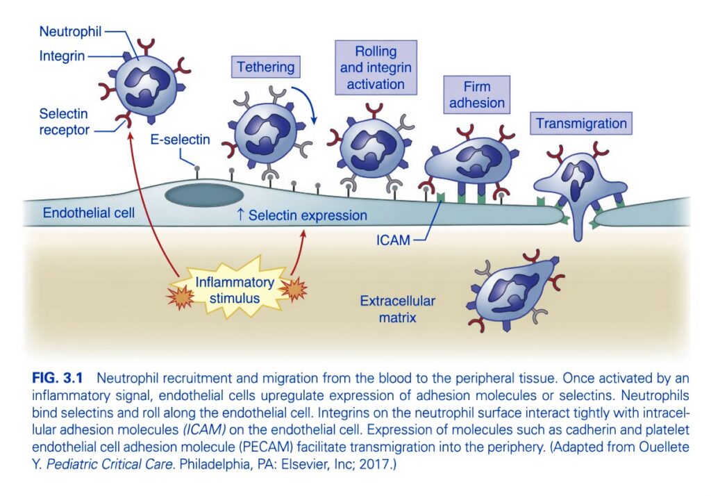
Neutrophil Recruitment and Migration (Figure 3.1)
Key Concept
- Purpose: To move neutrophils (the first immune cells) from the bloodstream to the site of infection or tissue injury.
- This process occurs in 5 simple sequential steps:
- Inflammatory stimulus
- Tethering
- Rolling and integrin activation
- Firm adhesion
- Transmigration (Diapedesis)
Step 1. Inflammatory Stimulus
What happens?
- Tissue injury or infection releases inflammatory signals.
- These signals activate the endothelial cells (cells lining the blood vessel).
Endothelial cell response
- Endothelial cells increase the expression of E-selectin (selectins) on their surface.
- They also express ICAM (Intercellular Adhesion Molecule).
Simple Concept
Injury tells the blood vessel:
Step 2. Tethering
What happens?
- A circulating neutrophil first makes weak contact with the endothelial surface.
Molecules involved
- E-selectin on endothelial cell
- Selectin receptor on neutrophil
Result
- The neutrophil is briefly attached instead of flowing away.
Easy Concept
Think of it as:
The neutrophil catches the blood vessel with one hand.
This attachment is weak and temporary.tep 3. Rolling and Integrin Activation
Rolling
- Because the attachment is weak, blood flow keeps pushing the neutrophil.
- It repeatedly attaches and detaches.
- Therefore it rolls along the vessel wall.
During rolling
Inflammatory signals activate:
- Integrins present on the neutrophil.
Before activation:
- Integrins are weak.
After activation:
- Integrins become strong adhesion molecules.
Easy Concept
Rolling allows the neutrophil to:
- Slow down
- Receive activation signals
- Prepare for strong attachment
Think of it as:
A moving car slowing down before parking.
Step 4. Firm Adhesion
What happens?
Activated integrins bind tightly to ICAM on the endothelial cell.
Molecules involved
Neutrophil
- Activated integrins
Endothelial cell
- ICAM
Result
- The neutrophil stops rolling.
- It becomes firmly attached to the blood vessel wall.
Easy Concept
Think of:
- Selectins = Velcro (weak grip)
- Integrins + ICAM = Super glue (strong grip)
Now the neutrophil cannot be washed away by blood flow.
Step 5. Transmigration (Diapedesis)
What happens?
- The neutrophil squeezes between adjacent endothelial cells.
- It leaves the blood vessel.
- It enters the surrounding tissue (extracellular matrix).
Molecules helping this step
The figure caption mentions:
- PECAM (Platelet Endothelial Cell Adhesion Molecule)
- Cadherins
These molecules help the neutrophil pass safely through the endothelial junctions.
Final destination
The neutrophil migrates toward:
- Infection
- Tissue injury
- Area of inflammation
Easy Concept
Think of:
The neutrophil exits the highway (blood vessel) through a small gate and enters the damaged tissue to fight microbes.
Molecules in Figure 3.1
| Molecule | Location | Function |
|---|---|---|
| E-selectin | Endothelial cell | Initiates weak attachment (tethering) and rolling |
| Selectin receptor | Neutrophil | Binds E-selectin |
| Integrin | Neutrophil | Produces strong adhesion after activation |
| ICAM | Endothelial cell | Binds activated integrins for firm adhesion |
| PECAM (mentioned in figure caption) | Between endothelial cells | Helps neutrophil pass through vessel wall |
| Cadherins (mentioned in figure caption) | Endothelial junctions | Assist transmigration |
Sequence to Remember
Inflammation
⬇
↑ E-selectin expression
⬇
Tethering
⬇
Rolling
⬇
Integrin activation
⬇
Firm adhesion (Integrin + ICAM)
⬇
Transmigration (PECAM-assisted)
⬇
Neutrophil enters infected tissue
Memory Trick
S → S → I → I → P
- S = Selectin → Tethering
- S = Selectin → Rolling
- I = Integrin activation
- I = ICAM binding (Firm adhesion)
- P = PECAM → Passage through vessel wall
Exam Pearls
- First leukocyte recruited in acute inflammation: Neutrophil
- Weak adhesion molecule: Selectin
- Strong adhesion molecule: Integrin
- Endothelial binding partner for integrin: ICAM
- Molecule facilitating diapedesis (transmigration): PECAM
- Rolling is mediated by: Selectins
- Firm adhesion is mediated by: Integrins binding ICAM
One-Line Summary
Inflammation activates endothelial cells, causing neutrophils to first tether via selectins, roll along the vessel wall, activate integrins, bind firmly to ICAM, and finally migrate between endothelial cells (via PECAM) into the injured tissue to destroy invading microbes.
Dendritic Cells
- Dendritic cells connect the innate and adaptive immune systems.
- They are the main professional antigen-presenting cells (APCs).
- They engulf and break down foreign proteins.
- These proteins are attached to MHC class I or MHC class II molecules.
- The antigen-MHC complex moves to the surface of the dendritic cell.
- The dendritic cell then travels to the lymph nodes and spleen.
- There, it activates naïve (resting) T cells.
- Naïve T cells become either:
- CD8+ cytotoxic T cells
- CD4+ helper T cells
- Extracellular proteins are broken down in lysosomes.
- They are presented with MHC class II.
- This activates CD4+ helper T cells.
- Intracellular proteins are broken down by the proteasome in the cytosol.
- They are presented with MHC class I.
- This activates CD8+ cytotoxic T cells.
- Some dendritic cells can present extracellular proteins through MHC class I by cross-presentation.
- Presentation of antigen with MHC starts the adaptive immune response.
- Dendritic cells also activate T cells by:
- CD80 and CD86 surface molecules.
- Producing IL-12.
- These costimulatory signals make dendritic cells the most efficient APCs.
- Macrophages and B cells are also APCs but are less efficient.
- Dendritic cells also process:
- Self-antigens.
- Harmless environmental antigens.
- Presentation of these antigens forms regulatory T cells.
- Regulatory T cells maintain immune tolerance and immune balance.
- Failure of this pathway can cause:
- Autoimmune diseases.
- Allergic reactions.
- Dendritic cells can produce:
- An active immune response against foreign antigens.
- A tolerant response to self-antigens.
- This dual function is important in cancer immunobiology.
- Tumor cells can escape the immune system by reducing dendritic cell function.
T Cell
- T cells and B cells are the main cells of the adaptive immune system.
- T cells mainly produce the cellular immune response.
- B cells mainly produce the humoral immune response.
- Both T and B cells recognize specific antigens.
- They rapidly multiply by clonal expansion.
- Both are essential for immune memory.
- T cell activation can be understood in three main steps.
- Many immune events occur together during this process.
- Mature dendritic cells present antigen-MHC complexes to naïve T cells.
- MHC class I presents intracellular antigens.
- This activates CD8+ cytotoxic T cells.
- MHC class II presents extracellular antigens.
- This activates CD4+ helper T cells.
- MHC class I is present on all nucleated cells.
- MHC class II is present only on APCs.
- PRR activation can change whether antigen is presented by MHC I or MHC II.
- TLR4 recognizes lipopolysaccharide from gram-negative bacteria.
- Surface activation of TLR4 increases cross-presentation.
- This increases MHC class I presentation and activates CD8+ T cells.
- After entering the endosome, TLR4 promotes MHC class II presentation.
- This mainly activates CD4+ helper T cells.
- Antigen-MHC presentation alone is not enough to activate T cells.
- Costimulatory molecules are also required.
- The most important are CD80/CD86 on dendritic cells binding to CD28 on T cells (Fig. 3.2).
- This lowers the activation threshold.
- It also increases IL-2 production.
- Cytokines are also required for complete T cell activation.
- The cytokines produced depend on the activated PRR.
- IL-12, IL-6, and TNF-α strengthen acute inflammation.
- They also influence T cell differentiation.
- IL-1 increases the acute-phase response.
- Type I interferon (IFN) promotes antiviral immunity.
- It also activates CD8+ cytotoxic T cells.
- IL-12 forms Th1 cells.
- IL-4 forms Th2 cells.
- IL-6 + TGF-β form Th17 cells.
- TGF-β alone can produce regulatory T cells when infection is absent.
- Complete T cell activation needs:
- Antigen-MHC presentation.
- Costimulation.
- Cytokines.
- Activated T cells produce different cytokines.
- Th1 cells fight intracellular pathogens.
- They produce IFN-γ.
- Th2 cells fight extracellular pathogens.
- They produce IL-4, IL-5, and IL-13.
- A balanced Th1 and Th2 response is important for normal immunity.
- Th17 cells respond to extracellular pathogens and fungi.
- They are commonly involved in autoimmune diseases.
- In chronic inflammation, Th17 cells may develop Th1-like features.
- Th17 cells produce IL-17.
- Regulatory T cells maintain immune memory and self-tolerance.
- They produce IL-10 and TGF-β.
- CD8+ cytotoxic T cells destroy virus-infected cells.
- They also produce IFN-γ.
- General anesthesia, surgery, blood transfusion, hypothermia, hyperglycemia, and postoperative pain reduce the T cell-dependent immune response.
- At the same time, ACTH and glucocorticoids increase.
- T cells help destroy circulating tumor cells.
- They also help prevent micrometastasis.
- After breast cancer surgery, regulatory T cells increased.
- This increase was associated with:
- Larger tumors.
- HER2 positivity.
- Shorter disease-free survival.
- Lower Th1 levels were also linked with:
- Larger tumor burden.
- HER2 positivity.
- This suggests postoperative immunosuppression may increase the risk of metastasis.
- It also supports research into postoperative immunomodulation.
B Cell
- B cells are the main cells of the humoral immune response.
- They produce antibodies (immunoglobulins, Ig).
- They also function as professional APCs.
- B cells first develop in the bone marrow.
- Their maturation depends on rearrangement of immunoglobulin genes.
- They undergo V(D)J recombination.
- Gene segments V, D, and J rearrange.
- This allows production of many different antibodies.
- B cells can recognize more than 5 × 10¹³ different antigens.
- Concept: Different gene combinations produce a very large number of antibodies.
- During V(D)J recombination, B cells pass through:
- Pro-B stage.
- Pre-B stage.
- Surface IgM marks the immature B cell stage.
- Immature B cells leave the bone marrow.
- They migrate to the spleen.
- In the spleen, they become:
- Naïve follicular B cells.
- Marginal zone B cells.
- Marginal zone B cells are the first defense against blood-borne microbes.
- They rapidly produce IgM without T cell help.
- Naïve follicular B cells are found in lymph nodes and blood.
- Their activation requires T cell help.
- Activated follicular B cells undergo class switching.
- They change from producing IgM to producing:
- IgG
- IgA
- IgE
- During class switching, further gene rearrangement occurs.
- This produces antibodies with higher affinity for the antigen.
- Memory B cells remain after an immune response.
- They remember the antigen.
- On re-exposure to the same antigen, they rapidly produce a strong immune response.
KEY CONCEPT
- Dendritic cells start the adaptive immune response by presenting antigens to T cells.
- T cell activation requires three things:
- Antigen-MHC presentation.
- Costimulation (CD80/CD86 → CD28) (Fig. 3.2).
- Cytokines.
- Th1 = Intracellular pathogens.
- Th2 = Extracellular pathogens & allergy.
- Th17 = Fungi & autoimmune diseases.
- Regulatory T cells = Immune tolerance.
- CD8+ T cells = Kill virus-infected cells.
- B cells produce antibodies and form memory B cells for faster future protection.
Conceptual Examples
- Virus infection → Dendritic cell → MHC I → CD8+ T cell → Kills infected cell.
- Bacterial infection outside cells → MHC II → CD4+ T cell → Helps B cells produce antibodies.
- Second exposure to the same germ → Memory B cells quickly produce high-affinity antibodies.
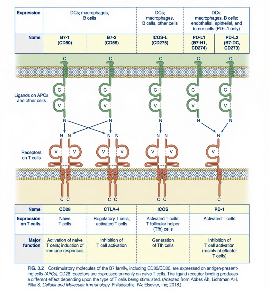
Figure 3.2 – Costimulatory Molecules of the B7 Family (Easiest Conceptual Summary)
Key Concept
T cells cannot be activated by antigen alone.
They also need a second signal (costimulatory signal) from Antigen-Presenting Cells (APCs).
Without this second signal:
- ❌ T cell becomes inactive (anergy)
- ❌ No proper immune response
With the correct second signal:
- ✅ T cell becomes activated
- ✅ Immune response starts
The Figure is Divided into Two Parts
Upper Part = Ligands (on APCs and other cells)
These molecules are present on:
- Dendritic cells (DCs)
- Macrophages
- B cells
- Some endothelial cells
- Some epithelial cells
- Tumor cells (PD-L1 only)
They act like keys.
Lower Part = Receptors (on T cells)
These are present on T cells.
They act like locks.
When the correct key fits into the correct lock,
the immune response is either:
- Activated
OR - Suppressed
There Are Five Important Ligands
1. B7-1 (CD80)
Expressed on
- Dendritic cells
- Macrophages
- B cells
It binds
It can bind to:
- CD28
- CTLA-4
This is very important because the same ligand can produce opposite effects depending on the receptor.
2. B7-2 (CD86)
Expressed on
- Dendritic cells
- Macrophages
- B cells
It also binds
- CD28
- CTLA-4
Exactly like CD80.
Easy Memory
CD80 and CD86 are the two B7 molecules.
Both can interact with
- CD28
- CTLA-4
3. ICOS-L (CD275)
Expressed on
- Dendritic cells
- Macrophages
- B cells
- Other cells
It binds only
ICOS
Function
It helps produce
- T follicular helper (Tfh) cells
These cells help B cells produce antibodies.
4. PD-L1 (B7-H1, CD274)
Expressed on
- Dendritic cells
- Macrophages
- B cells
- Endothelial cells
- Epithelial cells
- Tumor cells
Tumor cells commonly express PD-L1.
It binds
PD-1
Function
Suppresses T-cell activity.
Tumor cells use this mechanism to escape immune attack.
5. PD-L2 (B7-DC, CD273)
Expressed on
- Dendritic cells
- Macrophages
It binds
PD-1
Function
Also inhibits T-cell activation.
Now Understand the T-cell Receptors
1. CD28 (Activation Receptor)
Present on
Naïve T cells
Ligands
- CD80
- CD86
Function
Activates naïve T cells.
Starts the immune response.
Easy Concept
Think:
CD28 = Accelerator pedal
It turns the immune system ON.
2. CTLA-4 (Inhibitory Receptor)
Present on
- Regulatory T cells (Tregs)
- Activated T cells
Ligands
- CD80
- CD86
Exactly the same ligands as CD28.
Function
Stops T-cell activation.
Important Concept
Both CD28 and CTLA-4 bind the same B7 molecules.
But:
CD28 → Activation
CTLA-4 → Inhibition
Easy Concept
Think:
CD28 = Gas pedal
CTLA-4 = Brake pedal
Both use the same road (CD80/CD86).
3. ICOS
Present on
- Activated T cells
- T follicular helper (Tfh) cells
Ligand
ICOS-L
Function
Produces Tfh cells.
These cells help B cells make antibodies.
Easy Concept
ICOS is mainly important for
Helping B cells.
4. PD-1
Present on
Activated T cells
Ligands
- PD-L1
- PD-L2
Function
Suppresses T-cell activation.
Especially suppresses
Effector T cells.
Easy Concept
PD-1 is another brake.
It prevents excessive immune responses and protects against tissue damage.
Unfortunately, many tumors exploit this pathway to avoid being destroyed by T cells.
Understand Every Arrow in the Figure
CD80 (B7-1)
⬇
Can bind
- CD28 ✅
- CTLA-4 ✅
CD86 (B7-2)
⬇
Can bind
- CD28 ✅
- CTLA-4 ✅
ICOS-L
⬇
Binds only
ICOS
PD-L1
⬇
Binds only
PD-1
PD-L2
⬇
Binds only
PD-1
Complete Functional Summary
| Ligand (APC) | Receptor (T Cell) | Effect |
|---|---|---|
| B7-1 (CD80) | CD28 | Activates naïve T cells |
| B7-2 (CD86) | CD28 | Activates naïve T cells |
| B7-1 (CD80) | CTLA-4 | Inhibits T-cell activation |
| B7-2 (CD86) | CTLA-4 | Inhibits T-cell activation |
| ICOS-L (CD275) | ICOS | Produces T follicular helper (Tfh) cells |
| PD-L1 (CD274) | PD-1 | Inhibits activated T cells |
| PD-L2 (CD273) | PD-1 | Inhibits activated T cells |
Memory Tricks
1. CD28 = Start
CD28 starts the immune response.
2. CTLA-4 = Control
CTLA-4 controls (suppresses) T-cell activation.
3. PD-1 = Peace
PD-1 keeps T cells peaceful by reducing their activity.
4. ICOS = Immune Cooperation
ICOS helps T cells cooperate with B cells to produce antibodies.
Clinical Importance
Checkpoint inhibitors in cancer therapy
Many tumors express PD-L1, which binds PD-1 on activated T cells and switches them off, allowing the tumor to evade immune attack.
Modern immunotherapy blocks these inhibitory pathways:
- Anti-PD-1 antibodies block PD-1.
- Anti-PD-L1 antibodies block PD-L1.
- Anti-CTLA-4 antibodies block CTLA-4.
Blocking these “brakes” restores T-cell activity so the immune system can attack cancer cells more effectively.
Exam Pearls
- CD80 (B7-1) and CD86 (B7-2) bind both CD28 and CTLA-4.
- CD28 provides the major costimulatory signal required for activation of naïve T cells.
- CTLA-4 is an inhibitory checkpoint receptor that suppresses T-cell activation.
- ICOS-L binds ICOS and promotes T follicular helper (Tfh) cell development.
- PD-L1 and PD-L2 bind PD-1 and inhibit activated (especially effector) T cells.
- PD-L1 is commonly expressed by tumor cells, helping them evade immune surveillance.
One-Line Summary
B7 family ligands on antigen-presenting cells interact with specific receptors on T cells to either activate immunity (CD28), regulate antibody responses (ICOS), or suppress immune responses (CTLA-4 and PD-1), maintaining the balance between effective defense and prevention of excessive immune activation.
MADE BY SELF LEARNING CEO AND FOUNDER DR ASHRAF ( DR SHEEN )
