


- Innate immunity is the body’s first line of defense.
- It also helps start the adaptive immune response.
- Innate immunity includes:
- Physical barriers (skin lining/epithelial cells and mucus).
- Immune cells (neutrophils, dendritic cells, macrophages, and natural killer cells).
- Cytokines that control immune responses.
- Complement proteins.
- Earlier, innate immunity was thought to act the same way against all infections.
- New evidence shows that it can:
- Develop a type of memory.
- Protect against reinfection without T cells or B cells.
- Produce different responses depending on which PRR is activated first.
- Earlier, the self/nonself theory said the immune system responds only to foreign substances.
- This theory could not explain why the body usually does not attack:
- A developing baby (fetus).
- A growing cancer cell.
- The danger hypothesis is now more accepted.
- It says the immune system mainly responds to cell damage, not simply to foreign material.
- Damaged cells release danger signals called danger-associated molecular patterns (DAMPs) or alarmins (Fig. 3.3).
- Molecules coming from microbes are called pathogen-associated molecular patterns (PAMPs).
- The original danger hypothesis said:
- Severe cell injury causes necrosis.
- Cell contents leak out.
- Alarmins are released passively.
- Alarmins normally stay inside healthy cells.
- They are usually not released during apoptosis.
- Newer evidence shows that stressed cells can actively release alarmins even without necrosis.
- This happens by increasing alarmin production.
- IL-1α is a well-known alarmin.
- It detects chromatin damage.
- It sends this warning to nearby cells by releasing more IL-1α.
- Therefore, IL-1α can signal cell damage before the cell membrane breaks.
Toll-Like Receptors
- DAMPs are recognized by pattern recognition receptors (PRRs).
- PRRs are present:
- On the cell surface.
- Inside the cell.
- PRRs recognize specific PAMPs.
- PAMPs are essential parts of microbes.
- Because they are essential, microbes cannot easily change them to escape the immune system.
- The best-known PRRs are the toll-like receptors (TLRs).
- The Toll signaling pathway was first discovered in Drosophila melanogaster.
- The Toll protein activates NF-κB after recognizing lipopolysaccharide (LPS) from gram-negative bacteria.
- IL-1 also uses the NF-κB pathway.
- Finding that the IL-1 receptor (IL-1R) has a similar structure to the Toll protein greatly improved understanding of innate immune signaling.
- TLRs are proteins that pass through the cell membrane.
- Each TLR has:
- An outside ligand-binding part.
- An inside signaling part.
- TLRs are found:
- On the cell surface.
- Inside endosomes.
- When a DAMP binds to a TLR:
- Two TLRs join together (dimerization).
- This starts intracellular signaling pathways.
- Ten human TLRs have been identified.
- Each TLR recognizes different PAMPs.
- Each starts a different immune response.
- TLR4 mainly recognizes bacterial LPS.
- TLR1, TLR2, and TLR6 recognize other bacterial lipoproteins.
- TLR4 also recognizes the alarmins:
- HMGB1
- Heat shock protein 70
- TLR4 also helps cause sterile inflammation during ischemia-reperfusion injury.
- TLR3 recognizes double-stranded RNA.
- TLR7 and TLR8 recognize single-stranded viral RNA.
- TLRs activate the NF-κB and MAPK signaling pathways.
- These pathways increase production of inflammatory cytokines, especially:
- IL-1
- TNF-α
- These cytokines activate nearby innate immune cells.
- They also activate adaptive immune cells, including:
- Helper T cells.
- Cytotoxic T cells.
- Regulatory T cells.
- B cells.
KEY CONCEPT
- Innate immunity is the body’s first defense and also starts the adaptive immune response.
- DAMPs (alarmins) come from damaged body cells, while PAMPs come from microbes.
- PRRs, especially TLRs, recognize these danger signals.
- TLR4 mainly recognizes LPS and some alarmins.
- TLR activation causes release of IL-1 and TNF-α, which activate both innate and adaptive immunity.
Conceptual Examples
- A burn (no germs) → Damaged cells release DAMPs → TLRs detect them → Inflammation starts.
- Gram-negative bacterial infection → LPS (PAMP) activates TLR4 → IL-1 & TNF-α are released → Immune cells become active.
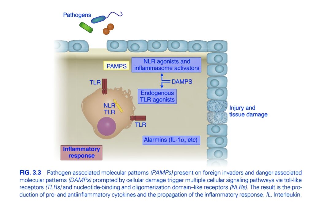
Figure 3.3 – How the Body Detects Infection and Tissue Damage (Exam Concept)
Key Concept
Your immune system constantly asks one simple question:
“Is this danger coming from a germ, or from damaged body cells?”
It detects both situations using special sensors called Pattern Recognition Receptors (PRRs).
These receptors recognize:
- PAMPs → Signals from pathogens (germs)
- DAMPs → Signals from damaged body cells
Both pathways ultimately produce inflammation.
Understand the Whole Figure First
The figure shows two different ways inflammation can begin.
Pathway 1 (Left Side)
Infection by pathogens
⬇
PAMPs
⬇
TLRs and NLRs recognize them
⬇
Inflammatory response
Pathway 2 (Right Side)
Tissue injury
⬇
Damaged cells release DAMPs
⬇
TLRs and NLRs recognize them
⬇
Inflammatory response
Part 1. Pathogens (Top Left)
The bacteria and other microbes shown at the top represent:
- Bacteria
- Viruses
- Fungi
- Parasites
These organisms carry molecules that are not normally found in human cells.
These molecules are called:
PAMPs
PAMP = Pathogen-Associated Molecular Patterns
Examples of PAMPs
- Bacterial lipopolysaccharide (LPS)
- Peptidoglycan
- Flagellin
- Viral RNA
- Unmethylated bacterial DNA
These molecules are common to many microbes and act as danger signals.
Part 2. TLR (Toll-Like Receptor)
The figure shows TLRs on the immune cell.
They are present:
- On the cell surface
- Inside the cell (endosomes)
Function
TLRs detect danger signals.
When they recognize PAMPs or some DAMPs, they activate the immune cell.
Easy Concept
Think of TLRs as:
Security cameras at the entrance and inside a building.
They constantly watch for danger.
Part 3. NLR (NOD-Like Receptor)
The figure also shows NLRs inside the immune cell.
Unlike TLRs,
NLRs stay inside the cell.
Function
They detect danger that has entered the cell.
They also help form the:
Inflammasome
Easy Concept
Think of NLRs as:
Indoor smoke detectors.
They detect danger inside the house.
Part 4. Tissue Injury (Right Side)
The figure shows damaged tissue.
When body cells are injured,
they release molecules that normally remain inside healthy cells.
These molecules signal:
“Something is wrong.”
These molecules are called:
DAMPs
DAMP = Damage-Associated Molecular Patterns
Examples of DAMPs
- ATP
- Uric acid crystals
- HMGB1
- Heat-shock proteins
- Mitochondrial DNA
These are self-derived danger signals, not microbial products.
Part 5. Endogenous TLR Agonists
Some DAMPs can directly activate TLRs.
These are called:
Endogenous TLR agonists
Meaning
They originate from our own damaged cells, not from microbes.
Easy Concept
Think of them as:
Broken pieces of your own house triggering the alarm system.
Part 6. NLR Agonists and Inflammasome Activators
Some DAMPs activate NLRs.
Activated NLRs assemble a protein complex called the:
Inflammasome
Function of the Inflammasome
The inflammasome activates inflammatory enzymes that produce powerful inflammatory cytokines, especially:
- IL-1β
- IL-18
These cytokines amplify inflammation.
Easy Concept
Think of the inflammasome as:
The emergency control room that switches on the full inflammatory response.
Part 7. Alarmins (IL-1α, etc.)
Damaged cells release:
Alarmins
The figure gives one example:
IL-1α
Function
Alarmins rapidly alert nearby immune cells that tissue damage has occurred.
They help initiate inflammation even before infection is confirmed.
Easy Concept
Imagine:
A fire alarm ringing throughout the neighborhood.
Everyone nearby becomes alerted.
Part 8. The Immune Cell
The central cell represents an innate immune cell, such as:
- Macrophage
- Dendritic cell
This cell contains:
- TLRs
- NLRs
Its job is to detect danger and coordinate the immune response.
Part 9. Final Result
Whether the danger comes from:
- Infection (PAMPs)
or - Tissue injury (DAMPs)
the outcome is the same:
Inflammatory Response
This includes:
- Cytokine release
- Recruitment of neutrophils and other leukocytes
- Increased blood flow
- Increased vascular permeability
- Elimination of microbes
- Removal of damaged tissue
- Initiation of tissue repair
Complete Flowchart
Pathogen Pathway
Pathogen
⬇
PAMPs
⬇
TLRs + NLRs recognize them
⬇
Immune cell activation
⬇
Cytokine production
⬇
Inflammation
Tissue Damage Pathway
Tissue injury
⬇
Damaged cells
⬇
DAMPs + Alarmins
⬇
TLRs + NLRs recognize them
⬇
Inflammasome activation
⬇
IL-1β, IL-18 and other inflammatory mediators
⬇
Inflammation
Difference Between PAMPs and DAMPs
| Feature | PAMPs | DAMPs |
|---|---|---|
| Source | Microbes (pathogens) | Damaged host cells |
| Meaning | Foreign danger signal | Self danger signal |
| Recognized by | TLRs and NLRs | TLRs and NLRs |
| Result | Inflammation | Inflammation |
Memory Tricks
PAMP
P = Pathogen
Think:
PAMP → Pathogen
DAMP
D = Damage
Think:
DAMP → Damaged cells
TLR
T = Toll
Think:
TLRs patrol the borders, detecting danger outside the cell and within endosomes.
NLR
Think:
NLRs guard the inside of the cell and can assemble the inflammasome.
Inflammasome
Think:
The immune system’s emergency alarm center that amplifies inflammation.
Exam Pearls
- PAMPs are conserved microbial molecules recognized by the innate immune system.
- DAMPs are endogenous molecules released from injured or dying cells.
- TLRs (Toll-like receptors) recognize both microbial and some endogenous danger signals.
- NLRs (NOD-like receptors) are intracellular receptors that can form the inflammasome.
- Inflammasome activation promotes production of IL-1β and IL-18.
- Alarmins (e.g., IL-1α) are released by damaged cells and rapidly initiate inflammation.
- Both infection and sterile tissue injury activate innate immunity through pattern-recognition receptors.
One-Line Summary
The innate immune system starts inflammation by using TLRs and NLRs to detect either microbial danger signals (PAMPs) from pathogens or damage signals (DAMPs and alarmins) from injured cells, leading to cytokine production, inflammasome activation, and a coordinated inflammatory response.
Cytokines
- Cytokines are small proteins that control the inflammatory response.
- They produce local and whole-body (systemic) effects.
- Each cytokine mainly causes either:
- Proinflammatory effects, or
- Antiinflammatory effects.
- They work by changing gene expression inside cells.
- They act by:
- Autocrine (same cell).
- Paracrine (nearby cells).
- Endocrine (distant cells through blood).
- Earlier, it was believed that:
- Early sepsis deaths were caused mainly by too much inflammation.
- Late sepsis deaths were caused by too much antiinflammatory activity, allowing infection to spread.
- New studies show that proinflammatory and antiinflammatory cytokines work together at the same time.
- Important cytokines and their functions are described below.
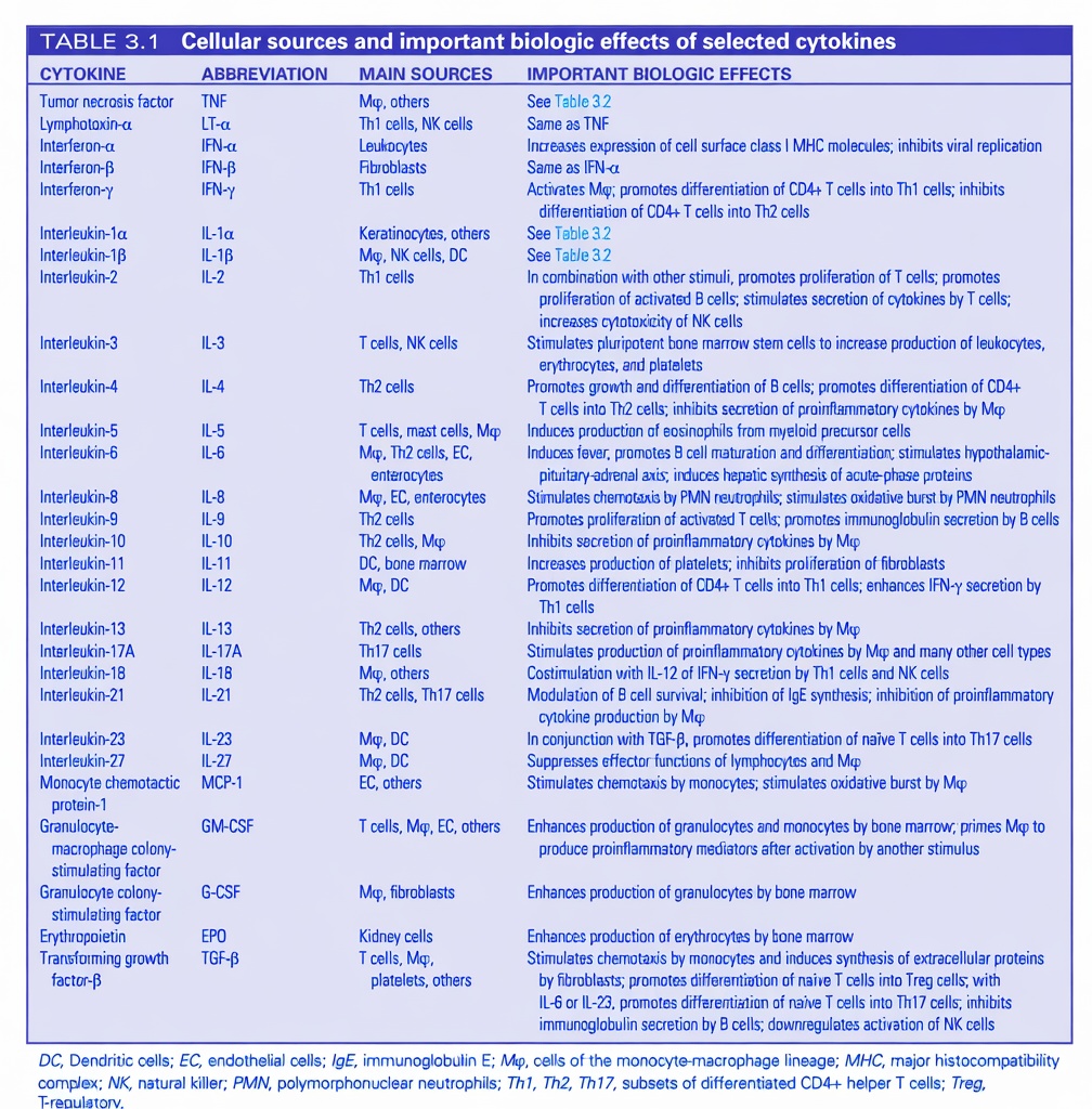
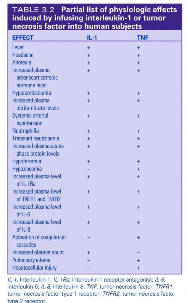
- A larger list is given in Tables 3.1 and 3.2.
- Main proinflammatory cytokines are:
- TNF-α
- IL-1
- IL-6
- IL-8
- IL-12
- IFN-γ
- TNF-α and IL-1β are hyperacute cytokines.
- They act within 1–2 hours after injury.
- IL-6 and IL-8 are subacute cytokines.
- They peak about 1–4 hours after injury.
- They stay in the blood longer than TNF-α and IL-1.
- Main antiinflammatory cytokines are:
- TGF-β
- IL-4
- IL-10
- During early inflammation, cytokines:
- Recruit PMN (neutrophil) leukocytes.
- Increase reactive oxygen species (ROS).
- Proinflammatory cytokines also promote blood clotting during trauma and infection.
TNF-α
- TNF-α is a 17-kDa protein.
- It is produced by:
- Innate immune cells.
- Adaptive immune cells.
- Fibroblasts.
- Along with IL-1, it is released quickly after infection or tissue injury.
- It starts rising within 30 minutes.
- Its half-life is 14–18 minutes.
- It reaches its highest level within 1–2 hours.
- TNF-α and IL-1 are involved in many inflammatory diseases.
- TNF-α acts through:
- TNFR1
- TNFR2
- sTNFR controls circulating TNF-α.
IL-1
- IL-1 is mainly produced by macrophages.
- It is also produced by adaptive immune cells and nonimmune cells.
- It acts through:
- IL-1R1
- IL-1R2
- IL-1Ra and soluble IL-1R2 regulate IL-1.
- IL-1 is also a hyperacute cytokine.
- Its half-life is about 10 minutes.
- Because TNF-α and IL-1 disappear quickly, they are poor markers for predicting injury severity or organ dysfunction.
- TNF-α and IL-1:
- Work together.
- Cause fever.
- Increase macrophage number, activity, and survival.
- They stimulate macrophages to release:
- IL-6
- IL-8
- Macrophage migration inhibitory factor.
- Lipid mediators.
- Reactive oxygen species.
- This increases inflammation.
- TNF-α increases:
- Endothelial adhesion molecules.
- Chemokines.
- Integrins on neutrophils.
- This helps immune cells enter tissues.
- TNF-α, IL-1, and complement promote the procoagulant state.
- They also activate the HPA axis and increase cortisol.
- Giving TNF-α or IL-1 alone can produce a condition similar to septic shock.
Interleukin-6
- IL-6 is a 21-kDa protein.
- It is a secondary cytokine.
- It is stimulated mainly by:
- IL-1
- TNF-α
- LPS
- It is produced by:
- Macrophages.
- Dendritic cells.
- Lymphocytes.
- Endothelial cells.
- Fibroblasts.
- Smooth muscle cells.
- IL-6 also causes fever.
- Its main function is the acute-phase response.
- The acute-phase response includes:
- Fever.
- Leukocytosis.
- Increased acute-phase proteins.
- The liver produces:
- CRP
- Complement proteins.
- Fibrinogen.
- Ferritin.
- CRP begins to rise about 8 hours after trauma.
- It peaks within 48 hours.
- IL-6 is a subacute cytokine.
- Persistent high IL-6 predicts a poor outcome in:
- SIRS.
- Sepsis.
- MODS.
- IL-6 also:
- Promotes blood clotting.
- Activates T cells and B cells.
- Helps produce new T and B cells.
- It may contribute to heart dysfunction during septic shock.
- IL-6 also has antiinflammatory effects.
- It reduces:
- TNF-α.
- IL-1.
- It increases:
- IL-1Ra.
- IL-10.
- TGF-β.
- It also stimulates prostaglandin E₂ release.
- IL-6 contributes to CARS, which occurs together with SIRS.
Interleukin-4
- IL-4 is an antiinflammatory cytokine.
- It is produced by:
- Mast cells.
- Basophils.
- Eosinophils.
- Th2 cells.
- It increases:
- IL-4.
- TGF-β.
- IL-10.
- It promotes Th2 cell formation.
- It inhibits Th1 cell formation.
- Therefore, it promotes the B-cell (humoral) immune response.
- It reduces the cell-mediated immune response.
- After central nervous system injury, IL-4-producing T cells appear.
- These cells protect neurons.
- They also improve recovery.
- Mice without IL-4 show poorer recovery after nervous system injury.
Interleukin-10
- IL-10 is a 35-kDa protein.
- It is produced by:
- Monocytes.
- Macrophages.
- Natural killer cells.
- Lymphocytes.
- IL-10 decreases:
- TNF-α.
- IL-1.
- IL-6.
- IFN-γ.
- It increases:
- IL-1Ra.
- sTNFR.
- It reduces:
- Phagocytosis.
- Antigen presentation by APCs.
- In LPS endotoxemia, IL-10 is protective.
- In polymicrobial sepsis, blocking IL-10 after 12 hours improved survival.
- Therefore, IL-10 can be helpful or harmful in sepsis.
- It may help convert early reversible sepsis into late irreversible sepsis.
Transforming Growth Factor-β
- TGF-β is a 25-kDa dimeric cytokine.
- Overall, it has antiinflammatory effects.
- It has three isoforms:
- TGF-β1.
- TGF-β2.
- TGF-β3.
- TGF-β regulates epithelial-to-mesenchymal transition (EMT).
- EMT is important for:
- Embryonic development.
- Tissue remodeling.
- Wound repair.
- TGF-β increases VEGF and promotes angiogenesis.
- It helps normal T-cell development.
- It prevents survival of autoreactive T cells.
- It decreases:
- IL-2 production.
- T-cell proliferation.
- It promotes regulatory T cells.
- It suppresses:
- IL-1.
- TNF-α.
- HMGB1.
- It increases:
- sTNFR.
- IL-1Ra.
- In early cancer, TGF-β suppresses tumor growth.
- In advanced cancer, tumor cells use TGF-β to:
- Grow.
- Invade.
- Metastasize.
- Through EMT, angiogenesis, and reduced inflammation, tumor cells escape immune attack.
KEY CONCEPT
- Cytokines are small proteins that control inflammation.
- TNF-α & IL-1 = First (hyperacute) cytokines → Fever + strong inflammation.
- IL-6 = Acute-phase cytokine → Produces CRP and is the best predictor of severe inflammation.
- IL-4, IL-10 & TGF-β = Main antiinflammatory cytokines.
- TGF-β helps wound healing, but in advanced cancer it can help tumors spread.
Conceptual Examples
- Bacterial injury → TNF-α & IL-1 rise first → Fever and inflammation start.
- Few hours later → IL-6 rises → Liver makes CRP.
- Healing phase → IL-10 & TGF-β reduce inflammation and help tissue repair.
MADE BYSELF LEARNING CEO AND FOUNDER DR SHEEN
