


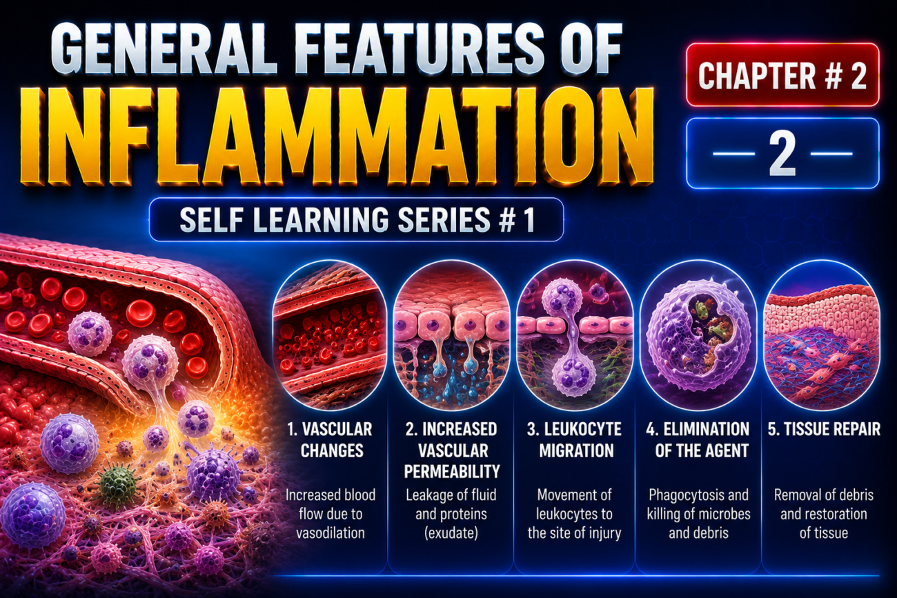
- Inflammation is a response of vascularized tissues to infection or tissue damage.
- It brings cells and molecules of host defense from the blood to the places where they are needed.
- The purpose is to eliminate the harmful cause of the injury.
- Although inflammation is often thought of as a harmful reaction, it is actually a protective response that is essential for survival.
- It helps remove the initial cause of cell injury, such as:
- Microbes
- Toxins
- It also removes the consequences of cell injury, such as:
- Necrotic cells
- Damaged tissues
- Inflammation also starts the process of repairing damaged tissues.
- Important defense mediators include:
- Phagocytic leukocytes
- Antibodies
- Complement proteins (Fig. 2.1)
- Most of these defense components normally circulate in the blood.
- While in the blood, they are separated from tissues and therefore cannot normally cause tissue damage.
- Infections and dead cells are usually located inside tissues, outside blood vessels.
- Inflammation brings leukocytes and proteins from the blood to:
- Foreign invaders, such as microbes
- Damaged or necrotic tissues
- The recruited cells and molecules are then activated.
- They remove the harmful or unwanted substances.
- Without inflammation:
- Infections would continue unchecked.
- Wounds would not heal.
- Injured tissues could remain as permanent, infected sores.
- Inflammation can be acute or chronic (Table 2.1).
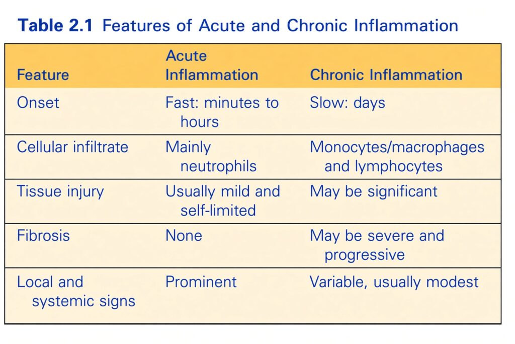
- Acute inflammation is the initial, rapid response to infection or tissue damage.
- It develops within minutes to hours and usually lasts for several hours to a few days.
- Its main features are:
- Leakage of fluid and plasma proteins, producing edema
- Accumulation of leukocytes, mainly neutrophils
- Neutrophils are also called polymorphonuclear leukocytes.
- When acute inflammation successfully removes the harmful stimulus, the reaction quickly subsides.
- If the harmful stimulus is not removed, the response can continue and progress to chronic inflammation.
- Chronic inflammation lasts for a longer time.
- It is associated with continuing:
- Tissue destruction
- Fibrosis, meaning deposition of connective tissue
- The external manifestations of inflammation are called the cardinal signs.
- They are:
- Heat — calor
- Redness — rubor
- Swelling — tumor
- Pain — dolor
- Loss of function — functio laesa
- The first four signs were described more than 2000 years ago by the Roman encyclopedist Celsus in De Medicina.
- Loss of function was added in the late 19th century by Rudolf Virchow, known as the “father of modern pathology.”
- These signs occur because of vascular changes and the recruitment and activation of leukocytes.
- Inflammatory reactions develop in steps called the five R’s:
- Recognition of the offending agent
- Recruitment of blood cells and proteins to the tissue
- Removal of the offending agent
- Regulation of the inflammatory reaction
- Repair of the injured tissue
- Although inflammation is normally protective, sometimes it itself becomes the cause of disease.
- In these situations, the damage caused by inflammation becomes the dominant feature.
- Inflammation caused by infections may produce local tissue damage and pain.
- Usually, these harmful effects disappear when the inflammation subsides, leaving little or no permanent damage.
- However, inflammation can become harmful when:
- It is directed against the body’s own tissues, as in autoimmune diseases
- It occurs against normally harmless environmental substances, as in allergies
- It continues for a long time, as in infections caused by microbes that are difficult to eliminate, such as Mycobacterium tuberculosis
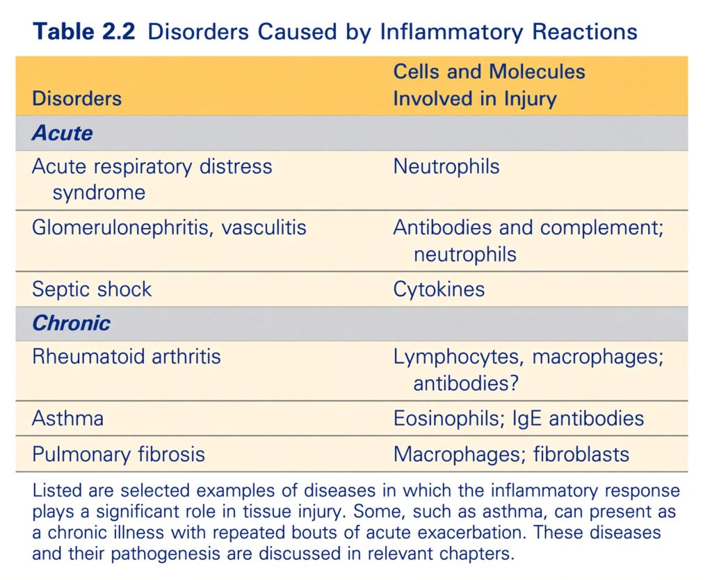
- These abnormal inflammatory reactions contribute to many chronic diseases, including:
- Rheumatoid arthritis
- Asthma
- Lung fibrosis (Table 2.2)
- Inflammation can also contribute to diseases mainly considered metabolic, degenerative, or genetic, such as:
- Atherosclerosis
- Type 2 diabetes
- Alzheimer disease
- Because inflammation can have many harmful effects, the lay press has dramatically called it “the silent killer.”
- Inadequate inflammation usually causes increased susceptibility to infections.
- Impaired inflammation can occur when production of leukocytes is reduced because the bone marrow is replaced by cancers, such as leukemias.
- It can also result from immunosuppressive drugs used to treat graft rejection and autoimmune diseases.
- Other conditions, such as malnutrition, can also impair inflammation.
- Rare inherited disorders can affect leukocyte function.
- These disorders help us understand how leukocytes normally respond.
- They are discussed in Chapter 5 in relation to immunodeficiency diseases.
- Once inflammation has removed the harmful agents, it subsides.
- At the same time, it starts the process of tissue repair.
- During tissue repair, injured tissue is replaced by:
- Regeneration of surviving cells
- Filling of remaining tissue defects with connective tissue (scarring)
KEY CONCEPT
Inflammation = a protective response that brings defense cells and molecules from blood to damaged tissue → removes the harmful cause → controls the response → repairs the tissue.
Conceptual examples:
- Microbe enters tissue → inflammation → leukocytes arrive → microbe removed → inflammation subsides → tissue repair
- Acute inflammation → minutes to hours → edema + neutrophils → short duration
- Persistent stimulus → chronic inflammation → continued tissue destruction + fibrosis
- Inflammation → heat + redness + swelling + pain + loss of function
- Five R’s → Recognition → Recruitment → Removal → Regulation → Repair
- Too little inflammation → increased susceptibility to infection
- Misdirected/excessive inflammation → tissue damage → diseases such as rheumatoid arthritis, asthma, and atherosclerosis
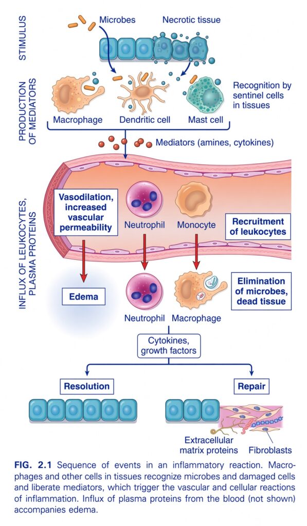
FIG. 2.1 — Sequence of Events in an Inflammatory Reaction
🧠 THE BIG IDEA
This figure shows the complete story of inflammation, from the moment tissue detects a problem until the tissue either returns to normal (resolution) or heals by repair.
The whole process can be remembered as:
Stimulus → Recognition → Mediators → Blood-vessel changes → Leukocyte recruitment → Elimination → Resolution or Repair
1. 🚨 STIMULUS — Something is wrong
At the very top, there are two major stimuli:
🦠 Microbes
Examples include bacteria and other infectious organisms.
💀 Necrotic tissue
Dead/damaged cells and tissues.
So inflammation can start because:
Something foreign is present OR something in the body has been damaged.
2. 👀 RECOGNITION — Sentinel cells detect the problem
The next step is:
Recognition by sentinel cells in tissues
What are sentinel cells?
They are cells that are already present in the tissue, waiting for danger.
The figure shows three important sentinel cells:
🟠 Macrophage
🟤 Dendritic cell
🟢 Mast cell
Think of them as:
Security guards permanently stationed in the tissue.
They detect:
- Microbes
- Damaged/necrotic cells
3. 🦠 MACROPHAGES
Macrophages recognize the danger and become activated.
They can:
- Detect microbes
- Detect damaged tissue
- Release inflammatory mediators
- Help remove microbes and dead cells
Think:
Macrophage = tissue security guard + cleanup cell
4. 🧬 DENDRITIC CELLS
Dendritic cells also recognize microbes and tissue damage.
They are important sentinel cells and participate in initiating immune responses.
For this figure, the key point is simply:
Dendritic cells recognize danger and participate in initiating inflammation.
5. 🟢 MAST CELLS
Mast cells are also already present in tissues.
When activated, they release important inflammatory mediators, particularly:
Amines such as histamine
These mediators help produce changes in local blood vessels.
6. 🧪 PRODUCTION OF MEDIATORS
After recognizing the problem, the sentinel cells release:
Inflammatory mediators
The figure specifically gives:
- Amines
- Cytokines
Think of mediators as:
Chemical messages that tell the surrounding tissue and blood vessels what to do.
7. 📢 MEDIATORS START THE INFLAMMATORY RESPONSE
The mediators act on nearby blood vessels and cells.
They produce two major vascular changes:
1. Vasodilation
2. Increased vascular permeability
These are extremely important.
8. 🔴 VASODILATION
Vasodilation = blood vessels become wider.
When vessels dilate:
More blood flows into the affected tissue.
This contributes to the classic inflammatory features such as:
- Redness
- Warmth
Think:
Vasodilation = open the blood-flow gates.
9. 💧 INCREASED VASCULAR PERMEABILITY
The blood vessels become more permeable.
This allows:
- Fluid
- Plasma proteins
to leave the bloodstream and enter the tissue.
The figure specifically notes that plasma proteins enter from the blood, although they are not drawn in the diagram.
10. 💦 EDEMA
When fluid moves out of the blood vessels and accumulates in the tissue:
Edema develops
So:
↑ Vascular permeability
↓
Fluid leaves blood vessels
↓
Fluid accumulates in tissue
↓
Edema
Easy memory:
Permeability ↑ → fluid out → edema
11. 🩸 NOW LEUKOCYTES ARE RECRUITED
The inflammatory mediators also cause:
Recruitment of leukocytes
Leukocytes = white blood cells
The figure shows:
Neutrophils
and
Monocytes
These cells leave the blood and enter the affected tissue.
12. 🟣 NEUTROPHILS
Neutrophils are shown first.
They are particularly important in acute inflammation.
They leave the bloodstream and move toward the site of injury/infection.
Think:
Neutrophils = rapid-response soldiers
They arrive quickly to attack microbes and help remove damaged material.
13. 🟠 MONOCYTES
Monocytes also leave the bloodstream.
Once they enter tissues, they can differentiate into:
Macrophages
So:
Monocyte → tissue → macrophage
Macrophages then help:
- Remove microbes
- Remove dead cells
- Produce cytokines and growth factors
14. 🦠 ELIMINATION OF MICROBES AND DEAD TISSUE
Now look at the right side of the blood vessel.
The recruited cells help with:
Elimination of microbes and dead tissue
This is one of the main purposes of inflammation.
The inflammatory response is not just about swelling.
Its goal is:
Remove the cause of injury and clear damaged tissue.
15. 🧹 MACROPHAGES = CLEANUP CREW
The macrophage shown in the tissue is engulfing material.
It helps remove:
- Microbes
- Dead cells
- Tissue debris
Think:
Neutrophils = rapid attack
Macrophages = attack + cleanup + coordination
16. 📢 CYTOKINES AND GROWTH FACTORS
After the inflammatory response, macrophages and other cells produce:
Cytokines
and
Growth factors
These chemical signals help determine what happens next.
The figure shows two possible outcomes:
Resolution
or
Repair
17. 🟢 RESOLUTION
Resolution means:
The inflammation settles down and the tissue returns toward its normal state.
Look at the bottom-left.
The epithelial cells become organized again.
Simple sequence:
Injury/infection
      ↓
Inflammation
      ↓
Cause eliminated
      ↓
Inflammatory response stops
      ↓
Normal tissue restored
      ↓
RESOLUTION
Think:
Resolution = clean up and return to normal.
18. 🛠️ REPAIR
Sometimes the original tissue cannot be completely restored.
Then the body uses:
Repair
The figure shows:
- Fibroblasts
- Extracellular matrix proteins
Fibroblasts produce extracellular matrix components that help rebuild damaged tissue.
Simple sequence:
Injury
  ↓
Inflammation
  ↓
Tissue damage remains
  ↓
Fibroblasts become involved
  ↓
Extracellular matrix is produced
  ↓
REPAIR
19. 🧱 FIBROBLASTS
Fibroblasts are important cells in tissue repair.
They produce:
Extracellular matrix proteins
These proteins provide structural support for the repaired tissue.
Think:
Fibroblasts = construction workers
They help build the framework of repaired tissue.
20. 🔄 RESOLUTION vs REPAIR
This is one of the most important comparisons.
| Resolution | Repair |
|---|---|
| Tissue returns toward normal | Damaged tissue is rebuilt |
| Minimal lasting structural damage | Structural replacement may occur |
| Normal tissue architecture can be restored | Fibroblasts and extracellular matrix are important |
| “Back to normal” | “Build/replace what was damaged” |
Easy memory:
Resolution = restore
Repair = rebuild
🔥 NOW FOLLOW EVERY ARROW IN THE FIGURE
STEP 1 — STIMULUS
Microbes OR necrotic tissue
↓
STEP 2 — RECOGNITION
Macrophages + dendritic cells + mast cells
↓
STEP 3 — MEDIATOR RELEASE
Amines + cytokines
↓
STEP 4 — BLOOD-VESSEL CHANGES
Vasodilation + increased vascular permeability
↓
STEP 5 — EDEMA
Fluid + plasma proteins leave blood vessels
↓
STEP 6 — LEUKOCYTE RECRUITMENT
Neutrophils + monocytes enter tissue
↓
STEP 7 — ELIMINATION
Microbes + dead tissue are removed
↓
STEP 8 — CYTOKINES + GROWTH FACTORS
↓
Two possible outcomes:
Resolution
→ tissue returns toward normal
Repair
→ fibroblasts + extracellular matrix rebuild damaged tissue
🧠 THE WHOLE FIGURE AS A SIMPLE STORY
Imagine a house security system:
🚨 Something dangerous enters:
Microbe / tissue damage
↓
👮 Security guards detect it:
Macrophage / dendritic cell / mast cell
↓
📢 They send alarms:
Inflammatory mediators
↓
🚪 Blood vessels open:
Vasodilation
↓
💧 The vessel walls become leaky:
Increased permeability → edema
↓
🪖 More soldiers arrive:
Neutrophils + monocytes
↓
🧹 They remove the enemy and debris:
Elimination
↓
🏠 The house is restored:
Resolution → return toward normal
OR
Repair → rebuild damaged structure
⭐ WHAT EACH CELL DOES
| Cell | Main role in this figure |
|---|---|
| Macrophage | Recognizes danger, releases mediators, removes microbes/dead tissue, produces cytokines/growth factors |
| Dendritic cell | Sentinel cell that recognizes danger |
| Mast cell | Sentinel cell that releases inflammatory mediators |
| Neutrophil | Rapidly recruited leukocyte that helps eliminate microbes |
| Monocyte | Recruited from blood; can become macrophage in tissues |
| Fibroblast | Produces extracellular matrix during repair |
🎯 MOST IMPORTANT CONCEPTS
1. Inflammation starts because of:
Microbes or necrotic tissue
2. Danger is detected by:
Macrophages, dendritic cells, mast cells
3. They release:
Amines + cytokines
4. These cause:
Vasodilation + increased vascular permeability
5. Result:
Edema + leukocyte recruitment
6. Main recruited cells shown:
Neutrophils + monocytes
7. Their job:
Eliminate microbes and dead tissue
8. Final outcomes:
Resolution OR repair
🔑 FINAL MEMORY LINE
Stimulus → Recognition → Mediators → Vasodilation/permeability → Edema → Leukocyte recruitment → Elimination → Resolution or Repair.
Even shorter:
“Detect → Signal → Open vessels → Recruit cells → Eliminate → Restore or Repair.”
CAUSES OF INFLAMMATION
- There are many causes of inflammation, but the following are the most common:
- Infections cause inflammation when the products of microbes are recognized by the body.
- Recognition of these microbial products produces different types of inflammatory reactions.
- Tissue necrosis can also cause inflammation.
- Tissue necrosis may result from:
- Ischemia — reduced blood flow
- Trauma
- Physical injury, such as thermal injury and irradiation
- Chemical injury, such as exposure to toxins
- Ischemia causes infarction in organs such as the heart and brain and in other tissues.
- Molecules released from necrotic cells can trigger inflammation even when there is no infection.
- This type of inflammation is called “sterile inflammation.”
- Foreign bodies can also cause sterile inflammation.
- Examples include:
- Sutures
- Tissue implants
- Immune reactions, also called hypersensitivity, can cause inflammation.
- These are reactions in which the normally protective immune system damages the person’s own tissues.
- Autoimmune diseases and allergies are examples of diseases caused by immune responses.
- In both conditions, inflammation is an important contributor to tissue injury (Chapter 5).
KEY CONCEPT
Main causes of inflammation = Infection + Tissue necrosis + Foreign bodies + Immune reactions.
Conceptual examples:
- Infection → microbial products recognized → inflammation
- Ischemia → tissue necrosis/infarction → inflammatory response
- Trauma/thermal injury/toxins → cell injury and necrosis → sterile inflammation
- Suture or tissue implant → foreign body → sterile inflammation
- Autoimmunity/allergy → immune system damages tissue → inflammation → tissue injury
RECOGNITION OF MICROBES AND DAMAGED CELLS
- The first step of inflammation is the recognition of microbes and dead (necrotic) cells.
- This recognition is carried out by cellular receptors and proteins circulating in the blood.
- All tissues contain resident cells whose main job is to:
- Detect foreign invaders or dead cells
- Ingest and destroy these harmful substances
- Start the inflammatory response
- The inflammatory response then brings cells and proteins from the blood to complete the removal process.
- The most important sentinel cells are:
- Tissue-resident macrophages
- Dendritic cells
- These cells have receptors that recognize microbial products in different parts of the cell:
- Cell surface → recognizes microbes in the extracellular space
- Endosomes → recognizes microbes that have been ingested
- Cytosol → recognizes certain microbes that may survive inside the cell
- The best-known receptors are Toll-like receptors (TLRs) (Chapter 5).
- When TLRs are activated, they cause production of cytokines.
- These cytokines trigger inflammation.
- Another sensing system consists of cytosolic NOD-like receptors (NLRs).
- When NLRs are activated, they recruit and activate a multiprotein complex called the inflammasome (Chapter 5).
- The inflammasome produces the biologically active cytokine interleukin-1 (IL-1).
- NLRs can recognize many different stimuli, including:
- Microbial products
- Leaked DNA, which indicates cell damage
- Decreased potassium levels inside the cytosol, which also indicate cell damage
- The cytokines produced during this process cause inflammation.
- The inflammation then helps eliminate the original stimulus, such as:
- Microbes
- Dead-cell debris
- If microbes pass through the tissue’s sentinel defense system and enter the bloodstream, they can then be recognized by plasma proteins.
- Important plasma proteins include:
- Antibodies
- Complement proteins
- These proteins can destroy microbes circulating in the blood.
- They can also be recruited to infected tissues, where they stimulate inflammatory reactions.
- With this recognition process understood, the next discussion focuses on acute inflammation, its mechanisms, and how it eliminates microbes and dead cells.
KEY CONCEPT
Recognition = Detect the danger → activate inflammatory signals → bring defense mechanisms → remove the harmful stimulus.
Conceptual examples:
- Microbe outside cell → surface TLR recognizes it → cytokines produced → inflammation
- Microbe ingested into endosome → TLR recognizes microbial products → inflammation
- Microbe inside cytosol → NLR detects it → inflammasome → IL-1 → inflammation
- Cell damage → leaked DNA/decreased cytosolic K⁺ → NLR activation → inflammation
- Microbe enters blood → antibodies/complement recognize it → microbe destruction + inflammatory response
- Overall: Recognition → inflammation → elimination of microbes/dead-cell debris
ACUTE INFLAMMATION
- Acute inflammation has three major components:
- Dilation of small blood vessels
- Increased permeability of the microvasculature
- Emigration of leukocytes from the microcirculation (see Fig. 2.1)
- Most of these changes occur in the postcapillary venules at the site of infection or tissue injury.
- The walls of these vessels can respond to stimuli.
- Their walls are also thin enough to allow fluid and proteins to pass through.
- Vasodilation slows down blood flow.
- This slowing of blood flow prepares the tissue for the next inflammatory reactions.
- Increased vascular permeability allows plasma proteins to leave the blood and enter the affected tissue.
- Transmigration moves leukocytes from inside the blood vessels into the area of infection or necrosis.
- Once in the affected tissue, leukocytes help:
- Destroy harmful agents
- Remove damaged material
- All these inflammatory reactions are caused by cytokines and other molecules.
- Together, these molecules are called inflammatory mediators.
- They are produced at the site of infection or necrosis.
KEY CONCEPT
Acute inflammation = Vessels open and become leaky + leukocytes leave the blood → reach the injured/infected tissue → destroy harmful agents and clean up the damage.
Conceptual sequence:
Infection/tissue injury → inflammatory mediators → vasodilation → increased vascular permeability → leukocyte transmigration → destruction of harmful agents + removal of damage
Vascular Reactions in Acute Inflammation
- Vasodilation is one of the earliest reactions in acute inflammation.
- It causes the visible redness (erythema) and warmth seen in most acute inflammatory reactions.
- The most important chemical mediator of vasodilation is histamine.
- Vasodilation is quickly followed by increased permeability of the microvasculature.
- This allows protein-rich fluid to move out of the blood vessels into the extravascular tissues.
- The escape of fluid, proteins, and blood cells from blood vessels into the interstitial tissue or body cavities is called exudation (Fig. 2.2).
- An exudate is fluid outside the blood vessels that:
- Has a high protein concentration
- Contains cellular debris
- An exudate indicates that the permeability of small blood vessels has increased, usually during inflammation.
- A transudate is different from an exudate.
- It has:
- Low protein content, mostly albumin
- Little or no cellular material
- Low specific gravity
- A transudate is essentially an ultrafiltrate of blood plasma.
- It forms because of an osmotic or hydrostatic imbalance across the vessel wall.
- It occurs without increased vascular permeability.
- It is usually not associated with inflammation (Chapter 3).
- Edema means an excess of fluid in the interstitial tissue or serous cavities.
- Edema can be either:
- Exudate
- Transudate
- Pus is a purulent exudate.
- It is an inflammatory exudate rich in:
- Leukocytes, mainly neutrophils
- Debris from dead cells
- Often microbes
- The main mechanism causing increased vascular permeability is contraction of endothelial cells.
- This contraction creates openings between endothelial cells.
- This mechanism is triggered by:
- Histamine
- Bradykinin
- Leukotrienes
- Other chemical mediators
- It occurs rapidly after exposure to these mediators, usually within 15–30 minutes.
- It is usually short-lived.
- In unusual situations, such as burns, increased vascular permeability can result from direct injury to endothelial cells.
- In these cases, leakage begins immediately after injury.
- The leakage continues for several hours until the damaged vessels become thrombosed or repaired.
- Loss of fluid and increased vessel diameter cause blood flow to slow down.
- They also increase the concentration of red blood cells inside small vessels.
- This increases the viscosity of blood.
- The affected small vessels become filled with red blood cells.
- This condition is called stasis.
- Microscopically, stasis appears as vascular congestion.
- Externally, it appears as localized redness of the affected tissue.
- In addition to blood vessel changes, lymph flow also increases.
- Increased lymph flow helps drain the edema fluid that accumulates because of increased vascular permeability.
- The lymphatic vessels may also become secondarily inflamed.
- Inflammation of lymphatic vessels is called lymphangitis.
- Clinically, lymphangitis appears as red streaks extending from the inflammatory focus along lymphatic channels.
- The draining lymph nodes may also become enlarged and painful.
- Enlargement occurs because of increased cellularity.
- The combination of these pathologic changes is called reactive or inflammatory lymphadenitis (Chapter 10).
KEY CONCEPT
Acute inflammation → vasodilation + increased vascular permeability → fluid/proteins leave vessels → edema + leukocytes enter tissue.
Conceptual examples:
- Histamine → vasodilation → increased blood flow → redness + warmth
- Histamine/bradykinin/leukotrienes → endothelial contraction → gaps → protein-rich exudate
- Inflammation → exudate → high protein + cellular debris
- Hydrostatic/osmotic imbalance → transudate → low protein + no inflammation
- Exudate + neutrophils + dead-cell debris ± microbes → pus
- Fluid loss + slow blood flow + increased RBC concentration → stasis → congestion + redness
- Inflammation → increased lymph flow → lymphangitis → red streaks → draining lymphadenitis
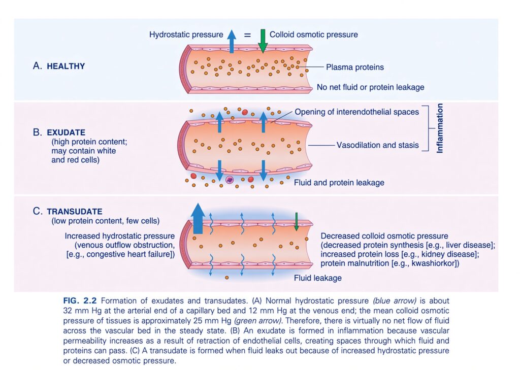
FIG. 2.2 — Formation of Exudates and Transudates
🧠 THE BIG IDEA
This figure explains why fluid leaves blood vessels and, most importantly, the difference between:
Exudate
→ fluid escapes because the vessel wall becomes leaky during inflammation
Transudate
→ fluid escapes because of a pressure imbalance, while the vessel wall itself is not primarily inflamed.
The easiest memory is:
EXUDATE = inflammation → leaky vessel → protein-rich fluid
TRANSUDATE = pressure problem → water-rich, protein-poor fluid
A. 🟢 HEALTHY BLOOD VESSEL
Look at the top panel.
The blood vessel is normal.
There are plasma proteins inside the vessel.
The figure shows two opposing forces:
🔵 Hydrostatic pressure
This pressure tends to:
Push fluid OUT of the blood vessel.
Think:
Hydrostatic pressure = PUSH fluid out
🟢 Colloid osmotic pressure
This is mainly produced by:
Plasma proteins
It tends to:
Pull/retain fluid IN the blood vessel.
Think:
Colloid osmotic pressure = PULL fluid in
⭐ THE BALANCE
The figure shows:
Hydrostatic pressure ≈ Colloid osmotic pressure
Therefore:
There is virtually no net fluid or protein leakage.
So in a healthy vessel:
🔵 Hydrostatic pressure
        OUT
         ↓
       [VESSEL]
         ↑
        IN
🟢 Colloid osmotic pressure
The two forces are approximately balanced.
1. What are the normal pressure values?
The caption gives:
Arterial end:
Hydrostatic pressure ≈ 32 mm Hg
Venous end:
Hydrostatic pressure ≈ 12 mm Hg
Tissue colloid osmotic pressure:
≈ 25 mm Hg
The exact numbers are less important here than understanding the direction of the forces.B. 🔴 EXUDATE
Now look at the middle panel.
This occurs during:
INFLAMMATION
The key event is:
Opening of interendothelial spaces
The endothelial cells pull apart/retract, creating gaps between them.
2. 🚪 What does “opening of interendothelial spaces” mean?
Endothelium
= cells lining the inside of blood vessels.
Normally:
The endothelial cells are closely joined.
During inflammation:
The endothelial cells retract, creating spaces between them.
Think of a wall made of bricks.
Normal:
🧱🧱🧱🧱🧱
No large gaps.
In inflammation:
🧱 ↔ 🧱 ↔ 🧱
Spaces open between the cells.
3. 💧 What passes through these spaces?
Because the vessel becomes more permeable:
Fluid can escape
AND
Plasma proteins can escape
AND sometimes:
White and red cells can also appear in the extravascular fluid.
Therefore:
Inflammation → increased vascular permeability → protein-rich fluid leaves vessel
4. 🧪 Why is EXUDATE protein-rich?
This is the key feature.
Because inflammation creates gaps in the endothelial barrier:
Large plasma proteins can escape from the blood into the tissue.
Therefore:
Exudate = high protein content
It may also contain:
- White blood cells
- Red blood cells
depending on the type and severity of vascular injury.
5. 🔴 VASODILATION AND STASIS
The middle panel also shows:
Vasodilation and stasis
Vasodilation
Blood vessels become wider.
Stasis
Blood flow becomes slower.
Together, these are important vascular changes during inflammation.
They contribute to:
Movement of fluid and proteins out of vessels and facilitate leukocyte recruitment.
6. 💦 EXUDATE → EDEMA
When fluid leaves the vessel and enters the tissue:
Edema develops
So:
Inflammation
     ↓
Vessel permeability ↑
     ↓
Protein + fluid escape
     ↓
Exudate
     ↓
Tissue swelling
⭐ EXUDATE IN ONE SENTENCE
Exudate is protein-rich inflammatory fluid produced because increased vascular permeability allows fluid and proteins to escape through endothelial gaps.
C. 🔵 TRANSUDATE
Now look at the bottom panel.
The figure describes:
Transudate
It has:
Low protein content and few cells
This is the major difference from exudate.
7. 🚨 TRANSUDATE IS NOT PRIMARILY CAUSED BY INFLAMMATION
This is extremely important.
In transudate:
The vessel wall is not made leaky by inflammatory endothelial injury.
Instead:
The balance of hydrostatic and colloid osmotic pressures is disturbed.
So fluid moves out even though there is no major inflammatory opening of endothelial gaps.
8. 🔵 CAUSE #1 — INCREASED HYDROSTATIC PRESSURE
Look at the left side of panel C.
Increased hydrostatic pressure
The figure gives:
Venous outflow obstruction
Example:
Congestive heart failure
How does this produce edema?Imagine fluid being pushed through a pipe.
If pressure inside the pipe increases:
More water is pushed outward.
So:
↑ Hydrostatic pressure
↓
More fluid pushed out of vessel
↓
Transudate
9. ❤️ CONGESTIVE HEART FAILURE
The figure gives congestive heart failure as an example.
In heart failure, venous pressure can increase.
Therefore:
↑ Venous pressure
↓
↑ Hydrostatic pressure
↓
Fluid moves out of vessels
↓
Edema / transudate
10. 🟢 CAUSE #2 — DECREASED COLLOID OSMOTIC PRESSURE
Look at the right side of panel C.
Decreased colloid osmotic pressure
Remember:
Plasma proteins help retain water inside blood vessels.
Therefore:
Less plasma protein → weaker inward pull → more fluid leaves the vessel.
11. WHY CAN PLASMA PROTEINS DECREASE?
The figure gives three examples.
① Decreased protein synthesis
Example:
Liver disease
The liver produces many plasma proteins, especially albumin.
Therefore:
Liver disease → ↓ protein synthesis → ↓ plasma colloid osmotic pressure
↓
Fluid leaves vessels
↓
Transudate
② Increased protein loss
Example:
Kidney disease
If excessive protein is lost in urine:
Protein loss ↑
↓
Plasma protein ↓
↓
Colloid osmotic pressure ↓
↓
Fluid moves out
③ Protein malnutrition
Example:
Kwashiorkor
Insufficient protein intake can reduce plasma proteins.
↓
Colloid osmotic pressure ↓
↓
Fluid leaves the circulation.
🧠 TRANSUDATE = PRESSURE PROBLEM
There are two major ways:
Hydrostatic pressure ↑
OR
Colloid osmotic pressure ↓
Both cause:
Fluid to move out of the blood vessel.
But the vessel does not become highly permeable to proteins as it does in inflammation.
Therefore:
Transudate = low protein + few cells
🔥 EXUDATE vs TRANSUDATE
| Feature | Exudate | Transudate |
|---|---|---|
| Main cause | Inflammation | Pressure imbalance |
| Vessel permeability | Increased | Relatively preserved |
| Endothelial gaps | Open | No inflammatory gaps |
| Protein | High | Low |
| Cells | May contain many cells | Few cells |
| Main mechanism | Fluid + protein escape | Mainly fluid movement |
| Examples | Inflammatory conditions | Heart failure, low plasma proteins |
🧠 THE EASIEST WAY TO DIFFERENTIATE THEM
🔴 EXUDATE
Think:
“EXIT because the vessel is damaged/leaky.”
INFLAMMATION
     ↓
Endothelial gaps open
     ↓
Protein escapes
     ↓
Fluid escapes
     ↓
EXUDATE
High protein
🔵 TRANSUDATE
Think:
“Pressure pushes water out.”
Pressure imbalance
       ↓
Fluid pushed out
       ↓
Vessel wall remains relatively intact
       ↓
TRANSUDATE
Low protein⭐ VERY IMPORTANT EXAM TRICK
If you see:
Inflammation + high-protein fluid
Think:
EXUDATE
If you see:
Heart failure + low-protein edema
Think:
TRANSUDATE
🧩 UNDERSTAND THE ARROWS IN EACH PANEL
🟢 A — HEALTHY
🔵 Hydrostatic pressure → pushes outward
🟢 Colloid osmotic pressure → pulls inward
Result:
Balance → no significant net leakage
🔴 B — EXUDATE
Inflammation
↓
Vasodilation + stasis
↓
Opening of interendothelial spaces
↓
Fluid + proteins leak out
↓
Protein-rich exudate
🔵 C — TRANSUDATE
Either:
↑ Hydrostatic pressure
OR
↓ Colloid osmotic pressure
↓
Fluid leakage
↓
Low-protein transudate
🎯 FINAL CONCEPT MAP
                    FLUID LEAVES VESSEL
                           │
              ┌────────────┴────────────┐
              ↓                         ↓
          EXUDATE                  TRANSUDATE
              ↓                         ↓
       INFLAMMATION               PRESSURE PROBLEM
              ↓                         ↓
     Endothelial gaps          ↑ Hydrostatic pressure
              ↓                  OR ↓ Oncotic pressure
       Protein escapes                 ↓
              ↓                   Mainly fluid escapes
      HIGH PROTEIN                     ↓
              ↓                   LOW PROTEIN
🔑 ONE-LINE MEMORY
EXUDATE = inflammation makes vessels leaky → protein-rich fluid.
TRANSUDATE = hydrostatic pressure rises or colloid osmotic pressure falls → protein-poor fluid.
Leukocyte Recruitment to Sites of Inflammation
- The movement of leukocytes from inside a blood vessel to the tissue is a multistep process controlled by adhesion molecules and cytokines.
- Normally, leukocytes move rapidly through small blood vessels.
- During inflammation, leukocytes must first stop inside the vessel and then move toward the harmful agent or damaged tissue outside the vessel.
- This process occurs in three main phases:
- Adhesion of leukocytes to the endothelium
- Transmigration of leukocytes through the vessel wall
- Movement toward the offending agent (Fig. 2.3)
- When blood flows from capillaries into postcapillary venules under normal laminar flow, red blood cells are mainly concentrated in the center of the vessel.
- This pushes leukocytes toward the vessel wall.
- Early in inflammation, blood flow becomes slower because of stasis.
- Because leukocytes are larger than red blood cells, they slow down and move closer to the endothelial surface.
- This movement toward the vessel wall is called margination.
- Being close to the vessel wall allows leukocytes to detect and respond to changes in the endothelium.
- When endothelial cells are activated by locally produced cytokines and other inflammatory mediators, they express adhesion molecules.
- Leukocytes attach loosely to these molecules.
- They repeatedly attach and detach, causing them to tumble along the endothelial surface.
- This process is called rolling.
- Eventually, the leukocytes stop at a particular point and adhere firmly to the endothelium.
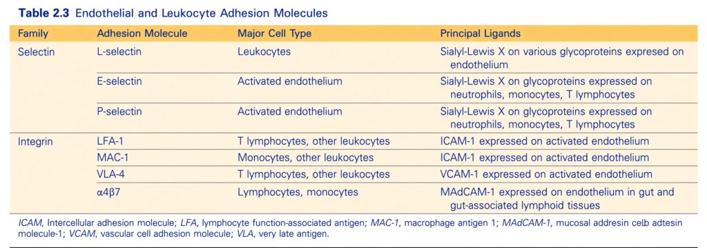
- The initial weak attachment and rolling are mediated by a family of proteins called selectins (Table 2.3).
- Selectins are receptors found on leukocytes and endothelial cells.
- Their extracellular part binds to carbohydrates, which explains the “lectin” part of the name.
- The ligands for selectins are sialic acid–containing oligosaccharides attached to glycoproteins.
- Some of these ligands are present on leukocytes, while others are present on endothelial cells.
- Endothelial cells express:
- E-selectin
- P-selectin
- The ligand for L-selectin
- Leukocytes express L-selectin.
- E-selectin and P-selectin are normally expressed at low levels or not at all on nonactivated endothelial cells.
- Their expression increases after stimulation by cytokines and other inflammatory mediators.
- Therefore, leukocyte binding occurs mainly at sites of infection or tissue injury, where these mediators are produced.
- In nonactivated endothelial cells, P-selectin is mainly stored inside intracellular membrane-bound vesicles called Weibel-Palade bodies.
- Within minutes of exposure to mediators such as histamine or thrombin, P-selectin moves to the cell surface.
- Similarly, E-selectin and the ligand for L-selectin are not normally expressed on resting endothelium.
- They are induced after stimulation by IL-1 and tumor necrosis factor (TNF).
- IL-1 and TNF are produced by:
- Tissue macrophages
- Dendritic cells
- Mast cells
- Endothelial cells
- These cells produce IL-1 and TNF after encountering microbes or dead tissues.
- Selectin-mediated interactions have low affinity and a fast off-rate.
- Therefore, flowing blood can easily break these weak connections.
- As a result, leukocytes repeatedly bind, detach, and bind again to the endothelium.
- These weak rolling interactions slow the leukocytes down.
- This gives leukocytes enough time to recognize additional adhesion molecules on the endothelial surface.
KEY CONCEPT
Leukocyte recruitment begins as:
Stasis → Margination → Selectins → Weak attachment → Rolling
Conceptual example:
Inflammation → blood flow slows → leukocytes move toward vessel wall (margination) → endothelial cells express selectins → leukocytes weakly attach → bind and detach repeatedly → rolling → leukocytes become ready for firm adhesion.
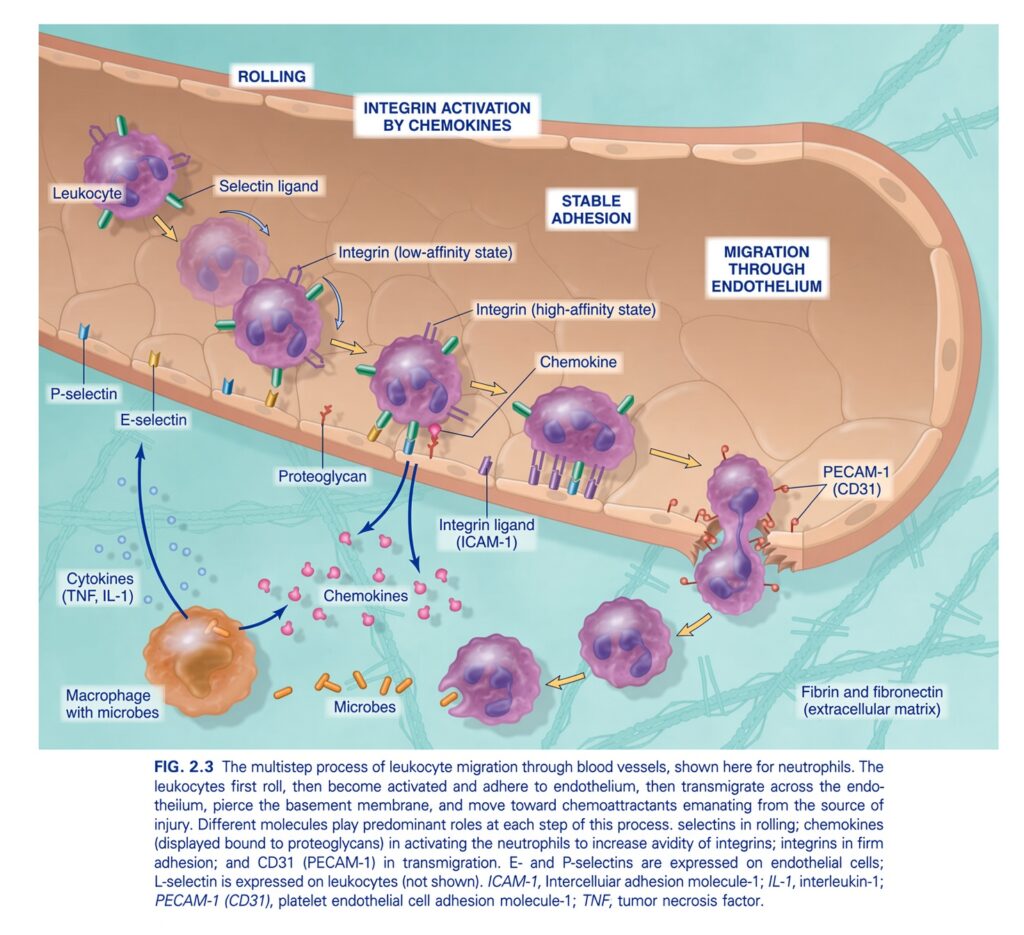
FIG. 2.3 — Multistep Process of Leukocyte Migration Through Blood Vessels
🧠 THE BIG IDEA
This figure shows how a leukocyte, especially a neutrophil, leaves the bloodstream and enters injured tissue.
The leukocyte does not simply jump out of the blood vessel.
It follows a specific sequence:
Rolling → Integrin activation → Stable adhesion → Transmigration → Chemotaxis
🧠 Super-easy memory:
ROLL → ACTIVATE → STICK → SQUEEZE → FOLLOW
1. 🚨 FIRST: INJURY ACTIVATES THE TISSUE
At the bottom-left, there is:
Macrophage with microbes
The macrophage recognizes the microbes and releases:
Cytokines
- TNF
- IL-1
These cytokines act on the nearby blood-vessel endothelium.
2. 🩸 CYTOKINES ACTIVATE THE ENDOTHELIUM
The cytokines cause endothelial cells to express adhesion molecules, especially:
E-selectin
P-selectin
These molecules are important for the first step:
Rolling
3. 🟣 STEP 1 — ROLLING
Look at the upper-left portion of the vessel.
The leukocyte is moving along the endothelial surface but is repeatedly:
Attaching → releasing → attaching → releasing
So instead of stopping immediately, it:
ROLLS along the vessel wall
Why does it roll?
Because of weak interactions between:
Selectins on endothelial cells
and
Selectin ligands on leukocytes
The figure shows:
- P-selectin
- E-selectin
- Selectin ligand
4. 🧲 SELECTINS = ROLLING MOLECULES
This is one of the most important exam concepts.
E-selectin
→ on endothelial cells
P-selectin
→ on endothelial cells
L-selectin
→ on leukocytes
They help leukocytes make weak, temporary attachments to the endothelium.
Therefore:
Selectins → Rolling
5. 🧠 WHY DOESN’T THE LEUKOCYTE STOP DURING ROLLING?
Because the selectin-leukocyte interactions are:
Weak and transient
They repeatedly break and reform.
Therefore the leukocyte:
Slows down but continues moving.
Think of it like a person running past a wall and repeatedly touching it with their hand.
Touch → release → touch → release
That is:
Rolling
6. 🩷 STEP 2 — CHEMOKINES ACTIVATE INTEGRINS
Now look toward the middle of the figure.
The endothelium displays:
Chemokines
The figure shows chemokines attached to:
Proteoglycans
These chemokines interact with receptors on the leukocyte.
This causes:
Integrin activation
7. 🔄 LOW-AFFINITY → HIGH-AFFINITY INTEGRINS
This is a very important visual part of the figure.
Initially:
Integrin = low-affinity state
The leukocyte is still rolling.
Then:
Chemokine binds to leukocyte
↓
Integrin changes shape/activation state
↓
Integrin = high-affinity state
Now the leukocyte can attach strongly to the endothelium.
🧠 Think of an integrin like a hand
Before chemokine activation:
✋ Weak grip
After activation:
✊ Strong grip
Therefore:
Chemokines convert leukocyte integrins from a low-affinity to a high-affinity state.
8. 🟢 STEP 3 — STABLE ADHESION
Now the leukocyte stops rolling.
Why?
Because activated integrins bind strongly to their endothelial ligands.
The figure specifically shows:
Integrin ligand — ICAM-1
So:
Leukocyte integrin
⬇️ binds strongly to
ICAM-1 on endothelial cells
↓
Stable adhesion
⭐ INTEGRINS = FIRM ADHESION
Remember this distinction:
Selectins
→ Rolling
Integrins
→ Firm/stable adhesion
This is a very high-yield concept.
9. 🔴 WHAT IS ICAM-1?
The figure labels:
ICAM-1
This is an adhesion molecule on endothelial cells.
It binds activated leukocyte integrins.
Therefore:
Integrin + ICAM-1 → strong attachment of leukocyte to endothelium
10. 🧍 THE LEUKOCYTE NOW STOPS
At this point the sequence has changed:
Before:
Rolling
The leukocyte is moving.
Now:
Stable adhesion
The leukocyte is firmly attached to the endothelial surface.
Think:
Selectins slow the leukocyte down → chemokines activate integrins → integrins make the leukocyte stop.
11. 🚪 STEP 4 — MIGRATION THROUGH THE ENDOTHELIUM
Now look toward the right side.
The figure labels:
Migration through endothelium
The leukocyte squeezes between endothelial cells.
This process is called:
Transmigration
or
Diapedesis
12. 🔵 PECAM-1 (CD31)
The figure specifically identifies:
PECAM-1 (CD31)
This molecule participates in the leukocyte’s movement across the endothelial junction.
So:
PECAM-1 (CD31) → transmigration
Easy memory:
PECAM = passage across endothelium
13. 🧱 THE LEUKOCYTE CROSSES THE BASEMENT MEMBRANE
After passing between endothelial cells, the leukocyte must move through the underlying tissue barrier, including the basement membrane.
It then enters the:
Extravascular tissue
14. 🧭 STEP 5 — CHEMOTAXIS
Once the leukocyte is outside the vessel, it needs to know:
Where exactly is the injury?
It follows a chemical trail.
This process is:
Chemotaxis
Chemotaxis = movement toward a chemical signal.
The injured tissue produces chemical signals.
The leukocyte detects the higher concentration and moves toward the source.
15. 🦠 THE LEUKOCYTE MOVES TOWARD THE MICROBES
Look at the bottom of the figure.
The neutrophils move toward:
Microbes
They follow the concentration gradient of chemotactic substances.
Think:
Chemicals act like a GPS signal guiding the leukocyte toward the infection.
🔥 NOW FOLLOW THE ENTIRE FIGURE
MICROBES / TISSUE INJURY
          ↓
Macrophage recognizes danger
          ↓
TNF + IL-1 released
          ↓
Endothelium activated
          ↓
E-selectin + P-selectin expressed
          ↓
       ROLLING
          ↓
Chemokines activate integrins
          ↓
Integrins:
LOW affinity → HIGH affinity
          ↓
Integrins bind ICAM-1
          ↓
   STABLE ADHESION
          ↓
PECAM-1 (CD31)
          ↓
TRANSMIGRATION
          ↓
Leukocyte enters tissue
          ↓
CHEMOTAXIS
          ↓
Moves toward microbes/injury
🧠 EACH MOLECULE HAS A JOB
| Molecule | Main job |
|---|---|
| E-selectin | Rolling |
| P-selectin | Rolling |
| L-selectin | Leukocyte adhesion/rolling interactions |
| Chemokines | Activate leukocyte integrins |
| Integrins | Stable/firm adhesion |
| ICAM-1 | Binds leukocyte integrins |
| PECAM-1 (CD31) | Transmigration through endothelium |
⭐ THE MOST IMPORTANT PAIRINGS
🟣 SELECTINS → ROLLING
Selectins = weak attachment
↓
Rolling
🟢 CHEMOKINES → INTEGRIN ACTIVATION
Chemokines
↓
Integrin low affinity → high affinity
🔵 INTEGRINS → STABLE ADHESION
High-affinity integrins
↓
ICAM-1
↓
Firm adhesion
🔴 PECAM-1 → TRANSMIGRATION
PECAM-1 (CD31)
↓
Leukocyte crosses endothelium
🟠 CHEMOTAXIS → DIRECTIONAL MOVEMENT
Chemotactic signals
↓
Leukocyte moves toward injury
🎯 THE 5 STEPS YOU MUST MEMORIZE
1. ROLLING
Selectins
↓
2. INTEGRIN ACTIVATION
Chemokines
↓
3. STABLE ADHESION
Integrins + ICAM-1
↓
4. TRANSMIGRATION
PECAM-1 (CD31)
↓
5. CHEMOTAXIS
Movement toward the source of injury
🧠 ONE VERY EASY STORY
Imagine a neutrophil is a soldier inside a blood vessel.
🚗 1. Rolling
Selectins say:
“Slow down!”
📢 2. Activation
Chemokines say:
“The infection is here!”
✋ 3. Stable adhesion
Integrins grab ICAM-1:
“STOP!”
🚪 4. Transmigration
PECAM-1 helps the leukocyte:
“Get through the vessel wall.”
🧭 5. Chemotaxis
Chemicals guide it:
“Go this way toward the microbes!”
🔑 FINAL MEMORY LINE
Selectins make leukocytes ROLL → chemokines ACTIVATE integrins → integrins cause STABLE ADHESION → PECAM-1 allows TRANSMIGRATION → chemotaxis guides leukocytes toward the injury.
ROLL → ACTIVATE → STICK → SQUEEZE → FOLLOW
Leukocyte Recruitment to Sites of Inflammation
- Firm adhesion of leukocytes to the endothelium is mediated by a group of leukocyte surface proteins called integrins (see Table 2.3).
- Integrins are transmembrane two-chain glycoproteins.
- They help leukocytes attach to the endothelium and help different cells attach to the extracellular matrix.
- Normally, integrins on leukocytes are present in a low-affinity form.
- Therefore, they do not firmly attach to their specific ligands until the leukocytes are activated by chemokines.
- Chemokines are cytokines that attract cells toward a particular site.
- Many cells produce chemokines at sites of inflammation.
- Chemokines bind to proteoglycans on endothelial cells.
- They then become present in high concentrations on the endothelial surface.
- When rolling leukocytes encounter these chemokines, the leukocytes become activated.
- Their integrins undergo structural changes and cluster together.
- As a result, integrins change from a low-affinity form to a high-affinity form.
- At the same time, other cytokines, especially TNF and IL-1, activate endothelial cells.
- TNF and IL-1 are also produced at sites of infection and tissue injury.
- They cause endothelial cells to increase the expression of integrin ligands.
- Therefore, two things happen at the same time:
- Integrins on leukocytes become high-affinity
- Integrin ligands on endothelium increase
- Together, these changes produce firm integrin-mediated attachment of leukocytes to the endothelium at the inflammatory site.
- The leukocytes therefore stop rolling.
- When integrins bind their ligands, they send signals into leukocytes.
- These signals cause cytoskeletal changes that stop the leukocytes and firmly attach them to the endothelium.
- The importance of leukocyte adhesion molecules is shown by mutations affecting integrins and selectin ligands.
- These mutations can cause recurrent bacterial infections because leukocytes cannot adhere properly and inflammation becomes defective.
- These disorders are called leukocyte adhesion deficiencies (Chapter 5).
- Drugs that block integrins are approved for treating some chronic inflammatory diseases, such as:
- Multiple sclerosis
- Inflammatory bowel disease
- After leukocytes become firmly attached to the endothelial surface, they move through the vessel wall.
- They mainly do this by squeezing between endothelial cells.
- This movement of leukocytes out of the blood vessel is called transmigration or diapedesis.
- PECAM-1, also called CD31, is an adhesion molecule present on both leukocytes and endothelial cells.
- PECAM-1 helps leukocytes pass through the endothelium.
- After crossing the endothelium, leukocytes pass through the basement membrane.
- They probably do this by secreting collagenases.
- They then enter the extravascular tissue.
- Their direction of movement within the tissue is controlled by locally produced chemokines.
- These chemokines create a diffusion gradient that leukocytes follow.
- After leaving the bloodstream, leukocytes move through the tissue toward the site of injury by a process called chemotaxis.
- Chemotaxis means movement along a chemical gradient.
- The most powerful chemoattractants include:
- Bacterial products, especially peptides with N-formylmethionine ends
- Cytokines, especially chemokines
- Complement components, especially C5a
- Leukotrienes
- These chemoattractants are produced in response to:
- Infections
- Tissue injury
- Immune reactions
- All these chemoattractants bind to G protein–coupled receptors on the surface of leukocytes.
- Signals from these receptors activate second messengers inside the leukocytes.
- These signals cause actin to polymerize at the front of the cell.
- They also cause myosin filaments to become localized at the back of the cell.
- The leukocyte moves by extending filopodia at the front.
- These extensions pull the back of the cell toward the direction of movement.
- Therefore, the leukocyte moves toward the inflammatory stimulus, following the direction of the locally produced chemoattractants.
KEY CONCEPT
Firm adhesion → Transmigration → Chemotaxis
Conceptual examples:
- Rolling leukocyte + chemokines → integrins activated → high-affinity integrins → firm adhesion
- TNF + IL-1 → endothelial cells express more integrin ligands → stronger leukocyte attachment
- Firm adhesion → PECAM-1/CD31 → leukocyte squeezes between endothelial cells → diapedesis
- Diapedesis → basement membrane crossed → leukocyte enters tissue
- Bacterial products/C5a/chemokines/leukotrienes → chemical gradient → chemotaxis
- Chemotaxis → actin at front + myosin at back → leukocyte moves toward inflammatory stimulus
Leukocyte Recruitment to Sites of Inflammation
- The type of leukocytes present in inflamed tissue depends on:
- The age/duration of the inflammatory response
- The type of stimulus
- In most forms of acute inflammation, neutrophils are the main cells during the first 6–24 hours.
- After this, neutrophils are usually replaced by monocytes during 24–48 hours (Fig. 2.4).
- Neutrophils predominate early for several reasons:
- They are more numerous in the blood than other leukocytes.
- They respond more rapidly to chemokines.
- They may attach more firmly to adhesion molecules that are rapidly produced on endothelial cells, especially P-selectin and E-selectin.
- After entering tissues, neutrophils are short-lived.
- They undergo apoptosis and disappear within a few days.
- Monocytes enter the tissue and develop into macrophages.
- Macrophages survive longer than neutrophils.
- Macrophages may also proliferate.
- Therefore, macrophages become the dominant cells in prolonged inflammatory reactions.
- However, this usual pattern of leukocyte infiltration has exceptions.
- In some infections, such as those caused by Pseudomonas bacteria, neutrophils continue to be recruited and remain the dominant cells for several days.
- In viral infections, lymphocytes may be the first cells to arrive.
- Some hypersensitivity reactions are dominated by:
- Activated lymphocytes
- Macrophages
- Plasma cells
- This pattern reflects the immune response.
- In allergic reactions and infections caused by certain parasites, eosinophils may be the main cell type.
- Understanding the molecular mechanisms of leukocyte recruitment and migration has provided many possible therapeutic targets for controlling harmful inflammation.
- Drugs that block TNF, an important cytokine involved in leukocyte recruitment, are very useful for treating chronic inflammatory diseases such as rheumatoid arthritis.
- Antibodies that block integrins are also used to control inflammatory diseases.
KEY CONCEPT
Typical acute inflammation:
0–24 hours → Neutrophils dominate → 24–48 hours → Monocytes arrive → Macrophages become dominant in prolonged inflammation.
Conceptual examples:
- Most acute inflammation → neutrophils first → monocytes later
- Neutrophils → short-lived → apoptosis → disappear
- Monocytes → macrophages → longer survival + proliferation → prolonged inflammation
- Pseudomonas infection → neutrophils remain dominant for several days
- Viral infection → lymphocytes may arrive first
- Allergy/parasites → eosinophils may dominate
- Chronic inflammatory disease → TNF blockade → can reduce harmful inflammation
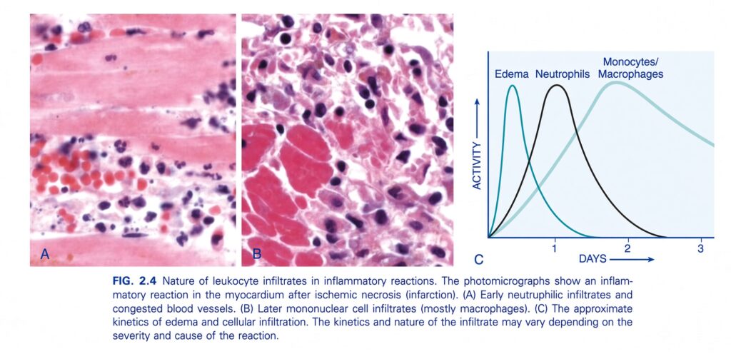
FIG. 2.4 — Nature of Leukocyte Infiltrates in Inflammatory Reactions
🧠 THE BIG IDEA
This figure shows how the inflammatory cells change with time, using inflammation in the myocardium after ischemic necrosis (myocardial infarction) as the example.
The most important pattern is:
Early → Edema + Neutrophils
Later → Monocytes/Macrophages
So remember:
NEUTROPHILS FIRST → MACROPHAGES LATER
A. 🔴 EARLY NEUTROPHILIC INFILTRATE
Look at A, the left microscopic image.
This represents the early stage of inflammation after myocardial infarction.
What can you see?
There are many cells with:
- Dark-staining nuclei
- Multilobed/segmented nuclei
These are:
Neutrophils
There are also many red blood cells inside congested blood vessels.
The caption specifically describes:
Early neutrophilic infiltrates and congested blood vessels
🟣 Why do neutrophils appear first?
Neutrophils are the body’s:
Rapid-response leukocytes
When tissue is suddenly injured:
Injury
↓
Inflammatory mediators are released
↓
Leukocytes are recruited
↓
Neutrophils arrive early
They quickly move into the damaged tissue to:
- Attack microbes
- Remove damaged material
- Participate in the acute inflammatory response
🩸 What does “congested blood vessels” mean?
The blood vessels contain an increased amount of blood.
So in image A you can see many:
Red blood cells
inside vessels.
This reflects vascular changes associated with acute inflammation.
B. 🟣 LATER MONONUCLEAR CELL INFILTRATE
Now look at B, the middle microscopic image.
This represents a later stage.
Instead of predominantly neutrophils, there are many:
Mononuclear cells
The caption tells us these are:🧹 What are macrophages doing?
Macrophages are important for:
- Removing dead cells
- Removing tissue debris
- Removing microbes
- Producing cytokines
- Helping coordinate healing
So after the initial neutrophil response:
Macrophages become increasingly important.
🧠 WHY DOES THE CELL TYPE CHANGE?
Think of inflammation as a two-stage cleanup team.
🚨 First responders:
Neutrophils
They arrive rapidly.
Their job:
Quickly respond to the injury.
🧹 Cleanup and repair team:
Monocytes → Macrophages
They arrive later.
Their job:
Clean up dead tissue and help coordinate resolution and repair.
C. 📈 THE GRAPH — MOST IMPORTANT PART
Now look at the graph on the right.
The graph shows how the activity of different inflammatory components changes over time.
X-axis:
DAYS
Moving right means:
More time has passed since the injury.
Y-axis:
ACTIVITY
Higher on the graph means:
Greater activity of that inflammatory component.
There are three curves:
- Edema
- Neutrophils
- Monocytes/Macrophages
1. 💧 EDEMA — FIRST AND FASTEST
Look at the blue/teal curve labeled:
Edema
It rises very quickly.
Then it falls quickly.
Meaning:
Edema develops very early after injury.
It reaches its peak before the neutrophil curve.
Then it rapidly decreases.
Why does edema happen early?
Inflammatory mediators cause:
Vasodilation + increased vascular permeability
↓
Fluid moves out of blood vessels
↓
Edema
Therefore:
Edema is an early vascular response to inflammation.
2. 🟣 NEUTROPHILS — EARLY CELLULAR RESPONSE
Look at the black curve.
It rises after edema begins.
It reaches its peak at approximately:
1 day
Then it gradually falls.
This represents:
Neutrophils are the predominant early leukocytes in acute inflammation.
🧠 Why do neutrophils peak early?
Because they are rapidly recruited from the bloodstream.
Think:
Neutrophils = emergency response team
They arrive quickly because the body needs an immediate cellular response.3. 🟢 MONOCYTES/MACROPHAGES — LATER RESPONSE
Now look at the green curve.
It rises more slowly.
It becomes prominent after the neutrophil response.
It reaches its peak around:
2 days
and remains elevated longer.
This represents:
Monocytes/macrophages become increasingly prominent during the later phase.
🔥 COMPARE THE THREE CURVES
💧 Edema
Fastest
↓
Peaks very early
↓
Falls quickly
🟣 Neutrophils
Early cellular response
↓
Peak around 1 day
↓
Then decline
🟢 Monocytes/Macrophages
Later cellular response
↓
Peak around 2 days
↓
Persist longer📊 THE GRAPH AS A SIMPLE TIMELINE
TIME AFTER INJURY

Immediately
     ↓
💧 EDEMA rises rapidly
     ↓
🟣 NEUTROPHILS rise
     ↓
Neutrophils peak ≈ 1 day
     ↓
Neutrophils decline
     ↓
🟢 MONOCYTES/MACROPHAGES rise
     ↓
Macrophages peak ≈ 2 days
     ↓
Macrophages remain active longer
🧠 WHY DOES THE MACROPHAGE CURVE LAST LONGER?
Because macrophages have roles beyond simply responding to the initial injury.
They help:
Clean up
Dead cells and tissue debris are removed.
Then they can produce:
Cytokines and growth factors
which influence:
- Resolution of inflammation
- Tissue repair
Therefore:
Macrophages are important not only for cleanup but also for what happens after the initial inflammatory response.
🔬 UNDERSTAND IMAGE A AND B TOGETHER
Image A = EARLY
You see predominantly:
Neutrophils
and:
Congested blood vessels
Therefore:
A = early acute inflammatory response
Image B = LATER
You see predominantly:
Mononuclear cells, mostly macrophages
Therefore:
B = later inflammatory response
⭐ THE MOST IMPORTANT CONCEPT
The inflammatory infiltrate is not made of the same cells throughout the entire process.
It changes with time.
Early:
Neutrophils
Later:
Monocytes → Macrophages
This is called a:
Change in the nature of the inflammatory infiltrate.
🧩 CONNECT THIS FIGURE WITH THE PREVIOUS FIGURE
The previous figure showed:
Leukocyte recruitment
Now this figure tells you:
Which leukocytes arrive when.
Previous figure:
Rolling → adhesion → transmigration → chemotaxis
↓
This figure:
Neutrophils arrive first
↓
Monocytes/macrophages become prominent later🎯 EXAM-IMPORTANT COMPARISON
| Feature | Neutrophils | Monocytes/Macrophages |
|---|---|---|
| Arrival | Early | Later |
| Main role | Rapid inflammatory response | Cleanup + coordination of later response |
| Graph peak | About 1 day | About 2 days |
| Duration | Relatively short | Longer |
| Typical association | Acute inflammation | Later phase of acute inflammation |
⚠️ IMPORTANT: THIS IS A GENERAL PATTERN
The graph represents an approximate pattern.
The caption specifically says:
The kinetics and nature of the infiltrate may vary depending on the severity and cause of the inflammatory reaction.
So don’t interpret the graph as an absolute rule that every inflammatory reaction will always have exactly the same timing.
🧠 EASIEST STORY
Imagine a building is damaged.
🚨 First:
Water quickly enters the damaged area.
= Edema
🚑 Then:
Emergency workers arrive quickly.
= Neutrophils
🧹 Later:
Cleanup workers arrive and stay longer.
= Monocytes/Macrophages
Therefore:
EDema → NEutrophils → MAcrophages
A useful sequence is:
E → N → M
Edema → Neutrophils → Monocytes/Macrophages
🔑 FINAL MEMORY
Acute inflammation begins with rapid vascular changes and edema, followed by an early neutrophil infiltrate. Later, monocytes enter the tissue and become macrophages, which become the predominant cells and persist longer.
One-line exam memory:
NEUTROPHILS FIRST, MACROPHAGES LATER.
Phagocytosis and Clearance of the Offending Agent
- Neutrophils and monocytes that have been recruited to a site of infection or cell death become activated.
- They are activated by:
- Products of microbes
- Products of necrotic cells
- Cytokines produced locally
- Activation produces several responses (eFig. 2.1).
- The most important responses for destroying microbes and removing dead tissue are:
- Phagocytosis
- Intracellular killing
Phagocytosis
- Phagocytosis means the ingestion of solid/particulate material by cells.
- The body’s most important phagocytic cells are:
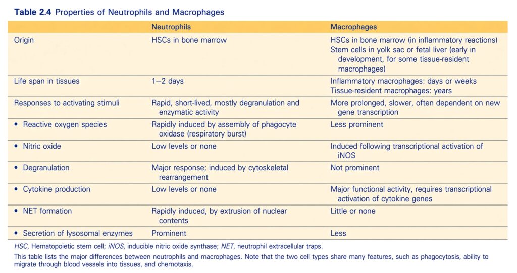
- Neutrophils
- Macrophages (Table 2.4)
- Neutrophils respond rapidly, but they are relatively short-lived.
- During inflammation, macrophages develop from blood monocytes and can survive for days or months.
- Some long-lived tissue-resident macrophages come from embryonic precursor cells that enter tissues early in life and remain there for years.
- Macrophage responses are generally slower but longer lasting.
- Neutrophils and macrophages can ingest microbes after recognizing them through phagocyte receptors.
- Examples of these receptors include:
- Mannose receptors, which recognize terminal mannose residues present in microbial glycoproteins
- Scavenger receptors
- Phagocytosis becomes much more efficient when microbes are coated with molecules called opsonins.
- Opsonins are molecules that help phagocytes recognize and ingest microbes.
- Phagocytes have specific receptors for these opsonins.
- Important opsonins include:
- Antibodies
- C3b, a cleavage product of complement
- Certain plasma lectins
- After a microbe binds to receptors on the phagocyte, the particle is engulfed.
- It enters a membrane-bound vesicle called the phagosome.
- The phagosome then fuses with lysosomes.
- This fusion causes lysosomal contents to be released into the phagolysosome (Fig. 2.5).
- During this process, neutrophils may also release their granule contents into the extracellular space.
KEY CONCEPT
Phagocytosis = Recognize → Bind → Engulf → Phagosome → Fuse with lysosome → Phagolysosome → Destroy/remove the material.
Conceptual examples:
- Microbe → phagocyte receptor → recognition → ingestion
- Microbe + opsonin → easier recognition → more efficient phagocytosis
- Antibody/C3b/plasma lectin → opsonization → phagocyte binding → ingestion
- Engulfed microbe → phagosome → lysosome fusion → phagolysosome → destruction
- Neutrophil → rapid response + short life
- Macrophage → slower response + longer survival → prolonged clearance
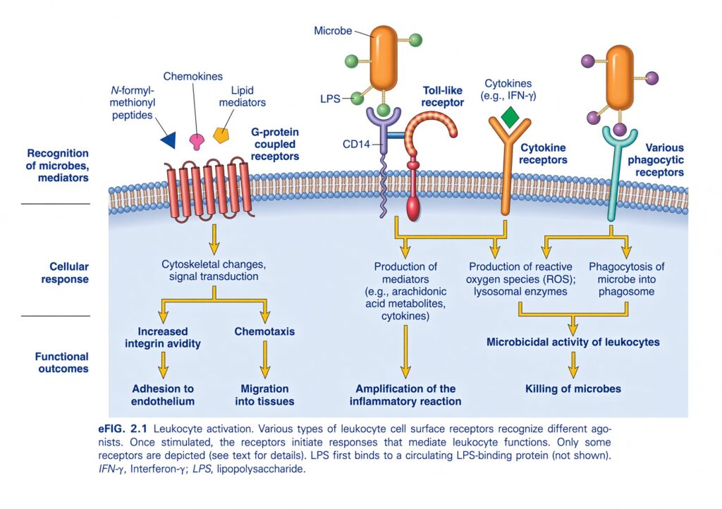
eFIG. 2.1 — Leukocyte Activation
🧠 THE BIG IDEA
This figure explains how leukocytes recognize danger and become activated.
A leukocyte has many different receptors on its surface. Each receptor recognizes a different signal, such as:
- Microbial products
- Chemokines
- Lipid mediators
- Cytokines
- Signals from microbes
When these receptors are activated, the leukocyte changes its behavior.
The whole figure in one line:
Danger signal → receptor activation → intracellular signaling → leukocyte response → inflammation + microbial killing
1. 🧩 FIRST UNDERSTAND THE THREE LEVELS
The figure is organized into three levels:
Recognition
The leukocyte detects a signal.
↓
Cellular response
The receptor sends a signal inside the leukocyte.
↓
Functional outcome
The leukocyte actually does something useful.
So:
RECOGNIZE → RESPOND → PERFORM
2. 🦠 WHAT CAN A LEUKOCYTE RECOGNIZE?
At the top of the figure, several different signals are shown.
N-formyl-methionyl peptides
These are peptides associated with microbes.
Chemokines
These are chemical signals that guide and activate leukocytes.
Lipid mediators
These are inflammatory lipid-derived signals.
Microbial products
The figure shows:
LPS
LPS = lipopolysaccharide, an important component of the outer membrane of many Gram-negative bacteria.
Cytokines
For example:
IFN-γ
Signals recognized by phagocytic receptors
These help leukocytes recognize material that can be engulfed.
3. 🧠 DIFFERENT RECEPTORS = DIFFERENT JOBS
The most important thing to understand is:
Not every receptor does the same thing.
Different receptors detect different danger signals and activate different leukocyte functions.
The figure shows four major receptor groups:
- G-protein-coupled receptors
- Toll-like receptors
- Cytokine receptors
- Phagocytic receptors
Let’s take them one by one.
4. 🔵 G-PROTEIN-COUPLED RECEPTORS
Look at the left side of the cell membrane.
The figure labels:
G-protein-coupled receptors
These receptors recognize signals such as:
- Chemokines
- N-formyl-methionyl peptides
- Lipid mediators
What happens after activation?
The receptor activates:
Cytoskeletal changes and signal transduction
This produces two important effects shown in the figure:
① Increased integrin avidity
② Chemotaxis
5. 🧲 INCREASED INTEGRIN AVIDITY
Remember from the previous figure:
Integrins help leukocytes stick firmly to the endothelium.
When leukocytes are activated:
Their integrins become better at binding.
This is called:
Increased integrin avidity
Therefore:
G-protein-coupled receptor activation
↓
Integrin activation
↓
Adhesion to endothelium
6. 🧭 CHEMOTAXIS
The same receptor signaling can also produce:
Chemotaxis
Chemotaxis means:
Directed movement of leukocytes toward the source of a chemical signal.
So:
Chemokine
↓
Leukocyte detects it
↓
Leukocyte moves toward higher concentration
↓
Migration into tissues
⭐ CONNECT THIS WITH FIG. 2.3
The previous figure showed:
Chemokines → integrin activation → stable adhesion
This figure explains part of how that activation occurs.
So:
Chemokines bind receptors → intracellular signaling occurs → integrins become high-affinity → leukocyte sticks to endothelium.
7. 🟠 TOLL-LIKE RECEPTORS
Now look at the middle portion.
The figure shows:
Toll-like receptor
These receptors recognize microbial products.
A major example shown is:
LPS
8. 🦠 LPS RECOGNITION
The figure also shows:
CD14
CD14 participates in recognition of LPS.
The simplified sequence is:
LPS from microbe
↓
LPS-binding system/CD14
↓
Toll-like receptor activation
↓
Intracellular signaling
↓
Production of inflammatory mediators
9. 🧪 WHAT DOES TOLL-LIKE RECEPTOR ACTIVATION PRODUCE?
The figure shows:
Production of mediators
Examples include:
- Arachidonic acid metabolites
- Cytokines
These mediators then:
Amplify the inflammatory reaction
🧠 Simple concept
Think of a Toll-like receptor as a:
Microbe alarm detector
It detects microbial components.
Then it tells the leukocyte:
“There is a microbe here—start the inflammatory response.”
10. 🟢 CYTOKINE RECEPTORS
Now look toward the center-right.
The figure shows:
Cytokine receptors
An example cytokine shown is:
IFN-γ
When cytokines bind to their receptors:
↓
The leukocyte becomes activated.
↓
It produces:
Reactive oxygen species (ROS)
and
Lysosomal enzymes
11. 💥 REACTIVE OXYGEN SPECIES — ROS
ROS are highly reactive molecules produced by activated leukocytes.
Their important function here is:
Microbial killing
Think:
ROS = chemical weapons used by activated leukocytes to damage and kill microbes.
12. 🧪 LYSOSOMAL ENZYMES
Leukocytes, especially phagocytes, contain lysosomal enzymes.
After activation, these enzymes contribute to:
Microbicidal activity
Therefore:
Cytokine receptor activation
↓
ROS + lysosomal enzymes
↓
Killing of microbes
13. 🟣 PHAGOCYTIC RECEPTORS
Look at the far right.
The figure labels:
Various phagocytic receptors
These receptors help leukocytes recognize material that should be engulfed.
Once activated:
Phagocytosis of microbe into phagosome
occurs.
14. 🫧 WHAT IS A PHAGOSOME?
A phagosome is a membrane-bound compartment formed when the leukocyte:
Engulfs a microbe.
Think of the leukocyte as a cell that can swallow a bacterium.
Microbe
   ↓
Recognition
   ↓
Engulfment
   ↓
Microbe enclosed
   ↓
PHAGOSOME
The microbe is then exposed to the cell’s killing mechanisms.
15. 🦠 PHAGOCYTOSIS + ROS + LYSOSOMAL ENZYMES
These processes work together.
Phagocytic receptors
↓
Microbe enters phagosome
ROS
↓
Damage/kill microbe
Lysosomal enzymes
↓
Digest/destroy microbial material
↓
Microbial killing🔥 NOW UNDERSTAND THE FOUR RECEPTOR GROUPS
① G-protein-coupled receptors
Recognize:
- Chemokines
- N-formyl-methionyl peptides
- Lipid mediators
↓
Cytoskeletal changes
↓
Integrin avidity ↑
↓
Adhesion to endothelium
AND
Chemotaxis
↓
Migration into tissues
② Toll-like receptors
Recognize:
- Microbial products
- LPS
↓
Mediator production
↓
Arachidonic acid metabolites + cytokines
↓
Amplification of inflammation
③ Cytokine receptors
Recognize:
- Cytokines such as IFN-γ
↓
ROS + lysosomal enzymes
↓
Microbial killing
④ Phagocytic receptors
Recognize:
- Microbial material suitable for engulfment
↓
Phagocytosis
↓
Microbe enters:
Phagosome
↓
Microbial killing
🧠 THE WHOLE FIGURE AS A SIMPLE STORY
Imagine a neutrophil as a soldier.
Step 1 — Find the enemy
The leukocyte detects:
Microbes / chemokines / inflammatory signals
↓
Step 2 — Activate
Receptors send signals into the cell.
↓
Step 3 — Stick to the blood vessel
Integrins become highly active.
↓
Step 4 — Move toward the infection
Chemotaxis guides the leukocyte.
↓
Step 5 — Recognize the microbe
Toll-like and phagocytic receptors detect microbial components.
↓
Step 6 — Swallow it
Phagocytosis forms a:
Phagosome
↓
Step 7 — Kill it
ROS + lysosomal enzymes
↓
Microbial killing
🔗 CONNECT THE FIGURE WITH LEUKOCYTE MIGRATION
You previously saw:
FIG. 2.3
Rolling → integrin activation → stable adhesion → transmigration → chemotaxis
This figure goes one step deeper and explains how leukocytes become activated to perform these functions.
Chemokines:
Chemokine receptor
↓
Signal transduction
↓
Integrin avidity ↑
↓
Stable adhesion
📊 HIGH-YIELD TABLE
| Receptor | Recognizes | Main response | Final effect |
|---|---|---|---|
| G-protein-coupled receptors | Chemokines, N-formyl-methionyl peptides, lipid mediators | Cytoskeletal changes, integrin activation, chemotaxis | Adhesion + migration |
| Toll-like receptors | Microbial products such as LPS | Mediator production | Amplifies inflammation |
| Cytokine receptors | Cytokines such as IFN-γ | ROS + lysosomal enzymes | Microbial killing |
| Phagocytic receptors | Microbial material | Phagocytosis | Microbial killing |
⭐ THE MOST IMPORTANT CONNECTIONS
🧲 Chemokines
→ G-protein-coupled receptors
→ Integrin activation
→ Adhesion
🧭 Chemokines
→ G-protein-coupled receptors
→ Chemotaxis
→ Migration into tissue
🦠 LPS
→ CD14 + Toll-like receptor
→ Mediator production
→ Amplification of inflammation
💥 Cytokines such as IFN-γ
→ Cytokine receptor
→ ROS + lysosomal enzymes
→ Microbial killing
🫧 Phagocytic receptors
→ Phagocytosis
→ Phagosome
→ Microbial killing
🎯 FINAL MEMORY MAP
                LEUKOCYTE
                    │
          ┌─────────┼─────────┐
          ↓         ↓         ↓
      CHEMOKINE     LPS     CYTOKINE
          ↓         ↓         ↓
        GPCR       TLR    Cytokine receptor
          ↓         ↓         ↓
      Integrins   Mediators   ROS +
      +           +           lysosomal
      chemotaxis  cytokines   enzymes
          ↓         ↓         ↓
     Adhesion +   ↑Inflam-   Microbial
     migration    mation     killing
                         \
                          \
                    PHAGOCYTIC
                     RECEPTORS
                          ↓
                    Phagocytosis
                          ↓
                      Phagosome
                          ↓
                  Microbial killing
🔑 ONE-LINE MEMORY
Leukocyte receptors detect different danger signals: GPCRs promote chemotaxis and integrin activation, Toll-like receptors promote inflammatory mediator production, cytokine receptors promote ROS and lysosomal enzyme production, and phagocytic receptors promote engulfment of microbes.
Intracellular Destruction of Microbes and Debris
- Microbes and ingested materials are destroyed inside phagocytes by:
- Reactive oxygen species (ROS), also called reactive oxygen intermediates
- Reactive nitrogen species, mainly derived from nitric oxide (NO)
- Lysosomal enzymes
- Normally, these potentially harmful substances are kept inside lysosomes.
- Phagocytosed materials are brought to these lysosomes.
- This keeps the potentially harmful substances separate from the cell’s cytoplasm.
- By keeping them separated, the phagocyte is protected from damage while it performs its normal function.
KEY CONCEPT
Phagocytosed material → lysosome → ROS + reactive nitrogen species + lysosomal enzymes → destruction of microbes and debris.
Conceptual example:
Microbe is engulfed → brought to lysosome → harmful killing substances remain contained → microbe is destroyed → phagocyte is protected from its own destructive substances.
ntracellular Destruction of Microbes and Debris
Reactive Oxygen Species
- Reactive oxygen species (ROS) are free radicals produced mainly in the phagolysosomes of neutrophils.
- When neutrophils are activated, a multicomponent enzyme called phagocyte oxidase (NADPH oxidase) is rapidly assembled in the membrane of the phagolysosome (Fig. 2.5B).
- This enzyme uses NADPH and transfers its reducing power to oxygen.
- Oxygen is converted into superoxide anion (O₂⁻).
- Superoxide is then converted into hydrogen peroxide (H₂O₂).
- H₂O₂ alone is not very effective at killing microbes.
- However, neutrophil azurophilic granules contain the enzyme myeloperoxidase (MPO).
- In the presence of a halide such as Cl⁻, MPO converts H₂O₂ into hypochlorite (ClO⁻).
- Hypochlorite is a powerful antimicrobial substance.
- It destroys microbes by:
- Halogenation — the halide becomes covalently attached to cellular components.
- Oxidation of proteins and lipids — including lipid peroxidation.
- The H₂O₂–MPO–halide system is the most effective bactericidal system of neutrophils.
- H₂O₂ can also be converted into the hydroxyl radical (•OH).
- The hydroxyl radical is another powerful destructive agent.
- These oxygen-derived free radicals can bind to and modify lipids, proteins, and nucleic acids.
- This damage helps destroy cells, including microbes.
- The production of ROS together with increased oxygen consumption is called the respiratory burst.
- Genetic defects that prevent normal ROS production cause an immunodeficiency disease called chronic granulomatous disease (Chapter 5).
Nitric Oxide
- Nitric oxide (NO) is a soluble gas produced from arginine by the enzyme nitric oxide synthase (NOS).
- NO also participates in microbial killing, especially in macrophages.
- Inducible NOS (iNOS) is increased in macrophages when microbial products and cytokines such as IFN-γ activate transcription of its gene (Fig. 2.5C).
- NO reacts with superoxide (O₂⁻) produced by phagocyte oxidase.
- This reaction produces the highly reactive molecule peroxynitrite (ONOO⁻).
- These nitrogen-derived molecules, like ROS, attack and damage the:
- Lipids
- Proteins
- Nucleic acids
of microbes.
Leukocyte Granule Contents
- Neutrophils have two main types of granules.
- These granules contain enzymes that can destroy microbes and dead tissue.
- However, these substances can also contribute to tissue damage.
- The smaller specific (secondary) granules contain:
- Lysozyme
- Collagenase
- Gelatinase
- Lactoferrin
- Plasminogen activator
- Histaminase
- Alkaline phosphatase
- The larger azurophil (primary) granules contain:
- Myeloperoxidase
- Bactericidal factors, such as defensins
- Acid hydrolases
- Neutral proteases, including:
- Elastase
- Cathepsin G
- Nonspecific collagenases
- Proteinase 3
- When neutrophils are activated, the contents of both types of granules are released.
- Phagocytic vesicles containing engulfed material can fuse with these granules and with lysosomes.
- This allows the ingested material to be destroyed inside the phagolysosomes by the actions of these enzymes.
- Macrophages also contain lysosomes filled with:
- Acid hydrolases
- Collagenase
- Elastase
- Phospholipase
- These enzymes can destroy ingested materials and cell debris.
- Activated neutrophils can also release chromatin components, including histones.
- These components form fibrillar networks called neutrophil extracellular traps (NETs) (eFig. 2.2).
- NETs bind and concentrate antimicrobial peptides and granule enzymes.
- This creates extracellular sites where microbes can be destroyed.
- During NET formation, the neutrophil loses its nucleus.
- This leads to death of the neutrophil.
- NETs have also been detected in the blood during sepsis.
- This occurs as a result of widespread neutrophil activation.
KEY CONCEPT
Microbe inside phagocyte → ROS + NO + lysosomal enzymes → microbial destruction.
Conceptual examples:
- NADPH oxidase → O₂ → superoxide → H₂O₂
- H₂O₂ + MPO + Cl⁻ → hypochlorite → powerful microbial killing
- H₂O₂ → hydroxyl radical → damage to microbial lipids, proteins, and nucleic acids
- NO + superoxide → peroxynitrite → microbial damage
- Phagosome + lysosome/granules → phagolysosome → enzymes destroy ingested material
- Activated neutrophil → chromatin + histones → NETs → extracellular microbial trapping and destruction
- Defective ROS production → chronic granulomatous disease
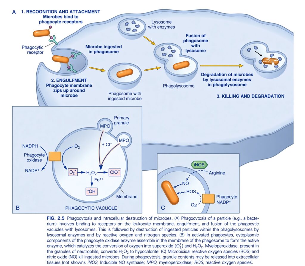
FIG. 2.5 — Phagocytosis and Intracellular Destruction of Microbes
🧠 THE BIG IDEA
This figure shows how a phagocyte, such as a neutrophil or macrophage, captures a microbe, swallows it, and then kills and digests it inside the cell.
The complete process is:
Recognition → Engulfment → Phagosome → Fusion with lysosome → Phagolysosome → Killing + degradation
The killing mainly occurs through:PHAGOCYTOSIS
1. 🔍 RECOGNITION AND ATTACHMENT
The microbe first comes into contact with the phagocyte.
The phagocyte has:
Phagocytic receptors
These receptors recognize and attach to the microbe.
So:
Microbe
↓
Phagocytic receptor
↓
Attachment
Think:
2. 🫳 ENGULFMENT
After attachment, the phagocyte membrane begins to:
Wrap around the microbe.
The membrane extends around the microbe from different sides.
Eventually, the microbe becomes completely surrounded.
The figure describes this as:
Phagocyte membrane zips up around microbe. 🫧 PHAGOSOME FORMS
Once the microbe is completely surrounded, it is enclosed inside a membrane-bound vesicle.
This vesicle is called:
Phagosome
So:
Microbe
   ↓
Attachment
   ↓
Membrane surrounds microbe
   ↓
Microbe enclosed
   ↓
PHAGOSOME
Important:
At this stage, the microbe is inside the phagocyte but has not yet been fully destroyed.
4. 🧪 LYSOSOME
The phagocyte already contains:
Lysosomes
Lysosomes contain digestive/killing enzymes.
Think of a lysosome as:
A small bag full of destructive enzymes.
5. 🔗 FUSION OF PHAGOSOME WITH LYSOSOME
The phagosome containing the microbe fuses with a lysosome.
Phagosome + Lysosome
↓
Phagolysosome
This is extremely important.
The phagolysosome is where the microbe is exposed to:
- Lysosomal enzymes
- ROS
- Other antimicrobial substances
6. 💥 KILLING AND DEGRADATION
Now the microbe is destroyed.
The figure shows:
Degradation of microbes by lysosomal enzymes
The microbe is broken down into smaller components.
So the overall sequence is:
Recognize → engulf → phagosome → fuse with lysosome → phagolysosome → kill → digest
B. PHAGOCYTIC VACUOLE — HOW ROS ARE PRODUCED
Now look at the lower-left box.
This part explains how the phagocyte produces reactive oxygen species.
7. ⚡ PHAGOCYTE OXIDASE
The figure shows an enzyme called:
Phagocyte oxidase
It uses:
NADPH
and:
O₂
to begin production of reactive oxygen species.
The important sequence shown is:
NADPH
↓
Phagocyte oxidase
↓
O₂ → O₂•⁻
The product:
O₂•⁻ = superoxide
8. 💥 SUPEROXIDE
Superoxide is a:
Reactive oxygen species (ROS)
It is highly reactive and contributes to microbial killing.
The figure then shows:
O₂•⁻ → H₂O₂
So superoxide can lead to formation of:
Hydrogen peroxide (H₂O₂)
9. 🧪 HYDROGEN PEROXIDE
The figure shows:
H₂O₂
Hydrogen peroxide is another reactive molecule involved in killing microbes.
But neutrophils can make an even more powerful antimicrobial substance using H₂O₂.
That is where:
MPO
comes in.
10. 🟣 MPO — MYELOPEROXIDASE
The figure shows:
MPO
inside the:
Primary granules
of neutrophils.
MPO uses:
H₂O₂ + Cl⁻
to produce:
ClO⁻
This represents hypochlorous acid/hypochlorite antimicrobial activity.
11. 🧠 WHY IS MPO IMPORTANT?
MPO converts hydrogen peroxide into a very powerful antimicrobial oxidant.
Think:
H₂O₂ is the starting weapon, and MPO helps convert it into a stronger weapon.
Therefore:
MPO → powerful microbial killing
This is especially important in neutrophils.
12. 🔥 HYDROXYL RADICAL
The figure also shows:
H₂O₂ → •OH
in the presence of:
Fe²⁺
The product:
•OH = hydroxyl radical
This is another highly reactive oxygen species that can damage microbes.
13. 🧨 THE MAIN ROS PATHWAY
The lower-left box can therefore be understood as:
NADPH + O₂
     ↓
Phagocyte oxidase
     ↓
Superoxide (O₂•⁻)
     ↓
H₂O₂
     ↓
 ┌───────────────┐
 ↓               ↓
MPO             Fe²⁺
 ↓               ↓
ClO⁻             •OH
These reactive substances help:
Kill the microbe
C. MICROBICIDAL REACTIVE OXYGEN SPECIES AND NITRIC OXIDE
The lower-right box shows another important killing mechanism.
There are two major components shown:
ROS
and
NO
14. 🟢 iNOS
The figure shows:
iNOS
This means:
Inducible nitric oxide synthase
It uses:
Arginine
to produce:
NO = nitric oxide
15. 🧪 NITRIC OXIDE
Nitric oxide participates in microbial killing.
The figure shows:
Arginine
↓
iNOS
↓
NO
NO can interact with reactive oxygen species.
↓
This contributes to:
Microbial killing
16. 💥 ROS + NO
The figure shows:
ROS
and
NO
acting around the microbe.
Together, reactive oxygen and nitrogen species create a strong antimicrobial environment.
So:
ROS + NO → damage and kill microbes
17. 🦠 WHAT ACTUALLY KILLS THE MICROBE?
The figure gives several mechanisms.
① Lysosomal enzymes
→ Digest microbial material
② Reactive oxygen species
→ Damage and kill microbes
③ MPO system
→ Generates powerful antimicrobial oxidants
④ Nitric oxide
→ Contributes to microbial killing
Therefore:
🔥 FOLLOW THE ENTIRE FIGURE FROM LEFT TO RIGHT
MICROBE
   ↓
1. Recognition
   ↓
Phagocytic receptor
   ↓
2. Attachment
   ↓
3. Engulfment
   ↓
Phagocyte membrane surrounds microbe
   ↓
4. PHAGOSOME
   ↓
5. Fusion with lysosome
   ↓
6. PHAGOLYSOSOME
   ↓
 ┌───────────────────────┐
 ↓                       ↓
Lysosomal enzymes       ROS + NO
 ↓                       ↓
Digestion               Microbial damage
 └───────────┬───────────┘
             ↓
      KILLING + DEGRADATION
             ↓
       Microbe destroyed
🧠 UNDERSTAND THE THREE PANELS
A = WHAT HAPPENS TO THE MICROBE
Recognition
↓
Engulfment
↓
Phagosome
↓
Fusion with lysosome
↓
Phagolysosome
↓
Killing and degradation
B = HOW ROS ARE GENERATED
NADPH + O₂
↓
Phagocyte oxidase
↓
Superoxide
↓
H₂O₂
↓
MPO + Cl⁻
↓
ClO⁻
Plus:
H₂O₂ + Fe²⁺
↓
•OH
C = HOW NO PARTICIPATES
Arginine
↓
iNOS
↓
NO
ROS
↓
Microbial killing
⭐ VERY IMPORTANT TERMS
| Term | Easy meaning |
|---|---|
| Phagocytosis | Cell captures and swallows a microbe |
| Phagocytic receptor | Receptor that helps recognize/attach to the microbe |
| Phagosome | Vesicle containing the engulfed microbe |
| Lysosome | Organelle containing digestive/killing enzymes |
| Phagolysosome | Phagosome fused with lysosome |
| Phagocyte oxidase | Enzyme system that generates ROS |
| ROS | Reactive oxygen species used for microbial killing |
| MPO | Myeloperoxidase; uses H₂O₂ and Cl⁻ to generate powerful antimicrobial oxidants |
| iNOS | Inducible nitric oxide synthase |
| NO | Nitric oxide; contributes to microbial killing |
🎯 THE MOST IMPORTANT CONNECTION
Phagosome ≠ Phagolysosome
This distinction is important.
Phagosome
= microbe has been swallowed.
Phagolysosome
= phagosome has fused with lysosome and the microbe is exposed to destructive substances.
So:
Swallow first → fuse with lysosome → kill and digest.
🧠 ONE EASY STORY
Imagine the phagocyte as a security guard with a garbage disposal system:
👀 1. Recognize
The guard identifies the intruder.
🤝 2. Attach
The guard grabs it.
🫳 3. Engulf
The guard swallows it.
🫧 4. Phagosome
The intruder is trapped inside a container.
🧪 5. Lysosome joins
A container full of destructive enzymes fuses with it.
💥 6. Kill
ROS, MPO, NO, and enzymes attack the microbe.
🧹 7. Degrade
The microbe is broken down.
🔑 FINAL MEMORY LINE
RECOGNIZE → ENGULF → PHAGOSOME → FUSE WITH LYSOSOME → PHAGOLYSOSOME → ROS/MPO/NO + ENZYMES → KILL → DEGRADE.
Leukocyte-Mediated Tissue Injury
- Leukocytes can cause injury to normal cells and tissues.
- This can happen during normal defense against microbes, especially when the microbes are difficult to eliminate, such as mycobacteria.
- Leukocytes can also cause tissue damage when the immune response is incorrectly directed against:
- Self-antigens, as in autoimmune diseases
- Normally harmless environmental antigens, as in allergic diseases
- Leukocyte-mediated tissue injury mainly occurs through the release of contents from granules and lysosomes.
- Some release of these substances normally occurs when activated leukocytes try to destroy microbes and other harmful substances.
- This process becomes excessive when phagocytes encounter materials that are difficult to ingest.
- Examples include antibodies deposited on indigestible flat surfaces.
- Phagocytosed substances such as urate and silica crystals can also damage the phagolysosome membrane.
- This damage can cause the release of harmful substances from the leukocytes.
- Harmful proteases released by leukocytes are normally controlled by antiproteases present in the blood and tissue fluids.
- The most important antiprotease is α₁-antitrypsin.
- α₁-antitrypsin is the major inhibitor of neutrophil elastase.
- If these antiprotease inhibitors are deficient, protease activity can continue for a long time.
- This occurs in patients with α₁-antitrypsin deficiency (Chapter 11).
- Although neutrophils and macrophages are important in acute inflammation, other cell types also have important roles.
- Some T cells, called Th17 cells, produce cytokines such as IL-17.
- IL-17 helps recruit neutrophils and stimulates the production of antimicrobial peptides that directly kill microbes.
- Without effective Th17 responses, people become more susceptible to fungal and bacterial infections.
- The skin abscesses that develop in these situations lack typical features of acute inflammation, such as warmth and redness.
- Eosinophils are particularly important in responses to helminthic parasites and in some allergic disorders.
- Mast cells and basophils are important cells in allergic reactions.
- Once acute inflammation has removed the offending stimulus, the inflammatory response subsides.
- This happens because:
- There is no further leukocyte recruitment.
- Inflammatory mediators are short-lived and decrease when their production stops.
- Neutrophils have short life spans.
KEY CONCEPT
Leukocytes protect tissues by destroying harmful agents, but their released enzymes and other substances can also damage normal tissue.
Conceptual examples:
- Persistent mycobacterial infection → continued leukocyte activity → tissue injury
- Autoimmune disease → leukocytes attack self-antigens → tissue damage
- Allergy → leukocytes respond to harmless environmental antigens → tissue injury
- Phagocyte + difficult-to-ingest material → excessive granule/lysosome release → tissue injury
- α₁-antitrypsin deficiency → uncontrolled neutrophil elastase → sustained protease activity
- Th17 → IL-17 → neutrophil recruitment + antimicrobial peptides
- Helminths/allergy → eosinophils important
- Acute inflammation ends → no further recruitment + mediators decline + neutrophils die → inflammation subsides
MEDIATORS OF INFLAMMATION
- The inflammatory reaction is started and controlled by chemicals called inflammatory mediators.
- These chemicals are produced at the site of inflammation.
- There are many different mediators, but understanding them is important because identifying these molecules has helped develop many effective anti-inflammatory drugs.
- The general properties of these mediators are described first, followed by the important individual molecules.
- Inflammatory mediators may come from:
- Cells at the site of inflammation
- Circulating precursors that become activated at the site of inflammation
- Cell-derived mediators can be produced in two main ways:
- They can be rapidly released from intracellular granules, such as amines.
- They can be newly synthesized (de novo) after stimulation, such as prostaglandins, leukotrienes, and cytokines.
- The main cells that produce mediators of acute inflammation are:
- Tissue macrophages
- Dendritic cells
- Mast cells
- Other cells can also produce inflammatory mediators, including:
- Platelets
- Neutrophils
- Endothelial cells
- Most epithelial cells
- Plasma-derived mediators, such as complement proteins, are produced mainly in the liver.
- They circulate in the blood as inactive precursors.
- When inflammation occurs, these precursors enter the inflammatory site and become activated.
- Their activation usually occurs through a series of proteolytic cleavages.
- Active inflammatory mediators are produced only when there is an appropriate stimulus.
- These stimuli include:
- Microbial products
- Substances released from necrotic cells
- This ensures that inflammation is activated only when and where it is needed.
- Most inflammatory mediators are short-lived.
- They are quickly:
- Broken down or inactivated by enzymes
- Removed (scavenged)
- Inhibited
- These built-in control mechanisms help prevent an excessive inflammatory reaction.
- The principal mediators of acute inflammation are summarized in Table 2.5.
KEY CONCEPT
Inflammatory mediators = chemical signals that start, control, and eventually stop inflammation.
Conceptual examples:
- Microbial product → local cells activated → inflammatory mediator released → inflammation
- Cell-derived mediator → macrophage/mast cell/etc. → rapid release or new synthesis
- Plasma-derived mediator → liver → inactive precursor in blood → activation at inflammation site
- Stimulus present → mediator activated → inflammation occurs
- Stimulus removed → mediators become inactive/are removed → inflammation decreases
- Most mediators are short-lived → helps prevent excessive inflammation
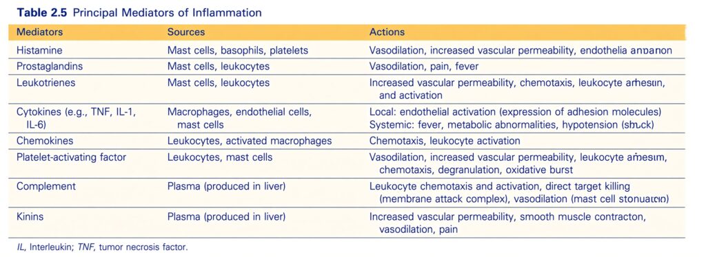
Vasoactive Amines: Histamine and Serotonin
- The major vasoactive amine is histamine.
- Histamine is stored as a preformed molecule in the granules of:
- Mast cells
- Blood basophils
- Platelets
- When these cells are activated, histamine is rapidly released.
- Therefore, histamine is one of the first mediators produced during inflammation.
- The richest source of histamine is mast cells.
- Mast cells are normally present in connective tissue near blood vessels.
- Mast cell degranulation and histamine release can occur in response to several stimuli.
- These include:
- IgE antibodies binding to mast cells, which causes immediate hypersensitivity (allergic) reactions (Chapter 5)
- Complement products called anaphylatoxins, especially C3a and C5a
- Physical injury, such as trauma, cold, or heat
- The exact mechanism by which physical injury causes histamine release is unknown.
- Antibodies and complement products bind to specific receptors on mast cells.
- This activates signaling pathways that cause rapid degranulation.
- Neuropeptides, such as substance P, can also trigger histamine release.
- Cytokines such as IL-1 and IL-8 may also trigger histamine release.
- Histamine causes:
- Dilation of arterioles
- Increased permeability of venules
- Its effects on blood vessels occur mainly when histamine binds to H1 receptors on microvascular endothelial cells.
- Common antihistamine drugs used to treat inflammatory conditions such as allergies work by binding to and blocking H1 receptors.
- Histamine can also cause contraction of some smooth muscles.
- However, leukotrienes are much more powerful and important in causing bronchial smooth muscle spasms, such as those occurring in asthma.
- Serotonin (5-hydroxytryptamine) is another preformed vasoactive mediator.
- It is present in platelets and certain neuroendocrine cells, such as cells in the gastrointestinal tract.
- Serotonin is a vasoconstrictor.
- However, its importance in inflammation is unclear.
KEY CONCEPT
Histamine = major early vasoactive amine → vasodilation + increased venular permeability.
Conceptual examples:
- Mast cell activation → histamine release → arteriolar dilation + venular permeability
- IgE + mast cell → degranulation → histamine → immediate allergic reaction
- C3a/C5a → mast cell activation → histamine release
- Histamine → H1 receptor → vascular effects
- Antihistamine → H1 receptor blocked → reduced histamine effects
- Asthma → leukotrienes are more important than histamine for bronchial smooth muscle spasm
- Serotonin → vasoconstriction → role in inflammation remains unclear
Arachidonic Acid Metabolites
- Prostaglandins and leukotrienes are lipid mediators produced from arachidonic acid (AA).
- They are made from AA present in cell membrane phospholipids.
- These mediators stimulate vascular and cellular reactions during acute inflammation.
- Arachidonic acid (AA) is a 20-carbon polyunsaturated fatty acid.
- AA is released from membrane phospholipids by cellular enzymes called phospholipases.
- The main enzyme involved is phospholipase A₂ (PLA₂).
- PLA₂ is activated by inflammatory stimuli, including:
- Cytokines
- Complement products
- Physical injury
- The substances produced from AA are called eicosanoids.
- The name eicosanoid comes from the Greek word eicosa, meaning 20.
- They are called eicosanoids because they are derived from 20-carbon fatty acids.
- Eicosanoids are produced by two major enzyme pathways:
- Cyclooxygenase (COX) pathway → prostaglandins
- Lipoxygenase (LOX) pathway → leukotrienes and lipoxins (Fig. 2.6)
- Eicosanoids bind to G protein–coupled receptors on many types of cells.
- Through these receptors, they can control almost every step of inflammation (Table 2.6).
KEY CONCEPT
Membrane phospholipids → PLA₂ → Arachidonic acid → two major pathways:
- COX → Prostaglandins
- LOX → Leukotrienes + Lipoxins
Conceptual example:
Inflammatory stimulus → PLA₂ activation → AA released from membrane → COX/LOX pathways → eicosanoids → vascular + cellular inflammatory reactions.
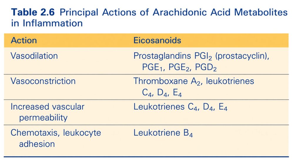
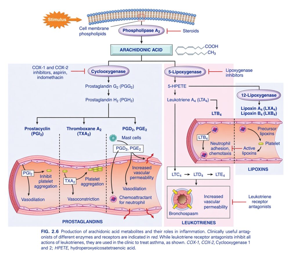
FIG. 2.6 — Production of Arachidonic Acid Metabolites and Their Roles in Inflammation
🧠 THE BIG IDEA
This figure shows how cell-membrane phospholipids are converted into arachidonic acid, and then how arachidonic acid is converted into two major groups of inflammatory mediators:
1. Prostaglandins
→ mainly through the cyclooxygenase (COX) pathway
2. Leukotrienes
→ mainly through the lipoxygenase pathway
There is also a third important group:
Lipoxins
→ help regulate/limit inflammatory leukocyte activity.
The easiest overall pathway is:
Membrane phospholipids → Arachidonic acid → COX pathway OR Lipoxygenase pathway
1. 🧱 STARTING POINT — CELL MEMBRANE PHOSPHOLIPIDS
At the top of the figure, the starting material is:
Cell membrane phospholipids
When a cell receives an inflammatory:
Stimulus
the enzyme:
Phospholipase A₂
is activated.
2. ✂️ PHOSPHOLIPASE A₂
Phospholipase A₂ acts on membrane phospholipids and releases:
ARACHIDONIC ACID
So:
Stimulus
   ↓
Cell membrane phospholipids
   ↓
Phospholipase A₂
   ↓
ARACHIDONIC ACID
🧠 Easy concept:
Phospholipase A₂ cuts arachidonic acid out of the cell membrane.
3. 💊 STEROIDS BLOCK THIS STEP
Look at the red inhibition mark beside:
Phospholipase A₂
The figure shows:
Steroids
So steroids inhibit the pathway very early, before arachidonic acid is released.
Easy memory:
Steroids → inhibit phospholipase A₂ → ↓ arachidonic acid metabolites
4. 🛣️ ARACHIDONIC ACID HAS TWO MAIN PATHWAYS
Once arachidonic acid is available, it can go in two major directions:
                  Arachidonic acid
                         │
             ┌───────────┴───────────┐
             ↓                       ↓
          COX pathway          Lipoxygenase pathway
             ↓                       ↓
      Prostaglandins              Leukotrienes
       + Thromboxane                + Lipoxins
🔵 COX PATHWAY — PROSTAGLANDINS
Look at the left side of the figure.
The enzyme is:
Cyclooxygenase
There are two major forms:
COX-1
COX-2
The figure shows drugs such as:
- Aspirin
- Indomethacin
acting as COX inhibitors.
5. 🧪 COX PRODUCES PGG₂ AND PGH₂
Arachidonic acid
↓
Cyclooxygenase
↓
Prostaglandin G₂ (PGG₂)
↓
Prostaglandin H₂ (PGH₂)
PGH₂ is then used to produce several important products.
6. 🌿 PGH₂ → PROSTACYCLIN
One pathway produces:
Prostacyclin (PGI₂)
The figure shows two major effects:
Vasodilation
and
Inhibition of platelet aggregation
So:
PGI₂ = dilates vessels + inhibits platelet aggregation
🧠 Easy memory:
PGI₂ = “I” inhibit platelets
7. 🩸 PGH₂ → THROMBOXANE A₂
Another pathway produces:
Thromboxane A₂ (TXA₂)
The figure shows:
Vasoconstriction
and
Platelet aggregation
So:
TXA₂ = constricts vessels + promotes platelet aggregation
🧠 Easy memory:
TXA₂ = “T” for Thrombosis/platelet aggregation
⭐ PGI₂ VS TXA₂
This is a very important comparison.
| PGI₂ (Prostacyclin) | TXA₂ (Thromboxane A₂) |
|---|---|
| Vasodilation | Vasoconstriction |
| Inhibits platelet aggregation | Promotes platelet aggregation |
Easy memory:
PGI₂ = prevents platelet sticking
TXA₂ = promotes platelet sticking
8. 🟠 PGD₂ AND PGE₂
PGH₂ can also produce:
PGD₂
PGE₂
The figure shows that these mediators contribute to:
Vasodilation
Increased vascular permeability
and:
Chemotaxis/attraction of neutrophils
So they participate in the inflammatory response.
9. 🔵 COX PATHWAY IN ONE MAP
achidonic acid
       ↓
Cyclooxygenase (COX)
       ↓
     PGG₂
       ↓
     PGH₂
       ↓
 ┌─────┼──────────┐
 ↓     ↓          ↓
PGI₂  TXA₂     PGD₂/PGE₂
 ↓     ↓          ↓
Vaso-  Vaso-    Vasodilation
dilation constriction
 ↓     ↓          +
↓      ↑         ↑ Vascular
platelet platelet permeability
aggregation aggregation
💊 COX INHIBITORS
The figure specifically shows:
COX-1 and COX-2 inhibitors
Examples:
- Aspirin
- Indomethacin
They inhibit:
Cyclooxygenase
Therefore:
🟣 LIPOXYGENASE PATHWAY — LEUKOTRIENES
Now move to the right side of the figure.
Arachidonic acid enters the:
Lipoxygenase pathway
The important enzyme shown is:
5-Lipoxygenase
10. 5-LIPOXYGENASE PATHWAY
Arachidonic acid
↓
5-Lipoxygenase
↓
5-HPETE
↓
Leukotriene A₄ (LTA₄)
LTA₄ then produces different leukotrienes.
11. 🟢 LTA₄ → LTB₄
One branch produces:
LTB₄
The figure shows LTB₄ causing:
Neutrophil adhesion
and:
Chemotaxis
So:
LTB₄ attracts and promotes recruitment of neutrophils.
🧠 Easy memory:
LTB₄ = brings neutrophils
12. 🟡 LTA₄ → LTC₄ → LTD₄ → LTE₄
Another branch produces:
LTC₄
↓
LTD₄
↓
LTE₄
These are called the cysteinyl leukotrienes.
The figure shows their major effects:
Bronchospasm
and:
Increased vascular permeability
13. 🫁 BRONCHOSPASM
Bronchospasm means:
Airway smooth muscle contracts, making the airways narrower.
So:
LTC₄ + LTD₄ + LTE₄
→ Bronchospasm
They are therefore important in airway inflammatory reactions such as asthma.
14. 💧 INCREASED VASCULAR PERMEABILITY
The same leukotrienes also increase:
Vascular permeability
So fluid can leave blood vessels more easily.
↓
Edema
⭐ LEUKOTRIENES — EASY COMPARISON
| Leukotriene | Main effect shown |
|---|---|
| LTB₄ | Neutrophil adhesion + chemotaxis |
| LTC₄ | Bronchospasm + ↑ vascular permeability |
| LTD₄ | Bronchospasm + ↑ vascular permeability |
| LTE₄ | Bronchospasm + ↑ vascular permeability |
🧠 Easy memory:
LTB₄ = Leukocyte recruitment
LTC₄, LTD₄, LTE₄ = Lung/airway contraction + leakage
15. 💊 LIPOXYGENASE INHIBITORS
The figure shows:
Lipoxygenase inhibitors
blocking the:
5-lipoxygenase pathway
Therefore:
5-lipoxygenase inhibition → decreased leukotriene production
16. 💊 LEUKOTRIENE RECEPTOR ANTAGONISTS
The figure also shows:
Leukotriene receptor antagonists
These work differently.
They do not necessarily stop leukotriene production.
Instead:
They block leukotrienes from acting on their receptors.
The figure notes their clinical use in:
Asthma
🟢 LIPOXINS
Now look at the upper-right part of the figure.
Arachidonic acid can also participate in formation of:
Lipoxins
The figure shows:
12-Lipoxygenase
producing:
Lipoxin A₄ (LXA₄)
and:
Lipoxin B₄ (LXB₄)
17. 🧠 WHAT ARE LIPOXINS DOING?
The figure shows:
Active lipoxins
in relation to:
- Neutrophils
- Platelets
Their important overall role is:
They help limit the inflammatory response, particularly by reducing neutrophil recruitment/activation.
So think of lipoxins as:
“Stop/slow down” signals for inflammation
🔥 NOW UNDERSTAND THE ENTIRE FIGURE
             STIMULUS
                 ↓
      Cell membrane phospholipids
                 ↓
        Phospholipase A₂
          ↑
       STEROIDS
       block here
                 ↓
         ARACHIDONIC ACID
                 │
       ┌─────────┴─────────┐
       ↓                   ↓
     COX                 5-Lipoxygenase
       ↓                   ↓
     PGG₂                5-HPETE
       ↓                   ↓
     PGH₂                 LTA₄
       │                   │
 ┌─────┼──────┐       ┌────┴─────┐
 ↓     ↓      ↓       ↓          ↓
PGI₂  TXA₂  PGD₂/    LTB₄      LTC₄
            PGE₂       ↓          ↓
 ↓     ↓      ↓    Neutrophil   LTD₄
Vaso-  Vaso-  Vaso-  adhesion      ↓
dilation constriction dilation +    LTE₄
 ↓     ↓      +      chemotaxis     ↓
↓platelet ↑platelet ↑permeability  Bronchospasm
aggregation aggregation              +
                                   ↑ permeability
🧠 THE DRUGS — WHERE THEY ACT
This is another very important part of the figure.
🟠 Steroids
Block:
Phospholipase A₂
↓
Reduce arachidonic acid release.
🔵 Aspirin + Indomethacin
Block:
COX enzymes
↓
Reduce prostaglandin/thromboxane production.
🟣 Lipoxygenase inhibitors
Block:
5-lipoxygenase
↓
Reduce leukotriene production.
🟢 Leukotriene receptor antagonists
Block:
Leukotriene receptors
↓
Prevent leukotriene effects.
⭐ THE MOST IMPORTANT PROSTAGLANDIN MEMORY
PGI₂
Vasodilation + ↓ platelet aggregation
TXA₂
Vasoconstriction + ↑ platelet aggregation
PGE₂ / PGD₂
Vasodilation + ↑ vascular permeability
⭐ THE MOST IMPORTANT LEUKOTRIENE MEMORY
LTB₄
Neutrophil chemotaxis + adhesion
LTC₄, LTD₄, LTE₄
Bronchospasm + ↑ vascular permeability
🧠 EASIEST WAY TO REMEMBER THE WHOLE FIGURE
Think of arachidonic acid as a raw material with two major factories:
🏭 COX factory
Produces:
Prostaglandins + Thromboxane
Main effects:
Vessel diameter + platelet behavior + inflammatory changes
🏭 Lipoxygenase factory
Produces:
Leukotrienes + Lipoxins
Main effects:
Neutrophil recruitment + bronchospasm + regulation of inflammation
🎯 HIGH-YIELD FLOW
Stimulus
↓
Phospholipase A₂
↓
Arachidonic acid
Then:
COX → PGI₂, TXA₂, PGD₂, PGE₂
OR
5-LOX → LTB₄, LTC₄, LTD₄, LTE₄
And:
12-LOX → Lipoxins
🔑 FINAL MEMORY BOX
Steroids block PLA₂.
Aspirin/indomethacin block COX.
PGI₂ dilates vessels and inhibits platelets.
TXA₂ constricts vessels and promotes platelets.
LTB₄ recruits neutrophils.
LTC₄/LTD₄/LTE₄ cause bronchospasm and increase vascular permeability.
Lipoxins help stop/limit neutrophil-driven inflammation.
ONE-LINE MASTER MEMORY:
PLA₂ → Arachidonic acid → COX = prostaglandins/TXA₂; 5-LOX = leukotrienes; 12-LOX = lipoxins.
Prostaglandins
- Prostaglandins (PGs) are produced by:
- Mast cells
- Macrophages
- Endothelial cells
- Many other cell types
- They are involved in the vascular and systemic effects of inflammation.
- Prostaglandins are produced by two enzymes called cyclooxygenases (COX):
- COX-1
- COX-2
- These two enzymes differ mainly in where and when they are expressed.
- COX-1 is produced in response to inflammatory stimuli.
- COX-1 is also normally present continuously in most tissues.
- In these tissues, COX-1 can perform important homeostatic functions.
- Examples include:
- Maintaining fluid and electrolyte balance in the kidneys
- Providing cytoprotection in the gastrointestinal tract
- COX-2, in contrast, is mainly induced by inflammatory stimuli.
- Therefore, COX-2 produces prostaglandins mainly during inflammatory reactions.
- COX-2 is normally low or absent in most healthy tissues.
- Prostaglandins are named according to their structural features.
- A letter identifies the type, such as:
- PGD
- PGE
- A subscript number, such as 1 or 2, indicates the number of double bonds in the molecule.
- The most important prostaglandins in inflammation are:
- PGE₂
- PGD₂
- PGF₂α
- PGI₂ (prostacyclin)
- TxA₂ (thromboxane A₂)
- Each of these is produced when a specific enzyme acts on an intermediate in the pathway.
- Some of these enzymes are present only in certain tissues and therefore have specific tissue functions.
- PGD₂ is the major prostaglandin produced by mast cells.
- PGE₂ is more widely distributed.
- PGD₂ and PGE₂ cause:
- Vasodilation
- Increased permeability of postcapillary venules
- Therefore, they increase exudation and the resulting edema.
- PGD₂ also acts as a chemoattractant for neutrophils.
- Platelets contain the enzyme thromboxane synthase.
- This enzyme produces TxA₂, the major eicosanoid in platelets.
- TxA₂ is:
- A powerful platelet-aggregating agent
- A powerful vasoconstrictor
- Vascular endothelial cells do not contain thromboxane synthase.
- Instead, they contain prostacyclin synthase.
- Prostacyclin synthase produces prostacyclin (PGI₂) and its stable end product PGF₁α.
- Prostacyclin is:
- A vasodilator
- A powerful inhibitor of platelet aggregation
- Therefore, prostacyclin helps prevent thrombus formation on normal endothelial cells.
- An imbalance between thromboxane and prostacyclin has been implicated as an early event in thrombosis of the coronary and cerebral arteries (Chapter 10).
- In addition to their local effects, prostaglandins also contribute to pain and fever.
- Pain and fever are two common systemic manifestations of inflammation.
KEY CONCEPT
Prostaglandins = AA-derived mediators → mainly produced through COX enzymes → regulate vascular reactions and contribute to pain and fever.
Conceptual examples:
- Inflammation → COX pathway → prostaglandins → vascular effects
- COX-1 → normally present in tissues → homeostatic functions
- COX-2 → induced by inflammation → inflammatory prostaglandins
- PGD₂ + PGE₂ → vasodilation + ↑ venular permeability → exudation + edema
- PGD₂ → neutrophil chemotaxis
- TxA₂ → platelet aggregation + vasoconstriction → promotes thrombosis
- PGI₂ → vasodilation + inhibits platelet aggregation → prevents thrombus formation
- Prostaglandins → pain + fever
Leukotrienes
- Leukotrienes are produced by leukocytes and mast cells.
- They are produced through the action of the enzyme lipoxygenase.
- Leukotrienes are involved in:
- Vascular reactions
- Smooth muscle reactions
- Leukocyte recruitment
- Leukotriene production occurs through multiple steps.
- The first leukotriene produced is LTA₄.
- LTA₄ can then give rise to:
- LTB₄
- LTC₄
- LTB₄ is produced by:
- Neutrophils
- Some macrophages
- LTB₄ is a powerful:
- Chemotactic agent
- Activator of neutrophils
- Therefore, LTB₄ helps attract and activate neutrophils.
- LTC₄ and its metabolites:
- LTD₄
- LTE₄
are produced mainly by mast cells.
- LTC₄, LTD₄, and LTE₄ cause:
- Strong vasoconstriction
- Bronchospasm, which is important in asthma
- Increased permeability of venules
KEY CONCEPT
Lipoxygenase → LTA₄ → LTB₄ or LTC₄
- LTB₄ → neutrophil chemotaxis + neutrophil activation
- LTC₄ → LTD₄ → LTE₄ → vasoconstriction + bronchospasm + ↑ venular permeability
Conceptual example:
Inflammation → lipoxygenase pathway → leukotrienes →
LTB₄ = “Bring + activate neutrophils”
LTC₄/LTD₄/LTE₄ = “Squeeze vessels + tighten bronchi + make venules leaky.”
Other Arachidonic Acid–Derived Mediators
- Lipoxins are also produced from arachidonic acid (AA) through the lipoxygenase pathway.
- Unlike prostaglandins and leukotrienes, lipoxins reduce inflammation.
- They reduce inflammation by:
- Inhibiting neutrophil chemotaxis
- Inhibiting neutrophil adhesion to the endothelium
- Therefore, reducing the recruitment of leukocytes
- Leukocytes, especially neutrophils, produce intermediate substances in the pathway that makes lipoxins.
- These intermediates are converted into lipoxins by platelets that interact with the leukocytes.
- Other anti-inflammatory mediators derived from AA have also been identified.
- These include substances called resolvins.
- They are called resolvins because they help resolve the active phase of acute inflammation.
- The role of these compounds in inflammation is still being actively studied.
KEY CONCEPT
AA → lipoxygenase pathway → lipoxins → ↓ neutrophil chemotaxis + ↓ endothelial adhesion → ↓ leukocyte recruitment → ↓ inflammation
Conceptual examples:
- Neutrophil chemotaxis ↓ → fewer neutrophils reach the inflammatory site
- Neutrophil adhesion ↓ → less leukocyte recruitment
- Lipoxins → suppress inflammation
- Resolvins → help resolve the active phase of acute inflammation
Pharmacologic Inhibitors of Prostaglandins and Leukotrienes
- The importance of eicosanoids in inflammation has led to the development of several anti-inflammatory drugs.
- Cyclooxygenase inhibitors include:
- Aspirin
- Other nonsteroidal anti-inflammatory drugs (NSAIDs), such as ibuprofen
- These drugs inhibit COX-1 and COX-2.
- Therefore, they decrease prostaglandin synthesis.
- This is why they are effective in treating pain and fever.
- Aspirin works by irreversibly inactivating cyclooxygenases.
- Selective COX-2 inhibitors were developed to target prostaglandins involved mainly in inflammation.
- However, COX-2 inhibitors may increase the risk of cardiovascular and cerebrovascular events.
- This may happen because they reduce endothelial production of prostacyclin (PGI₂).
- Prostacyclin normally has an antithrombotic effect.
- At the same time, COX-2 inhibitors leave the COX-1–mediated production of thromboxane A₂ (TxA₂) by platelets relatively intact.
- TxA₂ promotes platelet aggregation.
- Therefore, the balance between:
- PGI₂ → inhibits thrombosis
- TxA₂ → promotes platelet aggregation
can become disturbed.
- COX-2 inhibitors are now used mainly to treat arthritis and perioperative pain in patients who do not have cardiovascular risk factors.
- 5-lipoxygenase is not affected by NSAIDs.
- A drug that inhibits leukotriene production is zileuton.
- Zileuton is useful in the treatment of asthma.
- Corticosteroids are broad-spectrum anti-inflammatory drugs.
- They reduce the transcription of genes encoding:
- COX-2
- Phospholipase A₂
- Proinflammatory cytokines such as IL-1 and TNF
- iNOS
- Leukotriene receptor antagonists block leukotriene receptors.
- Therefore, they prevent the actions of leukotrienes.
- An example is zafirlukast.
- These drugs are used to treat:
- Allergic asthma
- Allergic rhinitis
KEY CONCEPT
Eicosanoid pathway → inflammation → drugs can block different points of the pathway.
Conceptual examples:
- Aspirin/NSAIDs → inhibit COX-1 + COX-2 → ↓ prostaglandins → ↓ pain + ↓ fever
- Aspirin → irreversible COX inhibition
- COX-2 inhibitors → ↓ inflammatory prostaglandins → useful in arthritis/perioperative pain
- COX-2 inhibition → ↓ PGI₂ while platelet TxA₂ remains → ↑ cardiovascular/cerebrovascular risk
- Zileuton → inhibits 5-lipoxygenase → ↓ leukotriene production → useful in asthma
- Corticosteroids → ↓ COX-2 + PLA₂ + IL-1 + TNF + iNOS → broad anti-inflammatory effect
- Zafirlukast → blocks leukotriene receptors → prevents leukotriene actions → allergic asthma + allergic rhinitis
MADE BY SELF LEARNING DR SHEEN
