


RENIN
- Renin is an enzyme released by the kidneys into the bloodstream.
- Injection of kidney extracts raises blood pressure because they contain renin.
- Renin works together with Angiotensin-Converting Enzyme (ACE) to produce angiotensin II.
Figure: Figure 38–6
- Renin is a glycoprotein.
- In humans, its molecular weight is 37,326.
- The renin molecule has two lobes (domains).
- The active site of renin is located in a deep cleft between these two lobes.
- Two aspartic acid residues are present in the active site:
- Position 104
- Position 292
- These two aspartic acid residues are essential for renin activity.
- Therefore, renin is classified as an aspartyl protease.
- Like many hormones, renin is first produced as a large preprohormone.
- Human preprorenin contains 406 amino acid residues.
- Removal of the 23-amino acid leader sequence produces prorenin, which contains 383 amino acid residues.
- Removal of the pro-sequence from prorenin produces active renin, which contains 340 amino acid residues.
- Prorenin has little or no biological activity.
- Some prorenin is converted into active renin inside the kidneys.
- Some prorenin is released directly into the bloodstream.
- Prorenin is also produced by other organs, including the ovaries.
- After removal of the kidneys (nephrectomy), blood prorenin levels decrease only slightly and may even increase.
- However, active renin levels fall almost to zero after nephrectomy.
- This shows that active renin is produced mainly, and probably only, by the kidneys.
- Very little prorenin is converted into active renin in the bloodstream.
- Prorenin is released continuously (constitutive secretion).
- Active renin is formed and stored in the secretory granules of the granular cells of the juxtaglomerular apparatus.
- These granular cells are the cells that produce renin.
- Active renin remains in the bloodstream for about 80 minutes or less (half-life).
- The only known function of active renin is to split angiotensin I from the amino-terminal end of angiotensinogen.
Figure: Figure 38–7
KEY CONCEPT
- Renin is an enzyme produced mainly by the kidneys. It is formed from preprorenin → prorenin → active renin. Active renin works with ACE to produce angiotensin II and its only known function is to convert angiotensinogen into angiotensin I, which is the first step in the renin–angiotensin system.
Conceptual Examples
- Example 1: When blood pressure falls, the kidneys release renin. Renin converts angiotensinogen → angiotensin I, and ACE then converts angiotensin I → angiotensin II, helping to restore blood pressure.
- Example 2: After both kidneys are removed (nephrectomy), active renin levels become almost zero, showing that the kidneys are the primary source of active renin.
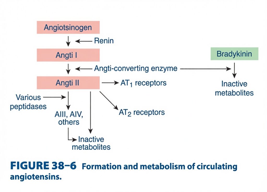
Figure 38-6: Formation and Metabolism of Circulating Angiotensins
Easiest & Most Conceptual Explanation FOR SELF LEARNERS
This figure explains how Angiotensin II is formed, how it works through its receptors, how it is broken down, and how ACE also breaks down bradykinin.
This figure is an extension of the Renin–Angiotensin–Aldosterone System (RAAS).
⭐ One-Line Concept
Renin forms Angiotensin I → ACE converts it into Angiotensin II → Angiotensin II acts mainly through AT₁ receptors → It is then broken down into smaller peptides and finally inactive metabolites. ACE also breaks down bradykinin.
First Understand the Whole Story
Angiotensinogen
        ↓ (Renin)
Angiotensin I
        ↓ (ACE)
Angiotensin II
      ↙       ↘
AT₁          AT₂
Receptors    Receptors
      ↓
Body Effects
      ↓
Peptidases
      ↓
Ang III → Ang IV
      ↓
Inactive Metabolites

ACE also breaks down:
Bradykinin
      ↓
Inactive Metabolites
Step 1: Angiotensinogen
At the top of the figure is:
Angiotensinogen
Produced by:
Liver
It is an inactive protein circulating in the blood.
Easy Concept
Think of angiotensinogen as:
Raw material waiting to be activated.
Step 2: Renin
Look at the arrow labeled:
Renin
Renin is released by the kidney.
Renin cuts angiotensinogen to produce:
Angiotensin I (Ang I)
Easy Memory
Renin starts the RAAS pathway.
Easy Concept
Renin acts like a pair of scissors.
Angiotensinogen
        ↓
      Renin
        ↓
Angiotensin I
Step 3: Angiotensin-Converting Enzyme (ACE)
The next arrow is labeled:
Angiotensin-Converting Enzyme (ACE)
ACE converts:
Angiotensin I
↓
Angiotensin II
Where is ACE found?
Mainly on the vascular endothelium, especially in the lungs.asy Concept
ACE is like a finishing machine.
It changes the inactive precursor into the active hormone.
Angiotensin I
       ↓
      ACE
       ↓
Angiotensin II
Step 4: Angiotensin II
This is the most important active hormone of the RAAS.
Nearly all major physiological effects are produced by Angiotensin II.
Angiotensin II Acts Through Two Receptors
The figure shows two receptors:
AT₁ Receptors
and
AT₂ Receptors
1. AT₁ Receptors (Most Important)
This is where most actions of Angiotensin II occur.
These include:
- Vasoconstriction
- Aldosterone secretion
- Sodium retention
- Water retention
- Increased blood pressure
- Increased sympathetic activity
Easy Memory
AT₁ = “Action Receptor”
Almost every important clinical effect is through AT₁.
Easy Concept
Think of AT₁ as the main switch.
When Angiotensin II presses this switch,
blood pressure rises.
2. AT₂ Receptors
The figure also shows:
AT₂ Receptors
These receptors are less important in normal adults.
They are involved in:
Easy Memory
AT₂ = Auxiliary receptor
Step 5: Breakdown of Angiotensin II
Look at the left side.
The figure shows:
Various Peptidases
These enzymes gradually break down Angiotensin II.
First into:
Angiotensin III (Ang III)
Then
Angiotensin IV (Ang IV)
Then eventually into:
Inactive Metabolites
Easy Concept
Imagine breaking a long chain into smaller and smaller pieces.
Eventually,
the pieces become useless.
Flow
Angiotensin II
      ↓
Ang III
      ↓
Ang IV
      ↓
Inactive Metabolites
What are Ang III and Ang IV?
They are breakdown products of Angiotensin II.
They still have some biological activity,
but they are much less potent than Angiotensin II.
Eventually,
they become inactive metabolites.
The Bradykinin Pathway
Look carefully at the right side.
The figure shows:
Bradykinin
↓
ACE
↓
Inactive Metabolites
Why is this Important?
ACE has two jobs.
Job 1
Convert:
Angiotensin I
↓
Angiotensin II
Job 2
Destroy:
Bradykinin
↓
Inactive metabolites
Easy Concept
ACE is a double-function enzyme.
It:
- Makes Angiotensin II
- Breaks down Bradykinin
What is Bradykinin?
Bradykinin is a natural substance that:
- Dilates blood vessels
- Lowers blood pressure
- Increases vascular permeability
Easy Memory
Bradykinin relaxes blood vessels.
Clinical Importance
Because ACE destroys bradykinin,
ACE inhibitors cause:
↑ Bradykinin
↓
Vasodilation
↓
Lower blood pressure
But increased bradykinin may also cause:
- Dry cough
- Angioedema
These are classic adverse effects of ACE inhibitors.
Complete Story of the Figure
Angiotensinogen
        ↓
      Renin
        ↓
Angiotensin I
        ↓
       ACE
        ↓
Angiotensin II
      ↙      ↘
AT₁         AT₂
      ↓
Main Physiological Effects
      ↓
Peptidases
      ↓
Ang III
      ↓
Ang IV
      ↓
Inactive Metabolites

ACE also:
Bradykinin
      ↓
Inactive Metabolites
Everyday Analogy
Imagine a factory.
Renin
Cuts raw material.
↓
ACE
Finishes the product.
↓
Angiotensin II
Becomes the final active worker.
↓
The worker presses switches (AT₁ receptors) to increase blood pressure.
After the job is complete,
recycling machines (peptidases) break the worker into smaller pieces until nothing active remains.
At the same time,
ACE also destroys bradykinin, a substance that normally relaxes blood vessels.
Easy Memory Trick
Remember: “RACE”
R = Renin starts the pathway
↓
A = Angiotensin I
↓
C = Converting Enzyme (ACE)
↓
E = Effective Hormone (Angiotensin II)
Then:
- AT₁ = Main actions
- AT₂ = Minor actions
- Peptidases = Breakdown
- ACE also destroys Bradykinin
High-Yield Exam Points
- Renin converts angiotensinogen into Angiotensin I.
- ACE (Angiotensin-Converting Enzyme) converts Angiotensin I into Angiotensin II, the principal active peptide of the RAAS.
- Angiotensin II exerts most of its physiological actions through AT₁ receptors, including vasoconstriction, aldosterone secretion, sodium retention, water retention, and increased blood pressure.
- AT₂ receptors have more limited roles in adults and are associated with cell growth regulation, tissue repair, vasodilation in some tissues, and fetal development.
- Various peptidases metabolize Angiotensin II into Angiotensin III (Ang III), Angiotensin IV (Ang IV), and ultimately inactive metabolites.
- ACE also degrades bradykinin into inactive metabolites.
- Because ACE normally breaks down bradykinin, ACE inhibitors increase bradykinin levels, contributing to vasodilation but also to the adverse effects of dry cough and angioedema.
KEY CONCEPT (Figure 38-6)
Figure 38-6 summarizes the formation, actions, and metabolism of circulating angiotensins. Renin released from the kidney converts angiotensinogen into Angiotensin I, which is then converted by Angiotensin-Converting Enzyme (ACE) into Angiotensin II, the major active hormone of the RAAS. Angiotensin II acts predominantly through AT₁ receptors, producing vasoconstriction, aldosterone secretion, sodium and water retention, and an increase in arterial blood pressure. It can also bind AT₂ receptors, which are involved mainly in tissue growth, repair, and developmental processes. Angiotensin II is progressively degraded by various peptidases into Angiotensin III, Angiotensin IV, and finally inactive metabolites. In addition to generating Angiotensin II, ACE degrades bradykinin, an endogenous vasodilator, making ACE a key regulator of both vasoconstrictor and vasodilator pathways.
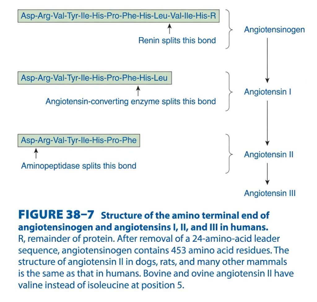
Figure 38-7: Structure of Angiotensinogen and Angiotensins I, II, and III
Easiest & Most Conceptual Explanation for SELF LEARNERS
This figure explains how enzymes cut (cleave) angiotensinogen step by step to produce Angiotensin I, Angiotensin II, and Angiotensin III.
The main concept is “one protein is gradually trimmed (Neat and tidy by cutting) into smaller active hormones.”
⭐ One-Line Concept
Angiotensinogen is a large protein. Renin cuts it to form Angiotensin I. ACE cuts Angiotensin I to form Angiotensin II. Aminopeptidase removes one more amino acid to form Angiotensin III.
First Understand the Whole Story
Large Protein (Angiotensinogen)
             │
        Renin cuts
             │
             ▼
     Angiotensin I (10 amino acids)
             │
         ACE cuts
             │
             ▼
     Angiotensin II (8 amino acids)
             │
     Aminopeptidase cuts
             │
             ▼
     Angiotensin III (7 amino acids)
Each enzyme removes a small piece, making the peptide shorter.tep 1: Angiotensinogen
At the top of the figure is:
Angiotensinogen
It is produced by the liver.
It is a large inactive protein.
The figure shows its amino-terminal sequence:
Asp–Arg–Val–Tyr–Ile–His–Pro–Phe–His–Leu–Val–Ile–His–RWhat does R mean?
The figure states:
R = Remainder of the protein
This means many more amino acids continue after this point.
Easy Concept
Imagine a long ribbon.
Only the first part is shown.
The remaining long ribbon is represented by R.
Step 2: Renin Splits Angiotensinogen
The arrow says:
Renin splits this bond
Renin is released from the kidney.
It cuts angiotensinogen at one specific peptide bond.
After this cut,
the first 10 amino acids are released.
These become:
Angiotensin I
Amino Acid Sequence of Angiotensin I
Asp–Arg–Val–Tyr–Ile–His–Pro–Phe–His–LeuImportant Point
Angiotensin I contains:
10 amino acids
It is therefore called a decapeptide.
Easy Memory
Renin makes the 10-amino-acid peptide.
Step 3: ACE Converts Angiotensin I into Angiotensin II
The figure says:
Angiotensin-Converting Enzyme splits this bond
ACE removes the last two amino acids from Angiotensin I.
Before ACE
Asp–Arg–Val–Tyr–Ile–His–Pro–Phe–His–Leu↓
ACE removes:
His–Leu↓
Remaining sequence:
Asp–Arg–Val–Tyr–Ile–His–Pro–PheThis is:
Angiotensin II
Important Point
Angiotensin II contains:
8 amino acids
It is called an octapeptide.
Easy Memory
ACE removes the last 2 amino acids.
10 → 8
Step 4: Aminopeptidase Forms Angiotensin III
The next arrow says:
Aminopeptidase splits this bond
This enzyme removes one amino acid from the beginning (N-terminus) of Angiotensin II.
Before
Asp–Arg–Val–Tyr–Ile–His–Pro–Phe↓
Remove:
Asp↓
Remaining:
Arg–Val–Tyr–Ile–His–Pro–PheThis becomes:
Angiotensin III
Important Point
Angiotensin III contains:
7 amino acids
It is therefore called a heptapeptide.
Easy Memory
Aminopeptidase removes the first amino acid, not the last.
Why Is It Called “Aminopeptidase”?
The enzyme removes amino acids from the:
Amino (N-terminal) end
Hence the name:
Aminopeptidase
Easy Number Trick
Remember these numbers:
| Molecule | Number of Amino Acids |
|---|---|
| Angiotensinogen | 453 amino acids (after removal of the 24-amino-acid leader sequence) |
| Angiotensin I | 10 |
| Angiotensin II | 8 |
| Angiotensin III | 7 |
What Happens at Each Step?
Step 1
Large inactive protein
↓
Renin cuts
↓
10-amino-acid peptide
Step 2
10 amino acids
↓
ACE removes 2 amino acids
↓
8 amino acids
Step 3
8 amino acids
↓
Aminopeptidase removes 1 amino acid
↓
7 amino acids
Easy Flow Diagram
Angiotensinogen
(453 amino acids)
        │
        │ Renin
        ▼
Angiotensin I
(10 amino acids)
        │
        │ ACE
        ▼
Angiotensin II
(8 amino acids)
        │
        │ Aminopeptidase
        ▼
Angiotensin III
(7 amino acids)
Everyday Analogy
Imagine carving a wooden stick.
First cut
A long stick becomes a smaller stick.
↓
Second cut
The smaller stick becomes even shorter.
↓
Third cut
One more small piece is removed.
The stick becomes shorter and shorter.
The enzymes do the same thing with angiotensin peptides.
What Is the Purpose of These Cuts?
Each enzyme changes the size and activity of the peptide.
- Angiotensinogen = inactive precursor
- Angiotensin I = inactive or minimally active precursor
- Angiotensin II = major active hormone of the RAAS
- Angiotensin III = active metabolite with weaker overall effects than Angiotensin II, but it can still stimulate aldosterone secretion
Species Note (Bottom of the Figure)
The figure states:
- After removal of a 24-amino-acid leader sequence, human angiotensinogen contains 453 amino acid residues.
- The structure of Angiotensin II is the same in humans, dogs, rats, and many other mammals.
- In cattle (bovine) and sheep (ovine), valine replaces isoleucine at position 5.
Easy Concept
Most mammals have almost the same Angiotensin II, with only minor species differences.
Easy Memory Trick
Remember “10 → 8 → 7”
- Angiotensin I = 10 amino acids
- Angiotensin II = 8 amino acids
- Angiotensin III = 7 amino acids
And remember the enzymes:
- Renin → forms Angiotensin I
- ACE → forms Angiotensin II
- Aminopeptidase → forms Angiotensin III
High-Yield Exam Points
- Angiotensinogen is a 453-amino-acid protein (after removal of the 24-amino-acid leader sequence) produced by the liver.
- Renin cleaves angiotensinogen to release Angiotensin I, a 10-amino-acid decapeptide.
- Angiotensin-Converting Enzyme (ACE) removes the terminal His–Leu dipeptide from Angiotensin I to produce Angiotensin II, an 8-amino-acid octapeptide.
- Angiotensin II is the principal biologically active peptide of the RAAS.
- Aminopeptidase removes the N-terminal aspartate (Asp) residue from Angiotensin II to produce Angiotensin III, a 7-amino-acid heptapeptide.
- Human Angiotensin II has the same amino acid sequence as that found in dogs, rats, and many other mammals.
- In bovine and ovine species, valine replaces isoleucine at position 5 of Angiotensin II.
KEY CONCEPT (Figure 38-7)
Figure 38-7 illustrates the stepwise enzymatic processing of angiotensin peptides. Renin cleaves the large precursor protein angiotensinogen to release Angiotensin I, a 10-amino-acid decapeptide. Angiotensin-Converting Enzyme (ACE) then removes the terminal His–Leu dipeptide to form Angiotensin II, the 8-amino-acid octapeptide that serves as the major active hormone of the renin–angiotensin–aldosterone system. Subsequently, aminopeptidase removes the N-terminal Asp residue to generate Angiotensin III, a 7-amino-acid heptapeptide that retains some biological activity, particularly in stimulating aldosterone secretion. This sequential trimming of the peptide chain demonstrates how specific enzymes generate progressively shorter angiotensin peptides with distinct physiological activities.
ANGIOTENSINOGEN
- Angiotensinogen is a protein that circulates in the blood.
- It is found in the α₂-globulin fraction of plasma.
Figure: Figure 38–6
- Angiotensinogen contains about 13% carbohydrate.
- It is made up of 453 amino acid residues.
- Angiotensinogen is synthesized in the liver.
- During its formation, it first contains a 32-amino-acid signal sequence.
- This signal sequence is removed inside the endoplasmic reticulum.
- The circulating level of angiotensinogen increases in response to:
- Glucocorticoids
- Thyroid hormones
- Estrogens
- Several cytokines
- Angiotensin II
KEY CONCEPT
- Angiotensinogen is a liver-produced plasma protein and the precursor of the renin–angiotensin system. It circulates in the α₂-globulin fraction of plasma and serves as the substrate on which renin acts to begin the formation of angiotensin II. Its blood level increases under the influence of several hormones and cytokines.
Conceptual Examples
- Example 1: The liver releases angiotensinogen into the bloodstream. When renin is released from the kidneys, it acts on angiotensinogen to produce angiotensin I, starting the renin–angiotensin system.
- Example 2: During pregnancy or estrogen therapy, estrogen increases angiotensinogen production by the liver, resulting in higher circulating levels of angiotensinogen.
ANGIOTENSIN-CONVERTING ENZYME & ANGIOTENSIN II
- Angiotensin-Converting Enzyme (ACE) is an enzyme that converts angiotensin I into angiotensin II.
- ACE removes two amino acids (histidyl-leucine) from angiotensin I.
- This converts the inactive angiotensin I into the active octapeptide angiotensin II.
Figure: Figure 38–7
- The same ACE enzyme also breaks down (inactivates) bradykinin.
Figure: Figure 38–6
- When ACE is blocked by ACE inhibitors, bradykinin is not broken down.
- As a result, bradykinin levels increase in the tissues.
- Bradykinin acts on B₂ receptors.
- This increased bradykinin is responsible for the dry cough that occurs in up to 20% of patients taking ACE inhibitors.
Clinical Box: Clinical Box 38–2
- Most ACE present in the bloodstream is located on endothelial cells (cells lining blood vessels).
- A large amount of angiotensin I is converted to angiotensin II as blood passes through the lungs.
- However, ACE is also present in many other tissues, so angiotensin II is produced throughout the body.
- ACE is an ectoenzyme (an enzyme attached to the outer surface of the cell membrane).
- ACE exists in two forms:
- Somatic ACE
- Germinal ACE
- Somatic ACE is found throughout the body.
- Germinal ACE is found only in postmeiotic spermatogenic cells and sperm cells (spermatozoa).
- Both forms have:
- One transmembrane domain
- A short cytoplasmic tail
- Somatic ACE is a 170-kDa protein.
- It has two similar extracellular domains.
- Each extracellular domain contains an active site.
- Germinal ACE is a 90-kDa protein.
- It has only one extracellular domain.
- It has only one active site.
- Both forms of ACE are produced from the same gene.
- The gene has two different promoters.
- These promoters produce two different messenger RNAs (mRNAs).
- In male mice without the ACE gene (ACE knockout mice):
- Blood pressure is lower than normal.
- Fertility is reduced.
- In female ACE knockout mice:
- Blood pressure remains normal.
- Fertility is not affected.
Figure: Figure 38–6, Figure 38–7
KEY CONCEPT
- ACE converts inactive angiotensin I into active angiotensin II and also breaks down bradykinin. Most ACE is present on vascular endothelial cells, especially in the lungs. ACE exists as somatic and germinal forms, both produced from the same gene. ACE inhibitors increase bradykinin levels, which commonly causes a dry cough.
Conceptual Examples
- Example 1: A patient takes an ACE inhibitor for hypertension. ACE cannot convert angiotensin I into angiotensin II, so blood pressure falls. At the same time, bradykinin accumulates, causing a persistent dry cough.
- Example 2: As blood flows through the lungs, endothelial ACE rapidly converts angiotensin I into angiotensin II, helping maintain normal blood pressure and fluid balance.
CLINICAL BOX 38–2
Pharmacologic Manipulation of the Renin–Angiotensin System
- The renin–angiotensin system can be controlled by different medicines.
- Some drugs decrease renin secretion.
- Examples include:
- Indomethacin (inhibits prostaglandin synthesis)
- Propranolol (β-adrenergic blocker)
- Some drugs directly inhibit renin activity.
- Examples include:
- Pepstatin
- Enalkiren
- These drugs prevent renin from converting angiotensinogen into angiotensin I.
- ACE inhibitors block the conversion of angiotensin I into angiotensin II.
- Examples include:
- Captopril
- Enalapril
- Some drugs block the action of angiotensin II at its receptors.
- Saralasin and similar drugs competitively block both:
- AT₁ receptors
- AT₂ receptors
- Losartan selectively blocks AT₁ receptors.
- PD-123177 and several other drugs selectively block AT₂ receptors.
CLINICAL CONCEPT CHECK 38–2
Case Summary
- Patient: Mrs. Johnson, 65-year-old woman
- Medical history: Hypertension
- Chief complaint: Chronic cough
- No history of:
- Smoking
- Allergies
- Asthma
- Recently started medication: Captopril
- Examination findings:
- No sinusitis
- No wheezing
Step-by-Step Interpretation
Step 1: Chronic cough
- The main symptom is a persistent chronic cough.
Step 2: Common causes are unlikely
- No smoking history.
- No asthma.
- No allergies.
- No sinus disease.
Step 3: Important clue
- The patient recently started taking captopril.
Step 4: Mechanism
- Captopril is an ACE inhibitor.
- ACE normally breaks down bradykinin.
- When ACE is inhibited:
- Bradykinin is not broken down.
- Bradykinin accumulates in the lungs.
- Bradykinin stimulates B₂ receptors.
- This produces a persistent dry cough.
Most Likely Diagnosis
ACE inhibitor–induced dry cough (caused by captopril).
Cause
- Captopril inhibits ACE.
- ACE inhibition prevents bradykinin breakdown.
- Accumulated bradykinin in the lungs stimulates B₂ receptors.
- This causes a chronic, persistent dry cough.
KEY CONCEPT
- Different drugs can block the renin–angiotensin system at different steps. Renin inhibitors block the formation of angiotensin I, ACE inhibitors block the formation of angiotensin II, and receptor blockers prevent angiotensin II from acting on its receptors. ACE inhibitors also increase bradykinin levels, which commonly causes a persistent dry cough.
Conceptual Examples
- Example 1: A patient starts captopril for hypertension and develops a dry cough a few weeks later because bradykinin accumulates after ACE is inhibited.
- Example 2: A patient who cannot tolerate the cough caused by captopril may be switched to losartan, which blocks AT₁ receptors without increasing bradykinin levels.
THE RENIN–ANGIOTENSIN SYSTEM
METABOLISM OF ANGIOTENSIN II
- Angiotensin II is broken down very quickly in the body.
- Its half-life in the bloodstream is about 1–2 minutes.
- Angiotensin II is broken down by different peptidase enzymes.
- An aminopeptidase removes the first amino acid (aspartic acid, Asp) from angiotensin II.
- This forms a seven-amino-acid peptide called angiotensin III.
- Angiotensin III still has physiological activity.
- Removal of another amino acid from angiotensin III forms a six-amino-acid peptide called angiotensin IV.
- Angiotensin IV also has some physiological activity.
- Most of the other peptide fragments formed during angiotensin breakdown are inactive.
- Aminopeptidase can also act on angiotensin I.
- It removes the first aspartic acid (Asp) residue from angiotensin I to form (des-Asp¹) angiotensin I.
- ACE can directly convert (des-Asp¹) angiotensin I into angiotensin III.
- Angiotensin-metabolizing enzymes are present in red blood cells and many body tissues.
- Angiotensin II is also removed from the bloodstream by trapping within the blood vessels of tissues other than the lungs.
Figure: Figure 38–7
- Renin is commonly measured by determining how much angiotensin I is produced after incubating a plasma sample.
- This test measures Plasma Renin Activity (PRA).
- Low PRA may occur because of low renin or low angiotensinogen.
- To avoid this problem, extra (exogenous) angiotensinogen is often added during testing.
- This allows measurement of Plasma Renin Concentration (PRC) instead of PRA.
- The normal Plasma Renin Activity (PRA) in a person lying down and eating a normal sodium diet is about 1 ng of angiotensin I produced per milliliter of plasma per hour.
- The normal plasma concentration of angiotensin II is about 25 pg/mL (approximately 25 pmol/L).
Figure: Figure 38–7
KEY CONCEPT
- Angiotensin II is rapidly broken down into angiotensin III and angiotensin IV by aminopeptidases. Angiotensin III and angiotensin IV retain some biological activity, while most other breakdown products are inactive. Renin activity is assessed by measuring angiotensin I production (PRA) or by measuring Plasma Renin Concentration (PRC) when additional angiotensinogen is supplied.
Conceptual Examples
- Example 1: After angiotensin II is formed, it remains in the blood for only 1–2 minutes before being converted into angiotensin III, which still has biological activity.
- Example 2: A patient has low Plasma Renin Activity (PRA). This may be due to low renin or low angiotensinogen. Adding extra angiotensinogen allows measurement of Plasma Renin Concentration (PRC) to accurately assess renin secretion.
THE RENIN–ANGIOTENSIN SYSTEM
ACTIONS OF ANGIOTENSINS
- Angiotensin I mainly acts as the precursor of angiotensin II.
- It has no other established physiological action.
- Angiotensin II causes constriction of arterioles (small arteries).
- This increases both systolic and diastolic blood pressure.
- Angiotensin II is one of the most powerful vasoconstrictors in the body.
- It is about 4–8 times more potent than norepinephrine in raising blood pressure (on a weight basis).
- The blood pressure–raising effect of angiotensin II is reduced in:
- Sodium (Na⁺)-depleted individuals
- Patients with liver cirrhosis
- Some other diseases
- In these conditions, blood levels of angiotensin II are already high.
- High angiotensin II levels decrease (down-regulate) angiotensin receptors on vascular smooth muscle.
- As a result, injected angiotensin II produces a smaller response.
- Angiotensin II directly stimulates the adrenal cortex to release aldosterone.
- The renin–angiotensin system is one of the main regulators of aldosterone secretion.
- Angiotensin II directly increases the release of norepinephrine from postganglionic sympathetic neurons.
- Angiotensin II contracts mesangial cells in the kidneys.
- This decreases the Glomerular Filtration Rate (GFR).
- Angiotensin II also acts directly on the renal tubules to increase sodium (Na⁺) reabsorption.
- Angiotensin II acts on the brain to reduce the sensitivity of the baroreceptor reflex.
- This further enhances its blood pressure–raising effect.
- Angiotensin II also stimulates thirst.
- It increases vasopressin (ADH) secretion.
- It also increases adrenocorticotropic hormone (ACTH) secretion.
- Angiotensin II cannot cross the blood–brain barrier.
- It produces these brain effects by acting on the circumventricular organs, which lie outside the blood–brain barrier.
- The area postrema mainly enhances the blood pressure response.
- The subfornical organ (SFO) and the Organum Vasculosum of the Lamina Terminalis (OVLT) mainly stimulate thirst (dipsogenic effect).
- The exact circumventricular organs responsible for increasing ADH and ACTH secretion are still uncertain.
- Angiotensin III [(des-Asp¹) angiotensin II] has about 40% of the blood pressure–raising effect of angiotensin II.
- Angiotensin III has 100% of the aldosterone-stimulating effect of angiotensin II.
- Although it was once thought that angiotensin III was the main natural stimulator of aldosterone, current evidence does not support this.
- Angiotensin III is now considered mainly a breakdown product that still has some biological activity.
- Angiotensin IV is also considered mainly a breakdown product with some biological activity.
- Some researchers suggest that angiotensin IV may have unique effects in the brain.
KEY CONCEPT
- Angiotensin II is the main active hormone of the renin–angiotensin system. It raises blood pressure by constricting blood vessels, stimulates aldosterone, increases sodium reabsorption, promotes norepinephrine release, decreases GFR, stimulates thirst, and increases ADH and ACTH secretion. Angiotensin III and angiotensin IV are mainly breakdown products that retain some biological activity.
Conceptual Examples
- Example 1: A patient with low blood pressure after dehydration releases angiotensin II, which constricts blood vessels, stimulates aldosterone and ADH, increases thirst, and conserves sodium and water, helping restore blood pressure.
- Example 2: A patient with chronic high levels of angiotensin II (such as in sodium depletion or cirrhosis) develops fewer angiotensin receptors on blood vessels, so an additional dose of angiotensin II produces a smaller increase in blood pressure.
Made BY SELF LEARNING CEO AND FOUNDER DR SHEEN
