


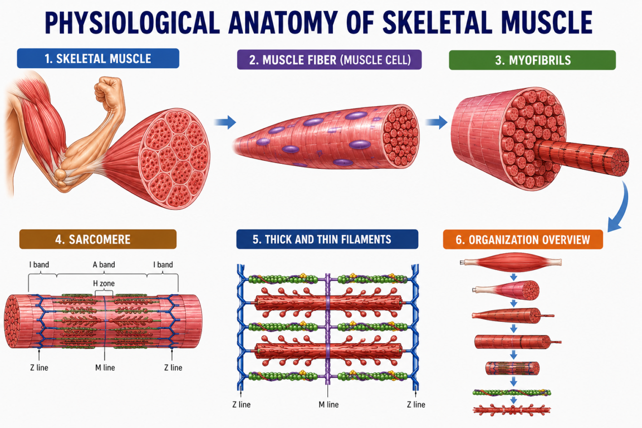
- Skeletal muscles are made up of many muscle fibers.
- Each muscle fiber has a diameter of about 10–80 micrometers (µm).
- Every muscle fiber is made of smaller subunits arranged inside it
- These subunits become progressively smaller from the whole muscle fiber to its internal structures.
- In most skeletal muscles, each muscle fiber extends from one end of the muscle to the other.
- This means that one muscle fiber usually runs the entire length of the muscle.
- About 98% of muscle fibers receive only one nerve ending.
- This single nerve ending is usually located near the middle of the muscle fiber.
- The nerve ending carries signals that stimulate the entire muscle fiber to contract.
KEY CONCEPT
- Skeletal muscle is composed of numerous muscle fibers measuring 10–80 µm in diameter.
- Each muscle fiber contains progressively smaller subunits.
- Most muscle fibers extend the entire length of the muscle.
- About 98% of muscle fibers are supplied by a single nerve ending.
- The nerve ending is usually located near the middle of the muscle fiber, allowing the entire fiber to contract.
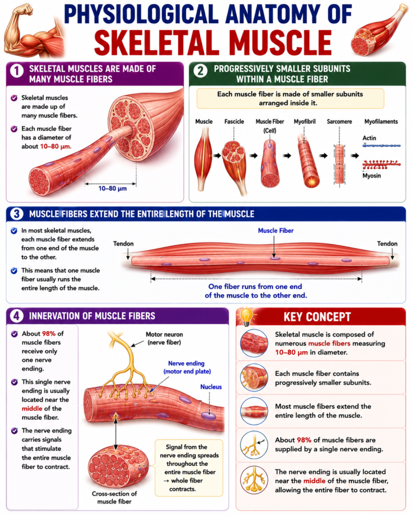
The Sarcolemma Is a Thin Membrane Enclosing a Skeletal Muscle Fiber
- The sarcolemma is a thin membrane that surrounds each skeletal muscle fiber.
- The sarcolemma has two main parts:
- A true cell membrane, called the plasma membrane.
- An outer coat made of a thin layer of polysaccharide material containing many thin collagen fibrils.
- At both ends of the muscle fiber, the outer layer of the sarcolemma fuses with a tendon fiber.
- The tendon fibers then join together in bundles.
- These bundles form the muscle tendons.
- The muscle tendons connect the muscles to the bones.
KEY CONCEPT
- The sarcolemma is the thin membrane that encloses a skeletal muscle fiber.
- It consists of the plasma membrane and an outer polysaccharide layer containing collagen fibrils.
- The outer layer of the sarcolemma joins with tendon fibers at both ends of the muscle fiber.
- Tendon fibers bundle together to form muscle tendons, which attach muscles to bones.
Myofibrils Are Composed of Actin and Myosin Filaments
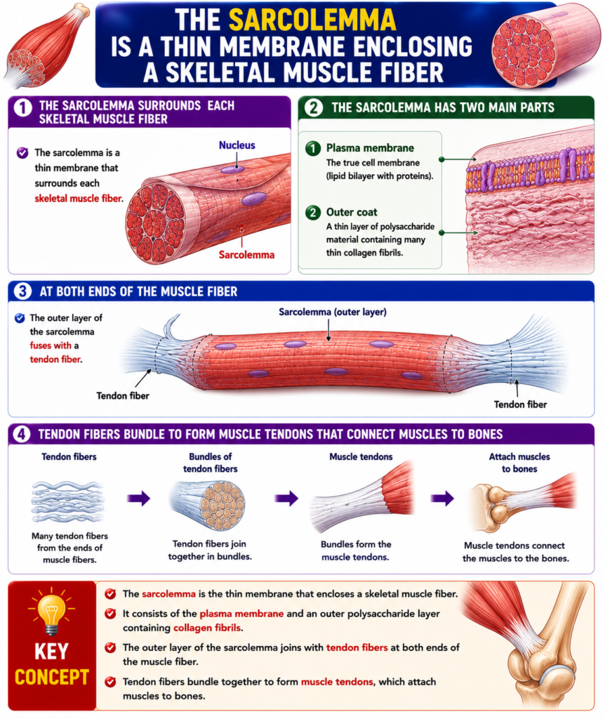
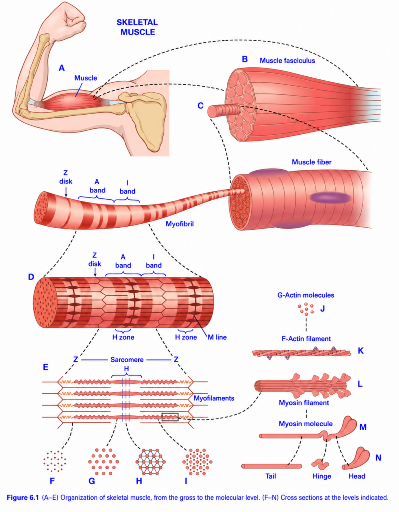
- Each muscle fiber contains several hundred to several thousand myofibrils (Fig. 6.1C).
- Each myofibril is made of about:
- 1500 myosin filaments.
- 3000 actin filaments (Fig. 6.1D–E).
- Actin and myosin are large protein molecules responsible for muscle contraction.
- In Fig. 6.1E–L:
- The thick filaments are myosin.
- The thin filaments are actin.
- The actin and myosin filaments partially overlap (interdigitate) (Fig. 6.1E).
- This overlapping produces alternating light and dark bands in the myofibril.
- The light bands contain only actin filaments.
- These are called I bands because they are isotropic to polarized light.
- The dark bands contain:
- Myosin filaments.
- The overlapping ends of the actin filaments.
- These are called A bands because they are anisotropic to polarized light.
- Small projections are present on the sides of the myosin filaments (Fig. 6.1E–L).
- These projections are called cross-bridges.
- Cross-bridges interact with actin filaments to produce muscle contraction.
- The ends of the actin filaments are attached to the Z disk (Fig. 6.1E).
- From the Z disk, the actin filaments extend in both directions and overlap with the myosin filaments.
- The Z disk is made of filamentous proteins different from actin and myosin.
- The Z disk extends across each myofibril.
- It also connects neighboring myofibrils across the entire muscle fiber.
- Therefore, the entire muscle fiber shows alternating light and dark bands.
- These bands give skeletal muscle and cardiac muscle their striated appearance.
- The portion of a myofibril between two successive Z disks is called a sarcomere (Fig. 6.1E).
- During muscle contraction (bottom of Fig. 6.4), the sarcomere length is about 2 µm.
- At this length:
- The actin filaments completely overlap the myosin filaments.
- The tips of the actin filaments just begin to overlap each other.
- At this sarcomere length, the muscle produces its greatest force of contraction.
KEY CONCEPT
- Each muscle fiber contains many myofibrils.
- Each myofibril is composed of thick myosin filaments and thin actin filaments.
- Partial overlap of actin and myosin forms the light I bands and dark A bands.
- Cross-bridges on myosin interact with actin to produce muscle contraction.
- Actin filaments are attached to the Z disk, and the region between two Z disks is called the sarcomere (Fig. 6.1E).
- At a sarcomere length of about 2 µm (Fig. 6.4), the muscle generates its maximum force of contraction.
Titin Filamentous Molecules Hold the Myosin and Actin Filaments in Place
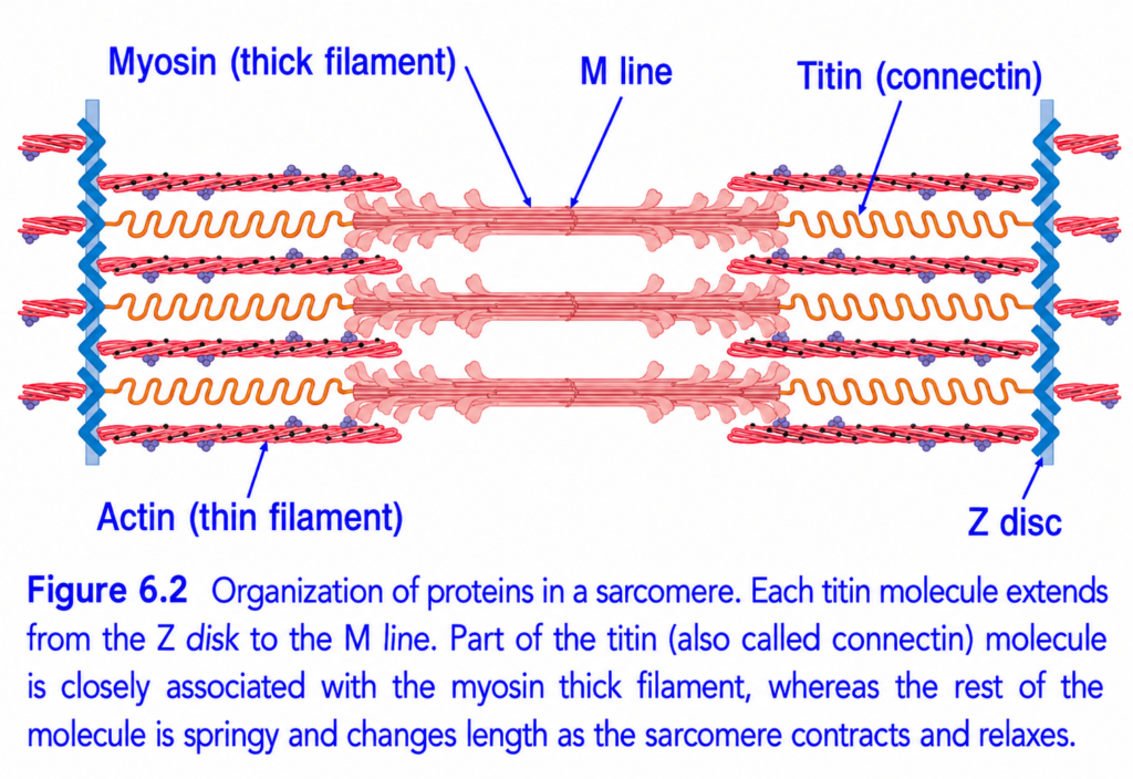
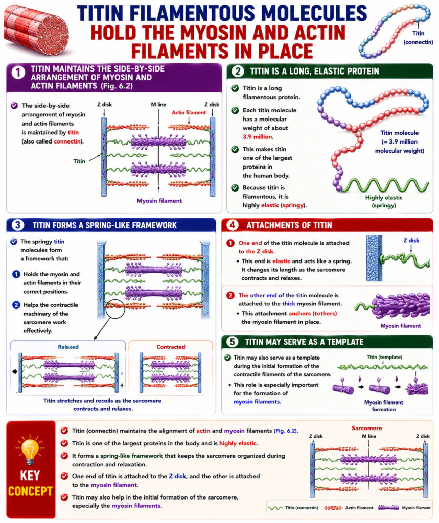
- The side-by-side arrangement of myosin and actin filaments is maintained by titin (also called connectin) (Fig. 6.2).
- Titin is a long filamentous protein.
- Each titin molecule has a molecular weight of about 3.9 million.
- This makes titin one of the largest proteins in the human body.
- Because titin is filamentous, it is highly elastic (springy).
- The springy titin molecules form a framework that:
- Holds the myosin and actin filaments in their correct positions.
- Helps the contractile machinery of the sarcomere work effectively.
- One end of the titin molecule is attached to the Z disk.
- This end is elastic and acts like a spring.
- It changes its length as the sarcomere contracts and relaxes.
- The other end of the titin molecule is attached to the thick myosin filament.
- This attachment anchors (tethers) the myosin filament in place.
- Titin may also serve as a template during the initial formation of the contractile filaments of the sarcomere.
- This role is especially important for the formation of myosin filaments.
KEY CONCEPT
- Titin (connectin) maintains the alignment of actin and myosin filaments (Fig. 6.2).
- Titin is one of the largest proteins in the body and is highly elastic.
- It forms a spring-like framework that keeps the sarcomere organized during contraction and relaxation.
- One end of titin is attached to the Z disk, and the other is attached to the myosin filament.
- Titin may also help in the initial formation of the sarcomere, especially the myosin filaments.
Sarcoplasm Is the Intracellular Fluid Between Myofibrils
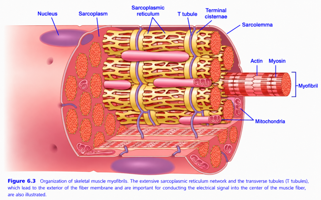
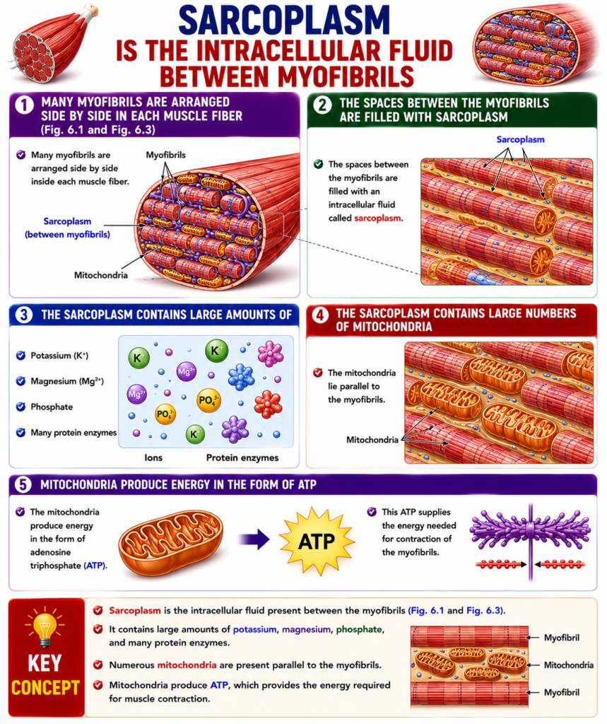
- Many myofibrils are arranged side by side inside each muscle fiber (Fig. 6.1 and Fig. 6.3).
- The spaces between the myofibrils are filled with an intracellular fluid called sarcoplasm.
- The sarcoplasm contains large amounts of:
- Potassium (K⁺).
- Magnesium (Mg²⁺).
- Phosphate.
- Many protein enzymes.
- The sarcoplasm also contains large numbers of mitochondria.
- The mitochondria lie parallel to the myofibrils.
- The mitochondria produce energy in the form of adenosine triphosphate (ATP).
- This ATP supplies the energy needed for contraction of the myofibrils.
KEY CONCEPT
- Sarcoplasm is the intracellular fluid present between the myofibrils (Fig. 6.1 and Fig. 6.3).
- It contains large amounts of potassium, magnesium, phosphate, and many protein enzymes.
- Numerous mitochondria are present parallel to the myofibrils.
- Mitochondria produce ATP, which provides the energy required for muscle contraction.
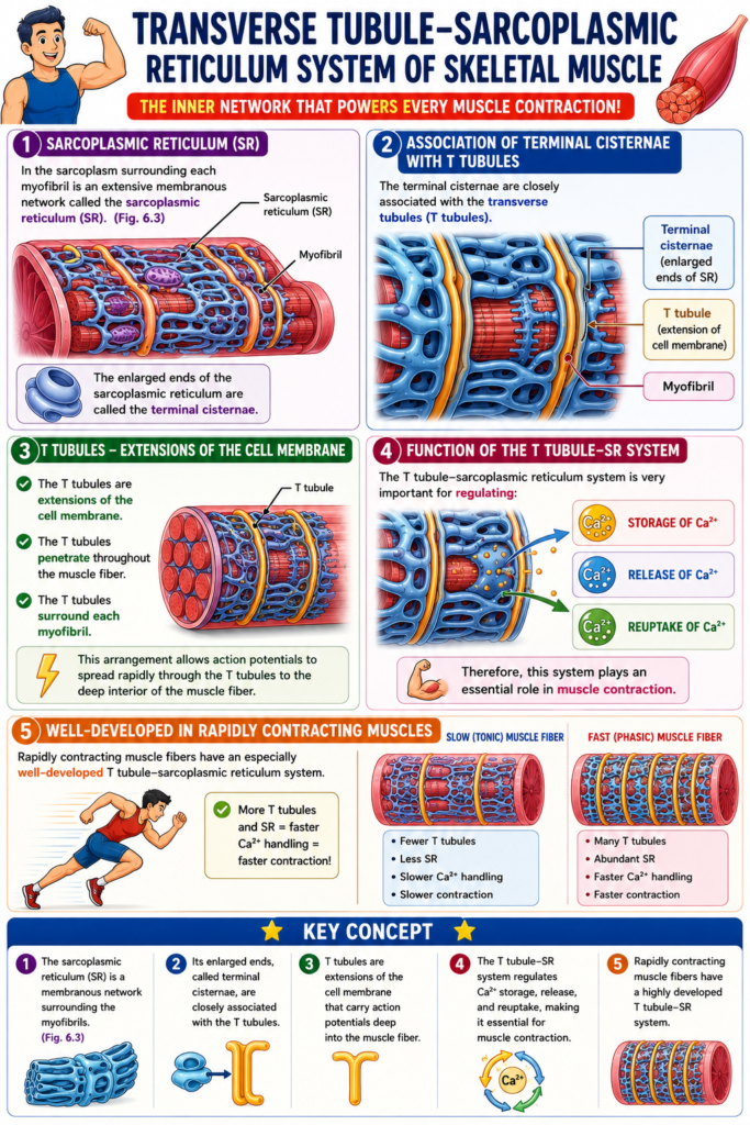
Transverse Tubule–Sarcoplasmic Reticulum System of Skeletal Muscle
- In the sarcoplasm surrounding each myofibril is an extensive membranous network called the sarcoplasmic reticulum (SR) (Fig. 6.3).
- The enlarged ends of the sarcoplasmic reticulum are called the terminal cisternae.
- The terminal cisternae are closely associated with the transverse tubules (T tubules).
- The T tubules are extensions of the cell membrane.
- The T tubules penetrate throughout the muscle fiber.
- The T tubules surround each myofibril.
- This arrangement allows action potentials to spread rapidly through the T tubules to the deep interior of the muscle fiber.
- The T tubule–sarcoplasmic reticulum system is very important for regulating:
- Storage of Ca²⁺.
- Release of Ca²⁺.
- Reuptake of Ca²⁺.
- Therefore, this system plays an essential role in muscle contraction.
- Rapidly contracting muscle fibers have an especially well-developed T tubule–sarcoplasmic reticulum system.
KEY CONCEPT
- The sarcoplasmic reticulum (SR) is a membranous network surrounding the myofibrils (Fig. 6.3).
- Its enlarged ends, called terminal cisternae, are closely associated with the T tubules.
- T tubules are extensions of the cell membrane that carry action potentials deep into the muscle fiber.
- The T tubule–SR system regulates Ca²⁺ storage, release, and reuptake, making it essential for muscle contraction.
- Rapidly contracting muscle fibers have a highly developed T tubule–SR system.
PREPARE AND MADE BY SELF LEARNING DR SHEEN

Thank you sir for making guyton eassy for us
Thanks too much
Easily understandable wording
thanks