


- The blood vessels of the gastrointestinal tract are part of a larger system called the splanchnic circulation (Fig. 63.6).
- The splanchnic circulation includes blood flow through:
- Gastrointestinal tract
- Spleen
- Pancreas
- Liver
- All the blood from the gut, spleen, and pancreas flows directly to the liver through the portal vein.
- Inside the liver, the blood passes through millions of tiny liver sinusoids.
- The blood then leaves the liver through the hepatic veins.
- The hepatic veins empty into the vena cava of the general circulation.
- Before entering the general circulation, the blood passes through the liver.
- This allows the reticuloendothelial cells lining the liver sinusoids to remove bacteria and other particles that enter the blood from the gastrointestinal tract.
- This process prevents harmful substances from reaching the rest of the body directly.
- Water-soluble, non-fat nutrients absorbed from the intestine, such as:
- Carbohydrates
- Proteins
- are carried to the liver through the portal vein.
- In the liver sinusoids, the reticuloendothelial cells and hepatic (liver) cells absorb and temporarily store 50%–75% of these nutrients.
- The liver cells also perform many chemical processing reactions on these nutrients.
- Most fats absorbed from the intestine do not enter the portal blood.
- Instead, they enter the intestinal lymphatic vessels.
- They then travel through the thoracic duct.
- Finally, they enter the systemic circulation, bypassing the liver.
KEY CONCEPT
- The splanchnic circulation (Fig. 63.6) includes the gut, spleen, pancreas, and liver.
- Blood from the gut, spleen, and pancreas first passes through the liver via the portal vein.
- In the liver:
- Reticuloendothelial cells remove bacteria and harmful particles.
- Hepatic cells store and process nutrients.
- 50%–75% of water-soluble nutrients are temporarily stored in the liver.
- Most fats bypass the liver by traveling through the intestinal lymphatics and thoracic duct into the systemic circulation.
Conceptual Examples
- Carbohydrates are absorbed from the intestine → Portal vein → Liver → Stored and processed by hepatic cells.
- Bacteria enter the portal blood from the intestine → Reticuloendothelial cells remove them in the liver → The rest of the body is protected.
- Dietary fat is absorbed → Intestinal lymphatics → Thoracic duct → Systemic circulation, without first passing through the liver.
REMEMBERING POINT
Temporary storage of absorbed water-soluble nutrients by the liver: 50%–75%.
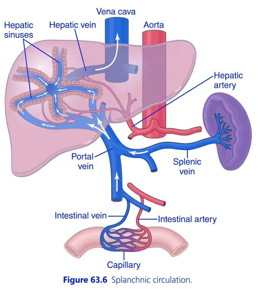
igure 63.6: Splanchnic Circulation
Easiest and Most Conceptual Explanation
⭐ One-Line Concept
The splanchnic circulation collects nutrient-rich blood from the intestines, spleen, pancreas (not shown), and stomach (not shown), sends it first to the liver through the portal vein, and then returns it to the heart through the hepatic veins and vena cava.
First Understand the Whole Story
Normally, blood from an organ returns directly to the heart.
The gastrointestinal organs are different.
Their blood first goes to the liver.
The liver:
- Processes nutrients
- Stores glucose
- Removes toxins
- Metabolizes drugs
- Filters bacteria
Only after this processing does the blood return to the heart.
What is Splanchnic Circulation?
The splanchnic circulation includes blood flow through:
- Intestines
- Stomach (not shown)
- Pancreas (not shown)
- Spleen
- Liver
The liver receives blood from two different sources:
- Portal vein
- Hepatic artery
Understanding Every Part of the Figure
1. Aorta (Red Vessel)
The aorta is the main systemic artery.
It supplies oxygenated blood to many organs.
From the aorta arise:
- Hepatic artery
- Intestinal artery
2. Intestinal Artery (Red)
The intestinal artery carries:
Oxygen-rich blood
↓
To the intestines.
Inside the intestine, it forms:
Capillaries
where oxygen and nutrients are exchanged.
3. Capillaries (Intestine)
These are tiny blood vessels.
Here:
- Oxygen enters intestinal cells.
- Nutrients absorbed from food enter the blood.
These nutrients include:
- Glucose
- Amino acids
- Vitamins
- Minerals
- Water
4. Intestinal Vein (Blue)
After absorbing nutrients,
Blood leaves the intestine through the:
Intestinal vein
Important point:
This blood is:
- Rich in nutrients
- Low in oxygen
It does not return directly to the heart.
Instead,
It flows toward the portal vein.
5. Splenic Vein (Blue)
The splenic vein drains blood from the:
Spleen
This blood joins the portal circulation.
(The pancreas also contributes veins to the portal system, although it is not shown.)
6. Portal Vein (Blue)
This is the most important vessel in the figure.
The portal vein carries:
- Nutrient-rich blood
- Venous blood from the gastrointestinal organs
↓
Directly to the liver.
Easy Concept
Think of the portal vein as a delivery truck carrying absorbed nutrients to the liver.
Why Doesn’t This Blood Go Directly to the Heart?
Because the liver must inspect everything absorbed from the intestine before it enters the systemic circulation.
The liver:
- Stores glucose as glycogen.
- Releases glucose when needed.
- Metabolizes drugs.
- Removes toxins.
- Processes amino acids.
- Produces plasma proteins.
- Helps remove bacteria entering from the gut.
7. Hepatic Artery (Red)
The liver has a second blood supply.
The hepatic artery delivers:
Oxygen-rich arterial blood
to liver tissue.
Important Point
The liver receives blood from:
Portal vein
Approximately 75% of hepatic blood flow.
- Rich in nutrients.
- Lower in oxygen than arterial blood.
Hepatic artery
Approximately 25% of hepatic blood flow.
- Rich in oxygen.
- Nourishes liver cells.
8. Hepatic Sinusoids
These are large, specialized capillaries inside the liver.
Here:
Blood from:
- Portal vein
- Hepatic artery
Mixes together.
What Happens in the Sinusoids?
Liver cells (hepatocytes):
- Remove toxins.
- Store nutrients.
- Metabolize drugs.
- Produce proteins.
- Process carbohydrates, fats, and proteins.
Easy Concept
The hepatic sinusoids act like a processing station where incoming blood is treated before leaving the liver.
9. Hepatic Vein
After the liver finishes processing the blood,
It leaves through the:
Hepatic vein
10. Vena Cava
The hepatic veins drain into the:
Inferior vena cava
The inferior vena cava returns blood to the:
Right atrium of the heart.
Following the Blood Step by Step
Oxygen Supply
Aorta
↓
Intestinal artery
↓
Intestinal capillaries
↓
Intestinal cells receive oxygen
Nutrient Absorption
Food is absorbed
↓
Nutrients enter intestinal capillaries
↓
Intestinal vein
↓
Portal vein
↓
Liver
↓
Hepatic sinusoids
↓
Hepatic vein
↓
Inferior vena cava
↓
Heart
Liver Oxygen Supply
Aorta
↓
Hepatic artery
↓
Hepatic sinusoids
↓
Mixes with portal blood
↓
Hepatic vein
↓
Inferior vena cava
↓
Heart
Why Does the Liver Have Two Blood Supplies?
Portal Vein
Provides:
- Nutrients
- Blood from the gastrointestinal tract
Main role:
Processing absorbed substances.
Hepatic Artery
Provides:
- Oxygen
Main role:
Keeping liver cells alive and functioning.
Understanding the White Arrows
The white arrows indicate the direction of blood flow:
- Intestine → Portal vein → Liver
- Hepatic artery → Liver
- Liver → Hepatic vein → Vena cava
Clinical Importance
Portal Hypertension
If pressure in the portal vein increases:
↓
Blood cannot pass easily through the liver.
↓
Portal hypertension develops.
This may lead to:
- Esophageal varices
- Ascites
- Splenomegaly
Liver Disease
If liver function is impaired:
- Nutrient metabolism decreases.
- Drug metabolism decreases.
- Detoxification decreases.
- Protein synthesis decreases.
Easy Story
Imagine a factory.
The intestines are the raw material supplier.
The portal vein is the delivery truck.
The liver is the processing factory.
After inspection and processing,
The finished product leaves through the hepatic vein and returns to the heart.
Understanding Every Label in the Figure
Aorta
- Main systemic artery.
- Supplies oxygenated blood to abdominal organs.
Intestinal Artery
- Carries oxygen-rich blood to the intestine.
Capillary
- Site of oxygen delivery and nutrient absorption.
Intestinal Vein
- Carries nutrient-rich venous blood from the intestine.
Splenic Vein
- Drains blood from the spleen into the portal system.
Portal Vein
- Carries nutrient-rich blood from the gastrointestinal organs to the liver.
- Provides about 75% of hepatic blood flow.
Hepatic Artery
- Supplies oxygen-rich blood to the liver.
- Provides about 25% of hepatic blood flow.
Hepatic Sinusoids
- Specialized liver capillaries.
- Portal venous blood and hepatic arterial blood mix here.
- Site of nutrient processing and detoxification.
Hepatic Vein
- Drains processed blood from the liver.
Vena Cava
- Returns blood from the liver to the heart.
High-Yield Summary Table
| Structure | Blood Carried | Main Function |
|---|---|---|
| Aorta | Oxygen-rich blood | Supplies abdominal organs |
| Intestinal artery | Oxygen-rich blood | Supplies intestine |
| Intestinal capillaries | Mixed during exchange | Nutrient absorption |
| Intestinal vein | Nutrient-rich, oxygen-poor blood | Drains intestine |
| Splenic vein | Venous blood from spleen | Joins portal circulation |
| Portal vein | Nutrient-rich venous blood | Delivers absorbed nutrients to the liver |
| Hepatic artery | Oxygen-rich blood | Supplies liver cells |
| Hepatic sinusoids | Mixed portal and arterial blood | Processing, metabolism, detoxification |
| Hepatic vein | Processed venous blood | Drains liver |
| Inferior vena cava | Venous blood | Returns blood to the heart |
Key Concept
The splanchnic circulation is a specialized vascular system in which nutrient-rich venous blood from the intestines, spleen, pancreas, and stomach is collected into the portal vein and delivered first to the liver instead of directly to the heart. The liver also receives oxygen-rich blood from the hepatic artery. Within the hepatic sinusoids, portal venous blood and hepatic arterial blood mix, allowing the liver to process nutrients, detoxify harmful substances, metabolize drugs, store glucose, and synthesize plasma proteins. The processed blood then leaves through the hepatic veins, enters the inferior vena cava, and finally returns to the heart.
ANATOMY OF THE GASTROINTESTINAL BLOOD SUPPLY
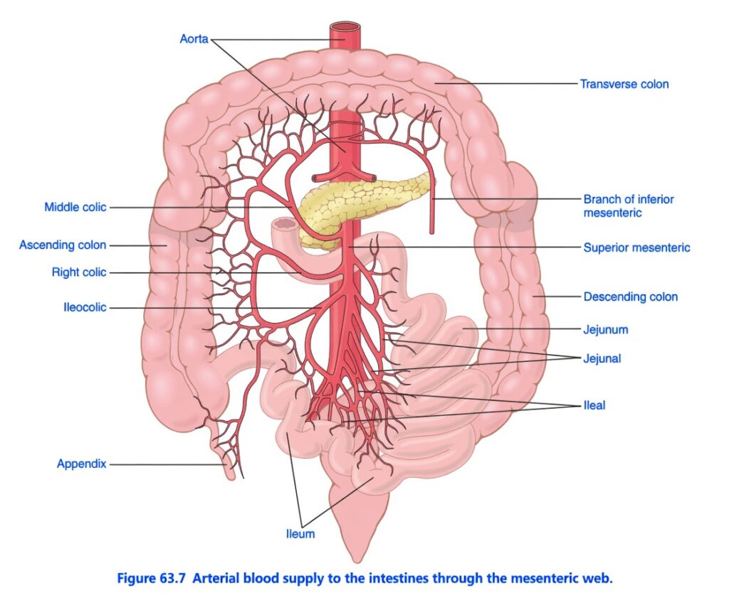
- Fig. 63.7 shows the general pattern of the arterial blood supply to the gastrointestinal tract.
- The superior mesenteric artery and inferior mesenteric artery supply the walls of the small and large intestines.
- These arteries supply the intestine through an arching arterial system.
- The celiac artery is not shown in the figure.
- The celiac artery supplies the stomach in a similar way.
- After entering the intestinal wall, the arteries branch into smaller arteries.
- These smaller arteries run around the intestine in both directions.
- The ends of these arteries meet on the side opposite the mesenteric attachment.
- From these circling arteries, even smaller arteries enter the intestinal wall.
- These small arteries spread:
- Along the muscle bundles
- Into the intestinal villi
- Into the submucosal vessels beneath the epithelium
- These blood vessels support the secretory and absorptive functions of the intestine.
- Fig. 63.8 shows the blood flow inside an intestinal villus.
- Each villus contains a small arteriole and a small venule.
- The arteriole and venule are connected by many looping capillaries.
- The walls of the arterioles are highly muscular.
- These muscular arterioles actively regulate the blood flow through each villus.
KEY CONCEPT
- Fig. 63.7 shows the arterial blood supply of the gastrointestinal tract.
- The superior and inferior mesenteric arteries supply the small and large intestines.
- The celiac artery supplies the stomach.
- Inside the intestinal wall, arteries divide into smaller branches that supply:
- Muscle
- Intestinal villi
- Submucosa
- Fig. 63.8 shows that each intestinal villus contains:
- One arteriole
- One venule
- Many looping capillaries
- Muscular arterioles regulate villus blood flow, helping support absorption and secretion.
Conceptual Examples
- Food is absorbed in the intestinal villi → Looping capillaries quickly carry nutrients into the blood.
- When absorption increases → Muscular arterioles dilate → More blood flows through the villi.
- The superior mesenteric artery supplies most of the small intestine, while the inferior mesenteric artery supplies much of the large intestine.
Figure 63.7: Arterial Blood Supply to the Intestines (Mesenteric Web)
Easiest and Most Conceptual Explanation
⭐ One-Line Concept
The aorta supplies oxygen-rich blood to the intestines through the mesenteric arteries. These arteries branch repeatedly to form a protective arterial network (mesenteric web) that ensures a continuous blood supply to the small and large intestines.
First Understand the Whole Story
The intestines constantly need oxygen and nutrients because they:
- Digest food.
- Absorb nutrients.
- Move food by peristalsis.
- Secrete digestive juices.
To perform these functions, they receive a rich blood supply from the aorta.
Instead of one straight artery, the blood vessels form a mesenteric web (arterial arcades). This branching network ensures that if one small branch is compressed during intestinal movement, nearby branches can continue supplying blood.
Understanding Every Part of the Figure
1. Aorta (Large Red Vessel)
This is the main artery of the body.
It carries:
- Oxygen-rich blood
- Nutrient-rich arterial blood
↓
It gives rise to the superior mesenteric artery and inferior mesenteric artery.
Easy Concept
Think of the aorta as the main highway carrying blood from the heart.
2. Superior Mesenteric Artery (SMA)
This is the major artery supplying the midgut.
It supplies:
- Lower part of the duodenum
- Jejunum
- Ileum
- Cecum
- Appendix
- Ascending colon
- Right two-thirds of the transverse colon
Easy Concept
The SMA supplies almost the entire small intestine and the first part of the large intestine.
3. Inferior Mesenteric Artery (IMA)
Only one branch is shown in this figure.
It supplies the hindgut:
- Left one-third of the transverse colon
- Descending colon
- Sigmoid colon (not shown)
- Upper rectum (not shown)
Easy Concept
The IMA supplies the last part of the large intestine.
4. Mesenteric Web (Arterial Arcades)
Notice the many branching red vessels.
These form the mesenteric arterial web (arcades).
This network:
- Distributes blood evenly.
- Provides alternate pathways (collateral circulation).
- Prevents ischemia if one small branch is temporarily compressed.
Easy Concept
Think of it as a road network with many alternate routes.
If one road is blocked, traffic can still reach its destination through another route.
5. Jejunal Arteries
These branches supply the:
Jejunum
Characteristics:
- Fewer arterial arcades.
- Longer straight arteries (vasa recta).
The jejunum absorbs:
- Most carbohydrates
- Proteins
- Fats
- Vitamins
Therefore, it needs a rich blood supply.
6. Ileal Arteries
These branches supply the:
Ileum
Characteristics:
- More arterial arcades.
- Shorter straight arteries.
The ileum absorbs:
- Vitamin B12
- Bile salts
- Remaining nutrients
7. Jejunum
The jejunum is the middle part of the small intestine.
Main function:
Maximum nutrient absorption.
It receives blood through the jejunal branches of the superior mesenteric artery.
8. Ileum
The ileum is the last part of the small intestine.
Main functions:
- Absorbs vitamin B12.
- Reabsorbs bile salts.
- Absorbs remaining nutrients.
Blood supply:
Ileal branches of the superior mesenteric artery.
9. Ileocecal Artery
This branch supplies:
- Terminal ileum
- Cecum
- Beginning of the large intestine
10. Right Colic Artery
Supplies:
Ascending colon
11. Middle Colic Artery
Supplies:
Transverse colon
(Mainly its proximal two-thirds.)
12. Branch of Inferior Mesenteric Artery
Supplies:
- Distal transverse colon
- Descending colon
This branch meets the middle colic artery, forming collateral circulation.
13. Ascending Colon
Receives blood mainly from:
- Right colic artery
- Ileocecal artery
14. Transverse Colon
Receives blood from:
- Middle colic artery
- Branches of the inferior mesenteric artery
This overlap provides a continuous blood supply.
15. Descending Colon
Supplied mainly by:
Branches of the inferior mesenteric artery.
16. Appendix
Blood supply:
Appendicular artery, a branch of the ileocecal artery (not separately labeled).
Why Does the Mesenteric Web Exist?
The intestines are always:
- Contracting
- Relaxing
- Twisting
- Moving food
A single straight artery could easily become compressed.
The arterial arcades ensure that blood can reach the intestine through multiple pathways.
Clinical Importance
Mesenteric Ischemia
If the superior mesenteric artery becomes blocked:
↓
Blood supply to a large portion of the intestine is reduced.
↓
This can cause:
- Severe abdominal pain
- Intestinal infarction
- Tissue necrosis
- Medical emergency
Collateral Circulation
Because of the arterial arcades:
A small blockage may not immediately stop blood flow, as nearby branches can compensate.
Easy Story
Imagine a city.
The aorta is the main highway.
The superior mesenteric artery is the major city road supplying most neighborhoods.
The inferior mesenteric artery supplies the remaining neighborhoods.
Instead of one road, there are many interconnected streets (arterial arcades), so if one street is blocked, traffic can still reach every house.
Understanding Every Label in the Figure
Aorta
- Main artery of the body.
- Carries oxygen-rich blood from the heart.
Superior Mesenteric Artery
- Supplies most of the small intestine and the proximal large intestine.
Branch of Inferior Mesenteric Artery
Jejunal Branches
- Supply the jejunum.
- Characterized by fewer arcades and longer vasa recta.
Ileal Branches
- Supply the ileum.
- Characterized by more arcades and shorter vasa recta.
Ileocecal Artery
- Supplies the terminal ileum, cecum, and beginning of the large intestine.
Right Colic Artery
- Supplies the ascending colon.
Middle Colic Artery
- Supplies most of the transverse colon.
Jejunum
- Major site of nutrient absorption.
Ileum
- Absorbs vitamin B12, bile salts, and remaining nutrients.
Ascending Colon
- Receives blood mainly from the right colic and ileocecal arteries.
Transverse Colon
- Receives blood from the middle colic artery and branches of the inferior mesenteric artery.
Descending Colon
- Receives blood mainly from branches of the inferior mesenteric artery.
Appendix
- Supplied by the appendicular artery, which arises from the ileocecal artery.
High-Yield Summary Table
| Structure | Blood Supply | Main Function |
|---|---|---|
| Aorta | Heart | Main source of arterial blood |
| Superior mesenteric artery | Aorta | Supplies midgut organs |
| Inferior mesenteric artery | Aorta | Supplies hindgut organs |
| Jejunal branches | Superior mesenteric artery | Supply the jejunum |
| Ileal branches | Superior mesenteric artery | Supply the ileum |
| Ileocecal artery | Superior mesenteric artery | Supplies terminal ileum, cecum, and appendix |
| Right colic artery | Superior mesenteric artery | Supplies the ascending colon |
| Middle colic artery | Superior mesenteric artery | Supplies the proximal two-thirds of the transverse colon |
| Inferior mesenteric branches | Inferior mesenteric artery | Supply the distal transverse and descending colon |
| Mesenteric arterial arcades | Interconnected arterial branches | Provide collateral circulation and continuous intestinal perfusion |
Key Concept
The arterial blood supply of the intestines originates from the aorta, mainly through the superior mesenteric artery and inferior mesenteric artery. These arteries divide into numerous branches that form mesenteric arterial arcades (the mesenteric web), creating multiple pathways for blood flow. The superior mesenteric artery supplies the jejunum, ileum, cecum, appendix, ascending colon, and proximal two-thirds of the transverse colon, whereas the inferior mesenteric artery supplies the distal transverse colon and descending colon. This extensive branching network ensures a continuous oxygen supply to the intestines even during constant intestinal movement and provides important collateral circulation if one arterial branch becomes obstructed.
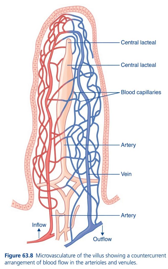
Figure 63.8: Microvasculature of the Intestinal Villus (Countercurrent Blood Flow)
Easiest and Most Conceptual Explanation
⭐ One-Line Concept
Each intestinal villus has an artery, a vein, a network of blood capillaries, and a central lacteal. Blood flows upward through the artery and downward through the vein, creating a countercurrent system that efficiently supports nutrient absorption but also makes the tip of the villus more vulnerable to low oxygen (ischemia).
First Understand the Whole Story
An intestinal villus is a finger-like projection that increases the surface area for absorption.
Each villus contains:
- One arteriole (artery) carrying oxygen-rich blood upward.
- Many blood capillaries where nutrients are absorbed.
- One venule (vein) carrying blood back downward.
- One central lacteal that absorbs fats.
The artery and vein run very close to each other, allowing oxygen to diffuse directly from the artery to the vein before reaching the tip. This arrangement is called countercurrent exchange.
Understanding Every Part of the Figure. Pink Outer Layer
This represents the intestinal epithelium.
Function
- Covers the villus.
- Contains absorptive epithelial cells.
- Nutrients pass through these cells into blood capillaries or the lacteal.
Easy Concept
Think of it as the absorbing skin of the villus.
2. Artery (Red Vessel)
Located near the base of the villus.
The arrow labeled Inflow shows that blood enters the villus through this artery.
The artery carries:
- Oxygen
- Glucose
- Amino acids
- Water
- Hormones
toward the top of the villus.
Easy Concept
The artery is the supply pipe bringing fresh blood into the villus.
3. Inflow Arrow
The upward arrow indicates:
Blood enters the villus from the intestinal circulation.
Easy Concept
This is the entry gate for oxygen-rich blood.
4. Blood Capillaries (Red and Blue Network)
These tiny vessels surround almost the entire villus.
Functions
They absorb:
- Glucose
- Amino acids
- Water
- Electrolytes
- Water-soluble vitamins
These nutrients enter the bloodstream through the capillary walls.
Easy Concept
The capillaries are the main nutrient-collection network.
5. Central Lacteal
The pale central tube is the central lacteal, a lymphatic vessel.
Function
It absorbs:
- Fatty acids
- Monoglycerides
- Chylomicrons
- Fat-soluble vitamins (A, D, E, K)
These fats enter the lymphatic system instead of the blood capillaries.
Easy Concept
Think of the lacteal as the fat-collection tube.
6. Vein (Blue Vessel)
Located beside the artery.
Blood flows downward through the vein.
It carries:
- Absorbed nutrients
- Carbon dioxide
- Deoxygenated blood
away from the villus.
Easy Concept
The vein is the drainage pipe carrying blood away after absorption.
7. Outflow Arrow
The downward arrow shows:
Blood leaves the villus through the vein.
Easy Concept
This is the exit gate for nutrient-rich blood.
8. Why Are the Artery and Vein So Close Together?
This is the most important concept in the figure.
The artery ascends while the vein descends immediately beside it.
Because they are so close:
- Oxygen diffuses from the artery into the nearby vein.
- This occurs before all the oxygen reaches the tip of the villus.
This process is called countercurrent oxygen exchange.
Easy Concept
Imagine two water pipes touching each other:
- One pipe carries hot water upward.
- The other carries cold water downward.
Heat transfers from one pipe to the other.
Similarly, oxygen transfers from the artery to the vein.
Countercurrent Arrangement
The artery and vein carry blood in opposite directions.
| Vessel | Direction |
|---|---|
| Artery | Upward (inflow) |
| Vein | Downward (outflow) |
Because blood flows in opposite directions, this is called a countercurrent arrangement.
Why Is This Important?
It helps maintain an efficient exchange of substances.
However, it also means that:
Some oxygen leaves the artery and enters the vein before reaching the villus tip.
Therefore:
- The base receives abundant oxygen.
- The tip receives comparatively less oxygen.
Clinical Importance
The tip of the villus is the first region affected when intestinal blood flow decreases.
Examples:
- Shock
- Severe dehydration
- Hypotension
- Mesenteric ischemia
Because oxygen is already partially lost by countercurrent exchange, the tip is especially susceptible to ischemic injury.
What Is Absorbed Where?
Blood Capillaries absorb:
- Glucose
- Amino acids
- Water
- Minerals
- Water-soluble vitamins
↓
These nutrients enter the portal circulation and travel to the liver.
Central Lacteal absorbs:
- Long-chain fatty acids
- Monoglycerides
- Chylomicrons
- Fat-soluble vitamins
↓
These enter the lymphatic system before reaching the bloodstream.
Easy Story
Imagine a tall building.
An elevator carrying supplies goes up (artery).
A second elevator carrying used materials comes down (vein).
Between them are many workers (capillaries) collecting nutrients from food.
In the center is a special fat-delivery tube (lacteal).
Because the two elevators travel close together, some oxygen is transferred from the upward-moving blood to the downward-moving blood before the upward blood reaches the top floor. Therefore, the top floor (villus tip) receives the least oxygen and is the first to be affected if blood flow falls.
Understanding Every Label in the Figure
Artery (Red)
- Carries oxygen-rich blood into the villus.
- Represents the inflow.
Inflow
- Entry of arterial blood into the villus.
Blood Capillaries
- Surround the villus.
- Absorb glucose, amino acids, water, electrolytes, and water-soluble vitamins.
Central Lacteal
- Lymphatic vessel in the center of the villus.
- Absorbs fats and fat-soluble vitamins.
Vein (Blue)
- Carries nutrient-rich, oxygen-poor blood away from the villus.
- Represents the outflow.
Outflow
- Exit of venous blood from the villus.
High-Yield Summary Table
| Structure | Function |
|---|---|
| Intestinal epithelium | Absorbs nutrients from the intestinal lumen |
| Artery (arteriole) | Brings oxygen-rich blood into the villus |
| Blood capillaries | Absorb glucose, amino acids, water, and electrolytes |
| Central lacteal | Absorbs fats and fat-soluble vitamins into the lymph |
| Vein (venule) | Carries nutrient-rich blood away from the villus |
| Countercurrent arrangement | Arterial and venous blood flow in opposite directions, allowing oxygen diffusion from artery to vein |
| Villus tip | Receives the least oxygen and is most vulnerable to ischemia |
Key Concept
Each intestinal villus contains an arteriole, a capillary network, a central lacteal, and a venule. Oxygen-rich blood enters through the artery, nutrients are absorbed into the capillaries, fats enter the central lacteal, and nutrient-rich blood leaves through the vein. Because the artery and vein run side by side with blood flowing in opposite directions, they form a countercurrent exchange system. Oxygen diffuses from the artery into the nearby vein before reaching the villus tip, making the tip of the villus relatively low in oxygen and therefore the most susceptible to ischemic injury during reduced intestinal blood flow.
GUT ACTIVITY AND METABOLIC FACTORS REGULATE GASTROINTESTINAL BLOOD FLOW
- Under normal conditions, the blood flow in each part of the gastrointestinal tract is directly related to the local activity.
- The blood flow in each layer of the gut wall also depends on how active that layer is.
- During active absorption of nutrients, the blood flow in the intestinal villi and nearby submucosa increases up to 8 times.
- Blood flow in the muscle layers of the intestinal wall also increases when gut movements (motor activity) increase.
- After a meal, the following activities increase:
- Motor activity
- Secretory activity
- Absorptive activity
- At the same time, gastrointestinal blood flow also increases greatly.
- After 2–4 hours, the blood flow gradually returns to the resting level.
KEY CONCEPT
- Gastrointestinal blood flow matches the level of local gut activity.
- More absorption → More blood flow to the villi and submucosa.
- More muscle activity → More blood flow to the muscle layer.
- After eating, motor, secretory, and absorptive activities all increase, so blood flow also increases.
- Blood flow returns to normal within 2–4 hours after the meal.
Conceptual Examples
- After eating food → Absorption increases → Blood flow to the villi increases up to 8-fold.
- Strong intestinal contractions → More blood is supplied to the intestinal muscles.
- About 2–4 hours after a meal → Digestion and absorption decrease → Blood flow returns to its resting level.
REMEMBERING POINT
Maximum increase in blood flow during active absorption:Up to 8-fold (8× the resting blood flow). Blood flow returns to resting level after:2–4 hours.
MECHANISMS OF INCREASED BLOOD FLOW DURING GASTROINTESTINAL ACTIVITY
- The exact reason for the increase in gastrointestinal blood flow during increased gut activity is not completely known.
- However, some mechanisms are known.
- First, several vasodilator substances are released from the intestinal mucosa during digestion.
- Most of these substances are peptide hormones, including:
- Cholecystokinin (CCK)
- Vasoactive intestinal peptide (VIP)
- Gastrin
- Secretin
- These hormones also control specific motor and secretory functions of the gastrointestinal tract.
- Second, some gastrointestinal glands release two kinins into the gut wall:
- Kallidin
- Bradykinin
- These kinins are released at the same time the glands secrete substances into the intestinal lumen.
- Kallidin and bradykinin are powerful vasodilators.
- They are believed to cause much of the increased blood flow (vasodilation) in the intestinal mucosa during secretion.
- Third, a decrease in oxygen concentration in the gut wall can increase intestinal blood flow by 50%–100%.
- During active gastrointestinal function, the metabolic rate of the mucosa and gut wall increases.
- This increased metabolism reduces the oxygen concentration in the gut wall.
- The lower oxygen level probably causes much of the vasodilation.
- The decrease in oxygen can also increase adenosine levels by up to 4 times.
- Adenosine is a well-known vasodilator.
- It may be responsible for much of the increase in gastrointestinal blood flow.
- Therefore, the increase in gastrointestinal blood flow during gut activity is probably caused by the combined effects of many of these factors, along with other mechanisms that have not yet been discovered.
KEY CONCEPT
- The exact mechanism of increased gastrointestinal blood flow is not fully understood.
- Three important known mechanisms are:
- Release of vasodilator peptide hormones (CCK, VIP, gastrin, secretin).
- Release of the kinins (kallidin and bradykinin).
- Reduced oxygen concentration, which increases adenosine and causes vasodilation.
- These mechanisms work together to increase blood flow during active digestion.
Conceptual Examples
- Food enters the intestine → CCK, VIP, gastrin, and secretin are released → Blood vessels dilate → Blood flow increases.
- Intestinal glands become active → Kallidin and bradykinin are released → Mucosal blood vessels dilate.
- During active absorption, oxygen is used rapidly → Oxygen level falls → Adenosine increases (up to 4-fold) → Blood flow increases further.
REMEMBERING POINTS
Decrease in oxygen concentration → Blood flow increases by approximately 50%–100%. Decrease in oxygen concentration → Adenosine increases by up to 4-fold.
Countercurrent Blood Flow in the Villi
- Fig. 63.8 shows that the arterial blood enters the villus while the venous blood leaves the villus in the opposite direction.
- The arterioles and venules lie very close to each other.
- Because of this arrangement, much of the oxygen diffuses directly from the arteriole into the nearby venule.
- Therefore, a large amount of oxygen does not reach the tip of the villus.
- As much as 80% of the oxygen may take this short-cut (short-circuit) route.
- As a result, this oxygen is not available for the metabolic needs of the villus.
- This type of blood flow is called a countercurrent mechanism.
- It is similar to the countercurrent mechanism in the vasa recta of the kidney medulla.
- Under normal conditions, this oxygen shunting does not harm the villi.
- However, in diseases where blood flow to the intestine is greatly reduced, such as circulatory shock, the oxygen reaching the tip of the villus becomes very low.
- This severe lack of oxygen can cause the tip of the villus or even the entire villus to undergo ischemic death.
- The damaged villus then breaks down (disintegrates).
- Therefore, in many gastrointestinal diseases, the villi become seriously damaged.
- This damage greatly reduces the intestine’s ability to absorb nutrients.
KEY CONCEPT
- Fig. 63.8 shows a countercurrent blood flow in the intestinal villi.
- Arterial and venous blood flow in opposite directions, and the vessels are very close together.
- Because of this, oxygen diffuses directly from arterioles to venules.
- Up to 80% of the oxygen may bypass the villus tissue.
- Normally, this does not damage the villi.
- During reduced intestinal blood flow (e.g., circulatory shock), the villus tips become severely oxygen-deficient.
- This can cause ischemic death of the villus, leading to poor intestinal absorption.
Conceptual Examples
- Normal blood flow → Some oxygen bypasses the villus → Enough oxygen still reaches the villus, so it functions normally.
- Circulatory shock → Blood flow falls greatly → Very little oxygen reaches the villus tip → Villus tip dies (ischemia).
- Damaged villi → Less nutrient absorption → Intestinal absorptive capacity decreases.
REMEMBERING POINTS
Maximum oxygen shunted directly from arteriole to venule:Up to 80%. Oxygen available to the villus: Approximately 20% or more, depending on blood flow conditions.
NERVOUS CONTROL OF GASTROINTESTINAL BLOOD FLOW
- Parasympathetic stimulation of the stomach and lower colon increases local blood flow.
- At the same time, it increases glandular secretion.
- This increase in blood flow is probably caused by the increased glandular activity, not by a direct effect of the parasympathetic nerves.
- In contrast, sympathetic stimulation has a direct effect on almost the entire gastrointestinal tract.
- It causes strong vasoconstriction (narrowing) of the arterioles.
- As a result, gastrointestinal blood flow decreases greatly.
- After a few minutes, the blood flow often returns close to normal if the sympathetic stimulation is not too strong.
- This recovery is called autoregulatory escape.
- During autoregulatory escape, local metabolic vasodilator mechanisms become active because of ischemia (reduced blood flow).
- These local vasodilator mechanisms overcome the sympathetic vasoconstriction.
- As a result, blood flow increases again toward normal.
- This restored blood flow supplies the nutrients needed by the gastrointestinal glands and smooth muscles.
KEY CONCEPT
- Parasympathetic stimulation:
- Increases glandular secretion.
- Increases local blood flow indirectly because gland activity increases.
- Sympathetic stimulation:
- Directly constricts gastrointestinal arterioles.
- Greatly decreases gastrointestinal blood flow.
- If sympathetic stimulation is not severe, autoregulatory escape occurs.
- During autoregulatory escape, local metabolic vasodilators produced during ischemia overcome sympathetic vasoconstriction.
- Blood flow then returns close to normal, supplying the gastrointestinal glands and muscles.
Conceptual Examples
- Parasympathetic stimulation during digestion → More glandular secretion → Blood flow increases to support secretion.
- Stress activates the sympathetic nervous system → Arterioles constrict → Blood flow to the gut decreases.
- A few minutes later (if sympathetic stimulation is mild) → Local vasodilators produced during ischemia dilate the vessels → Blood flow returns near normal (autoregulatory escape).
Importance of Sympathetic Nervous Depression of Gastrointestinal Blood Flow When Other Parts of the Body Need Extra Blood Flow
- An important function of sympathetic vasoconstriction in the gastrointestinal tract is to temporarily reduce blood flow to the gut.
- This allows more blood to be sent to other organs when they need it most.
- During heavy exercise, the skeletal muscles and heart require more blood.
- Therefore, sympathetic stimulation decreases gastrointestinal and other splanchnic blood flow for a short time.
- During circulatory shock, the body’s vital organs, especially the brain and heart, are at risk because of reduced blood supply.
- In this situation, sympathetic stimulation can reduce splanchnic blood flow to a very low level.
- This reduced blood flow can continue for many hours, helping preserve blood for the vital organs.
- Sympathetic stimulation also causes strong vasoconstriction of the large intestinal and mesenteric veins.
- This reduces the volume of blood stored in these veins.
- As a result, a large amount of blood is pushed into the general circulation.
- In hemorrhagic shock or other low blood volume states, this mechanism can provide about 200–400 mL of extra blood.
- This extra blood helps maintain the general circulation.
KEY CONCEPT
- Sympathetic stimulation reduces gastrointestinal blood flow when other organs need more blood.
- During heavy exercise, blood is redirected from the gut to the skeletal muscles and heart.
- During circulatory shock, blood is redirected from the splanchnic circulation to the brain and heart.
- Sympathetic stimulation also constricts the intestinal and mesenteric veins.
- This pushes 200–400 mL of stored blood into the general circulation, helping maintain blood volume.
Conceptual Examples
- Heavy exercise → Sympathetic activity increases → Gut blood flow decreases → More blood reaches the heart and working muscles.
- Hemorrhagic shock → Sympathetic stimulation constricts intestinal and mesenteric veins → 200–400 mL of blood enters the circulation → Blood pressure is better maintained.
- Severe circulatory shock → Splanchnic blood flow falls greatly → Available blood is preserved for the brain and heart.
REMEMBERING POINT
Extra blood supplied to the circulation during hemorrhagic shock: 200–400 mL.
GASTROINTESTINAL MICROBIOTA
- The human gastrointestinal tract contains trillions of microorganisms.
- These microorganisms are collectively called the microbiota.
- The microbiota plays an important role in maintaining normal body balance (homeostasis).
- The term microbiome is often used in the same way as microbiota.
- However, the microbiome also includes the genomes (genetic material) of the microbiota.
- At birth, the gastrointestinal tract is almost sterile.
- After birth, microorganisms quickly colonize the gut.
- These microorganisms live in symbiosis with each other and with the host.
- The gut microbiota of infants is influenced by several factors, including:
- Mode of birth (vaginal delivery or cesarean section)
- Type of feeding (breast milk or formula)
- Use of antibiotics
- After breastfeeding stops and the baby starts a more varied diet, the gut microbiota rapidly becomes more diverse.
- Eventually, the gut contains about 400–1000 different species of bacteria and other microorganisms.
- In healthy humans, most gut microorganisms are strictly anaerobic.
- About 90% belong to the Bacteroidetes and Firmicutes phyla.
- Each of these phyla contains many different types of bacteria.
- The number of microorganisms increases gradually along the gastrointestinal tract.
- There are relatively few microorganisms in the stomach.
- There are very large numbers in the colon.
- The average number of bacteria is:
- Stomach and duodenum: 10¹–10³ bacteria per gram of contents
- Jejunum and ileum: 10⁴–10⁷ bacteria per gram
- Colon: 10¹¹–10¹² microorganisms per gram
- The types of microorganisms also differ in different parts of the gastrointestinal tract.
- This variation is due to differences in:
- pH
- Mucus thickness
- Bile acids
- Immune factors
- Transit time of intestinal contents
- Other factors
KEY CONCEPT
- The gastrointestinal microbiota consists of trillions of microorganisms that help maintain homeostasis.
- The microbiome includes the microbiota and their genetic material.
- The gut is almost sterile at birth, but microorganisms rapidly colonize it after birth.
- Infant microbiota is influenced by:
- Birth method
- Diet
- Antibiotic use
- After a varied diet begins, the gut contains 400–1000 different microbial species.
- About 90% of gut microbes belong to Bacteroidetes and Firmicutes.
- Microbial numbers increase from the stomach to the colon.
Conceptual Examples
- Baby born vaginally and breastfed → Gut microbiota develops after birth → Microbial diversity gradually increases.
- As solid foods are introduced → More microbial species colonize the intestine.
- Colon → Contains the highest number of microorganisms, making it the main site of gut microbiota.
Different microbial species in the adult gut:400–1000 species. Major bacterial phyla:About 90% = Bacteroidetes + Firmicutes. Average bacterial concentration:
- Stomach & duodenum: 10¹–10³ bacteria/g
- Jejunum & ileum: 10⁴–10⁷ bacteria/g
- Colon: 10¹¹–10¹² microorganisms/g
Think of the gastrointestinal (GI) tract as a long road from the mouth → stomach → small intestine → colon.
As food moves along this road, the number of bacteria keeps increasing.
1. Different Microbial Species = 400–1000 Species
- Species means different types (kinds) of bacteria.
- An adult intestine contains about 400–1000 different kinds of microorganisms.
- It is like a large city where hundreds of different families live together.
Easy Example
🏙️ One city
- Family A
- Family B
- Family C
- Family D
- …
- Hundreds of different families
➡️ Similarly, your intestine contains 400–1000 different bacterial species.
2. About 90% = Bacteroidetes + Firmicutes
Imagine 100 bacteria inside your intestine.
Out of these:
- 🟢 About 90 bacteria belong to only two major groups:
- Bacteroidetes
- Firmicutes
- 🔵 Only about 10 bacteria belong to many other groups.
Easy Memory Trick
100 bacteria
⬇️
90 bacteria
- Bacteroidetes
- Firmicutes
10 bacteria
- Other bacterial groups
👉 Remember: The intestine is mostly filled with these two major bacterial families.
3. Bacterial Concentration Along the GI Tract
The farther food moves through the intestine, the more bacteria are present.
| Part of GI tract | Number of bacteria | Easy understanding |
|---|---|---|
| Stomach & Duodenum | 10¹–10³/g | Very few bacteria |
| Jejunum & Ileum | 10⁴–10⁷/g | Moderate number |
| Colon | 10¹¹–10¹²/g | Extremely large number |
Step-by-Step Understanding
Stomach & Duodenum
10¹–10³ bacteria/g
This means:
- 10¹ = 10 bacteria
- 10² = 100 bacteria
- 10³ = 1,000 bacteria
➡️ So each gram contains about
10 to 1,000 bacteria
Why so few?
Because the stomach contains strong acid, which kills many bacteria.
Jejunum & Ileum
10⁴–10⁷ bacteria/g
This means:
- 10⁴ = 10,000
- 10⁵ = 100,000
- 10⁶ = 1,000,000
- 10⁷ = 10,000,000
➡️ So each gram contains about
10 thousand to 10 million bacteria
Why more?
- Acid becomes weaker.
- More nutrients are available.
- Bacteria can grow more easily.
Colon
10¹¹–10¹² bacteria/g
This means:
- 10¹¹ = 100,000,000,000
- 10¹² = 1,000,000,000,000
➡️ Each gram contains about
100 billion to 1 trillion bacteria
This is the highest bacterial concentration in the whole GI tract.
Why?
- Food stays longer.
- Plenty of nutrients remain.
- Very little oxygen is present.
- This environment is ideal for bacterial growth.
Easy Flow Chart
Mouth
⬇️
Few bacteria
⬇️
Stomach
➡️ 10–1,000 bacteria/g
⬇️
Jejunum & Ileum
➡️ 10,000–10 million bacteria/g
⬇️
Colon
➡️ 100 billion–1 trillion bacteria/g
➡️ Highest number of bacteria
Easy Story
Imagine you are traveling through three towns.
🏜️ Town 1 (Stomach)
Very few people live here.
➡️ 10–1,000 bacteria
⬇️
🏘️ Town 2 (Small Intestine)
Many more people live here.
➡️ 10,000–10 million bacteria
⬇️
🏙️ Town 3 (Colon)
This is a huge city packed with people.
➡️ 100 billion–1 trillion bacteria
Super Easy Memory Trick
“As Food Goes Down → Bacteria Go Up.”
Stomach
⬇️ Few
Small Intestine
⬇️ More
Colon
⬇️ Maximum
KEY CONCEPT
- Adult intestine contains 400–1000 different microbial species.
- About 90% belong to Bacteroidetes and Firmicutes.
- Bacteria increase from the stomach to the colon:
- Stomach & Duodenum: 10–1,000 bacteria/g
- Jejunum & Ileum: 10,000–10 million bacteria/g
- Colon: 100 billion–1 trillion bacteria/g
- The colon contains the largest number of bacteria because it provides the best environment for bacterial growth.
Functions of the Gastrointestinal Microbiota
- The large and diverse microbiota in the gut performs many important body functions.
- One major function is protecting the body against harmful (pathogenic) microorganisms.
- Gut bacteria produce antimicrobial substances.
- They also compete with harmful microbes for:
- Nutrients
- Places to attach to the intestinal lining
- This prevents harmful microbes from colonizing the intestine.
- Because the intestine contains huge numbers of bacteria, the intestinal immune system has developed special protective mechanisms.
- These mechanisms prevent excessive bacterial growth.
- They also stop bacteria from crossing the intestinal barrier.
- Important protective mechanisms include:
- Mucus production
- Antimicrobial proteins
- Immunoglobulin A (IgA)
- If bacteria cross the intestinal barrier because of a “leaky gut,” they are usually destroyed before reaching the rest of the body.
- However, if the gut microbiota changes from a healthy balance to an unhealthy balance (dysbiosis), many diseases may develop.
- Dysbiosis is associated with:
- Gastrointestinal disorders
- Systemic diseases
- Dysbiosis also increases the risk of infection by harmful microorganisms.
- The gut microbiota also helps regulate:
- Digestion
- Extraction of nutrients from food
- Vitamin production
- Hormonal functions throughout the body
- Modification and removal of certain toxins and drugs
- Regulation of bone density
- Many other body functions
- The amount of fat, protein, and fiber in the diet affects the composition of the gut microbiota.
- Changes in the microbiota produce microbial metabolites.
- These metabolites help regulate:
- Energy metabolism
- Other physiological functions
- Their effects may occur through:
- Immune-dependent mechanisms
- Immune-independent mechanisms
- A high-calorie Western-style diet is associated with:
- Gut dysbiosis
- Inflammation
- In severe cases, this may cause:
- Leaky gut
- Movement of gut bacteria into the liver
- Liver inflammation
- Nonalcoholic steatohepatitis (NASH)
- The importance of a healthy microbial balance is also seen during long-term antibiotic treatment.
- Chronic antibiotic use often causes:
- Diarrhea
- Gut dysbiosis
- These changes may continue for several weeks even after stopping antibiotics.
- The exact mechanisms by which the gut microbiota produces its beneficial or harmful effects are still not completely understood.
- This remains an active area of scientific research.
KEY CONCEPT
- The gut microbiota protects against harmful bacteria by:
- Producing antimicrobial substances
- Competing for nutrients and attachment sites
- The intestinal immune system protects the gut using:
- Mucus
- Antimicrobial proteins
- IgA
- Leaky gut allows bacteria to cross the intestinal barrier, but they are usually destroyed before spreading through the body.
- Dysbiosis (unhealthy microbiota) increases the risk of:
- Gut diseases
- Systemic diseases
- Infections
- The microbiota also helps with:
- Digestion
- Nutrient absorption
- Vitamin production
- Hormonal regulation
- Drug and toxin metabolism
- Bone health
- Diet strongly influences the gut microbiota.
- High-calorie Western diets and long-term antibiotics can cause dysbiosis.
Conceptual Examples
- Good bacteria occupy the intestine → Harmful bacteria cannot find space or nutrients → Infection is prevented.
- Healthy diet with adequate fiber → Healthy microbiota → Better digestion and vitamin production.
- Long-term antibiotics → Many beneficial bacteria are destroyed → Diarrhea and dysbiosis develop.
- High-calorie Western diet → Dysbiosis → Leaky gut → Bacteria reach the liver → Liver inflammation (NASH).
Easy Flow Chart
Healthy microbiota
⬇️
Produces antimicrobial substances + Competes with harmful bacteria
⬇️
Protects the intestine from infection
⬇️
Supports digestion, vitamin production, nutrient extraction, and hormone regulation
OR
Poor diet / Long-term antibiotics
⬇️
Dysbiosis (unhealthy microbiota)
⬇️
Leaky gut + Increased harmful bacteria
⬇️
Gut diseases + Systemic diseases + Liver inflammation (NASH)
MADE BY EASIEST AND SELF LEARNING CEO AND FOUNDER DR SHEEN
