- There are two adrenal glands, each weighing about 4 g.
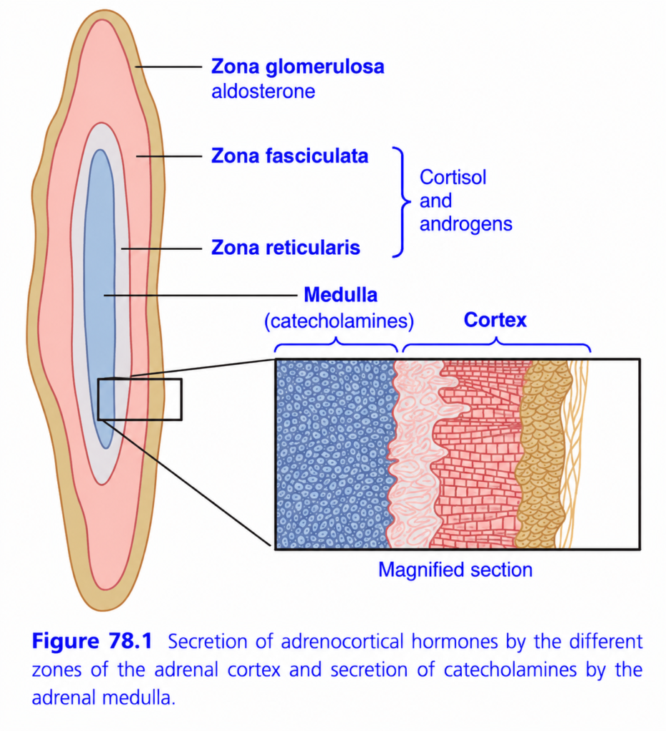
- Each adrenal gland lies at the upper pole of a kidney (Fig. 78.1).
- Each gland has two major parts: adrenal medulla and adrenal cortex.
- The adrenal medulla forms the central 20% of the gland.
- It is functionally connected to the sympathetic nervous system.
- Sympathetic stimulation causes the medulla to release epinephrine and norepinephrine.
- These hormones produce effects that are almost the same as direct sympathetic nerve stimulation throughout the body.
- The adrenal cortex produces a different group of hormones called corticosteroids.
- All corticosteroids are made from cholesterol.
- They have similar chemical structures, but small structural differences give them different important functions.
KEY CONCEPT
Adrenal gland = Medulla + Cortex
- Medulla → epinephrine + norepinephrine → sympathetic-type effects.
- Cortex → corticosteroids → made from cholesterol.
CONCEPTUAL EXAMPLES
- Sympathetic stimulation → adrenal medulla → epinephrine/norepinephrine.
- Cholesterol → adrenal cortex → corticosteroid hormones.
CORTICOSTEROIDS: MINERALOCORTICOIDS, GLUCOCORTICOIDS, AND ANDROGENS
- The adrenal cortex produces two major types of hormones: mineralocorticoids and glucocorticoids.
- It also produces small amounts of sex hormones, especially androgens.
- Adrenal androgens normally have only a small role, but excessive production can cause masculinizing effects.
- Mineralocorticoids mainly affect extracellular-fluid electrolytes, especially sodium and potassium.
- Glucocorticoids have important effects that increase blood glucose.
- Glucocorticoids also strongly affect protein and fat metabolism.
- More than 30 steroids have been identified from the adrenal cortex.
- The two most important for normal human endocrine function are:
- Aldosterone → principal mineralocorticoid
- Cortisol → principal glucocorticoid
KEY CONCEPT
Adrenal cortex → Mineralocorticoids + Glucocorticoids + Androgens
- Aldosterone → Na⁺/K⁺ and extracellular electrolytes
- Cortisol → ↑ blood glucose + protein/fat metabolism
- Androgens → minor normal role; excess → masculinizing effects
CONCEPTUAL EXAMPLES
- Aldosterone → mineral balance
- Cortisol → glucose metabolism
- Excess adrenal androgens → masculinizing effects
SYNTHESIS AND SECRETION OF ADRENOCORTICAL HORMONES
- The adrenal cortex has three distinct layers (Fig. 78.1):
- Zona glomerulosa → about 15% of cortex → mainly secretes aldosterone.
- Zona fasciculata → about 75% and widest layer → mainly secretes cortisol and corticosterone, plus small amounts of adrenal androgens and estrogens.
- Zona reticularis → inner layer → mainly secretes adrenal androgens, plus small amounts of estrogens and glucocorticoids.
- Zona glomerulosa is the only adrenal region capable of significant aldosterone secretion because it contains aldosterone synthase.
- Aldosterone secretion is mainly stimulated by angiotensin II and increased extracellular K⁺.
- Zona fasciculata secretion is mainly controlled by ACTH through the hypothalamic-pituitary axis.
- Zona reticularis secretion is also regulated by ACTH, although other pituitary factors may contribute.
- Aldosterone and cortisol are controlled independently.
- Angiotensin II mainly increases aldosterone and affects the zona glomerulosa.
- ACTH mainly increases cortisol and adrenal androgens and affects the zona fasciculata and reticularis.
- All adrenal steroid hormones are made from cholesterol.
- About 80% of the cholesterol used for steroid synthesis comes from circulating LDLs.
- LDL enters adrenal cells through receptor-mediated endocytosis and releases cholesterol for hormone synthesis.
- ACTH increases LDL receptors and enzymes that release cholesterol from LDL.
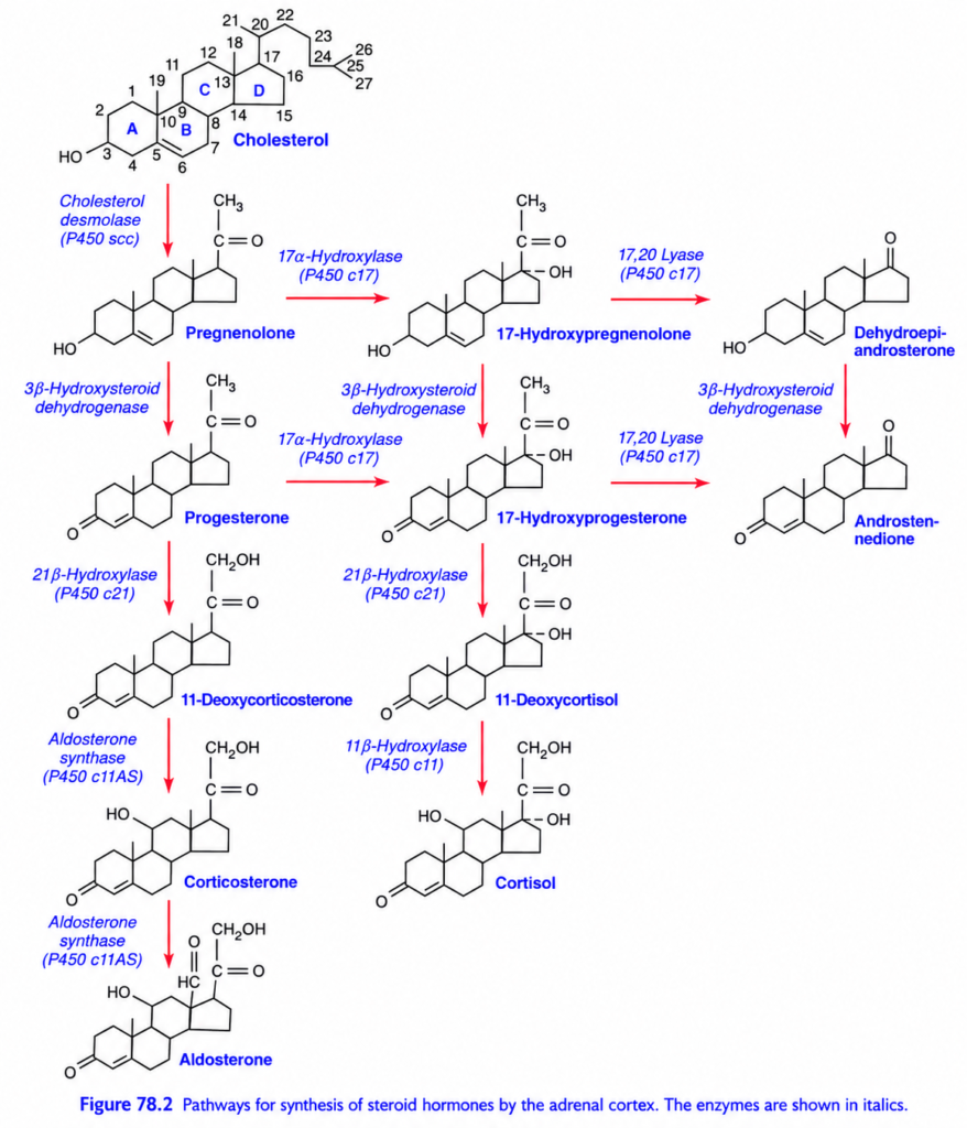
- Cholesterol enters the mitochondria and is converted by cholesterol desmolase into pregnenolone.
- This is the rate-limiting step in adrenal steroid formation (Fig. 78.2).
- Both ACTH and angiotensin II stimulate conversion of cholesterol to pregnenolone.
- Formation of aldosterone, cortisol, and adrenal androgens occurs mainly in the mitochondria and endoplasmic reticulum.
- Each step requires a specific enzyme.
- A defect in even one enzyme can greatly change which adrenal steroids are produced and in what amounts.
- For example, 21β-hydroxylase deficiency prevents normal cortisol formation.
- Low cortisol → CRH ↑ → ACTH ↑ → adrenal cortex grows, especially the fasciculata and reticularis.
- The enlarged cortex produces excess steroids such as androstenedione, which can be converted into active androgens.
- Therefore, 21β-hydroxylase deficiency can cause excessive masculinizing hormones.
- The major mineralocorticoid is aldosterone.
- The major glucocorticoid is cortisol.
- Important mineralocorticoids:
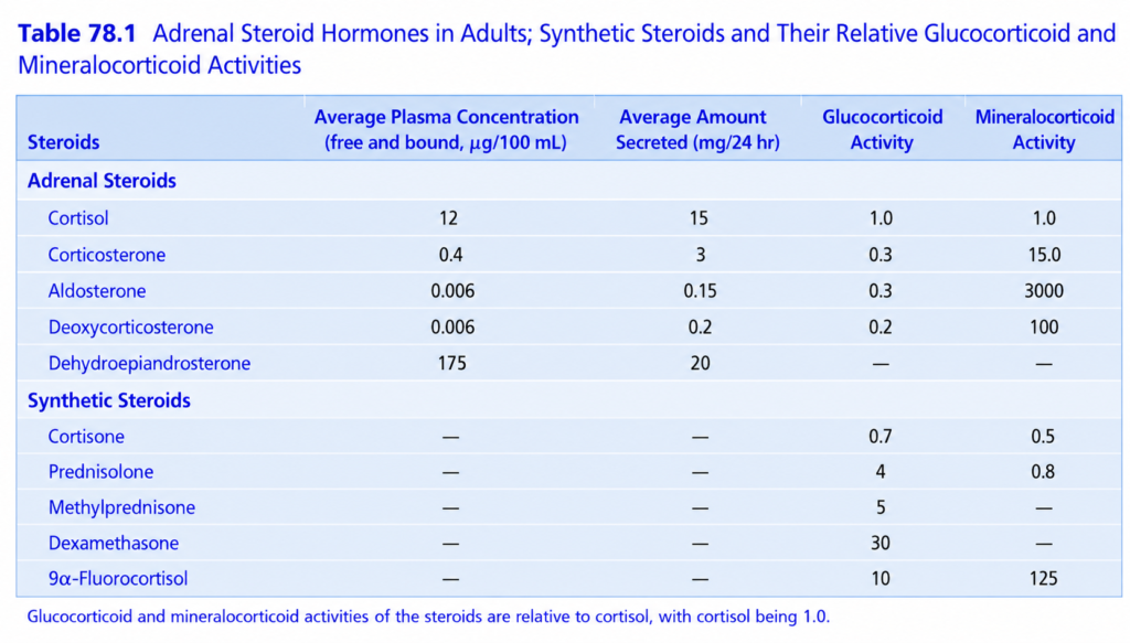
- Aldosterone → very potent; about 90% of mineralocorticoid activity.
- Deoxycorticosterone → about 1/30 as potent as aldosterone.
- Corticosterone → slight activity.
- 9α-Fluorocortisol → synthetic; slightly more potent than aldosterone.
- Cortisol and cortisone → slight mineralocorticoid activity.
- Important glucocorticoids:
- Cortisol → very potent; about 95% of glucocorticoid activity.
- Corticosterone → about 4% of total activity and much less potent.
- Cortisone → almost as potent as cortisol.
- Prednisone → synthetic; about 4× cortisol potency.
- Methylprednisone → about 5× cortisol potency.
- Dexamethasone → about 30× cortisol potency.
- Some corticosteroids have both glucocorticoid and mineralocorticoid activity.
- Cortisol normally has some mineralocorticoid activity, so excessive cortisol can also produce mineralocorticoid effects.
- Dexamethasone has very strong glucocorticoid activity with almost no mineralocorticoid activity.
- About 90–95% of cortisol in plasma is bound to proteins, mainly cortisol-binding globulin (transcortin) and some albumin.
- This protein binding slows cortisol removal, giving cortisol a half-life of about 60–90 minutes.
- About 60% of aldosterone is protein-bound and 40% is free.
- Therefore, aldosterone has a shorter half-life of about 20 minutes.
- Protein binding acts as a reservoir, reducing rapid changes in free hormone levels and helping distribute hormones uniformly.
- Adrenal steroids are mainly metabolized in the liver.
- They are converted mainly into inactive compounds by conjugation with glucuronic acid and, to a lesser extent, sulfates.
- About 25% of these conjugates leave through bile and feces.
- The rest enter blood, are filtered by the kidneys, and are excreted in urine.
- Liver disease slows hormone inactivation, while kidney disease reduces excretion of inactive conjugates.
- Normal plasma aldosterone is about 6 ng/100 mL, with secretion about 150 μg/day.
- Aldosterone levels vary greatly with sodium and potassium intake.
- Plasma cortisol averages about 12 μg/100 mL, with secretion about 15–20 mg/day.
- Cortisol secretion varies during the day: it is higher in the early morning and lower in the evening.
KEY CONCEPT
Adrenal cortex → 3 zones → different hormones
Glomerulosa → Aldosterone → Angiotensin II + K⁺
Fasciculata → Cortisol → ACTH
Reticularis → Androgens → mainly ACTH
Cholesterol → pregnenolone → adrenal steroid hormones
CONCEPTUAL EXAMPLES
- Angiotensin II ↑ → zona glomerulosa → aldosterone ↑.
- ACTH ↑ → zona fasciculata/reticularis → cortisol + androgens ↑.
- Cholesterol → mitochondria → pregnenolone → steroid hormones.
- 21β-hydroxylase deficiency → cortisol ↓ → ACTH ↑ → adrenal cortex enlargement + androgen excess.
- Cortisol → mostly protein-bound → slower removal.
- Aldosterone → more free hormone → shorter half-life.