


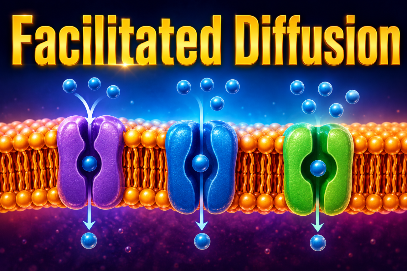
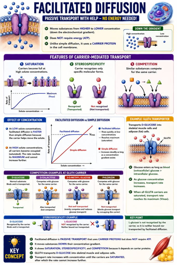
- Facilitated diffusion moves substances from higher concentration to lower concentration (down the electrochemical gradient).
- It does not require energy (ATP).
- Unlike simple diffusion, it uses a carrier protein in the cell membrane.
- Because it uses a carrier, it shows the features of carrier-mediated transport:
- Saturation – carriers become full at high solute concentrations.
- Stereospecificity – the carrier recognizes only specific molecular forms.
- Competition – similar substances can compete for the same carrier.
- At low solute concentrations, facilitated diffusion is faster than simple diffusion because the carrier helps move the solute.
- At high solute concentrations, all carriers become occupied (saturated).
- After saturation, the transport rate cannot increase further and reaches its maximum.
- In contrast, simple diffusion continues as long as a concentration gradient exists.
- A good example is the movement of D-glucose into skeletal muscle cells and adipose (fat) cells by the GLUT4 transporter.
- GLUT4 belongs to the GLUT family of glucose transporters.
- Glucose continues to enter the cell as long as blood (extracellular) glucose concentration is higher than intracellular glucose concentration.
- As glucose concentration increases, the transport rate also increases.
- When all GLUT4 carriers become saturated, the glucose transport rate reaches its maximum.
- Other sugars such as D-galactose, 3-O-methyl glucose, and phlorizin compete with glucose for the same carrier.
- These substances reduce glucose transport by binding to the carrier.
- Some competing substances are also transported, such as D-galactose.
- Others only occupy the carrier and block glucose transport, such as phlorizin.
- L-glucose is not recognized by the carrier.
- Therefore, L-glucose is neither bound nor transported by facilitated diffusion.
- Other examples of facilitated diffusion include urea
- transporters (UTs) and organic cation transporters.
KEY CONCEPT
- Facilitated diffusion is passive transport that uses carrier proteins but does not require ATP.
- It moves substances down their concentration gradient.
- It shows saturation, stereospecificity, and competition because it depends on carrier proteins.
- GLUT4 transports D-glucose into skeletal muscle and adipose cells.
- Transport increases until the carriers are saturated, after which the rate cannot increase further.
Primary Active Transport
- In primary active transport, one or more solutes move against the electrochemical gradient (uphill).
- Solutes move from low concentration to high concentration.
- This movement requires energy because it is moving uphill.
- The energy is supplied by ATP (adenosine triphosphate).
- ATP is broken down into ADP (adenosine diphosphate) and Pi (inorganic phosphate).
- Breaking ATP releases energy from its terminal high-energy phosphate bond.
- The released phosphate attaches to the transport protein.
- This starts a cycle of phosphorylation and dephosphorylation.
- When ATP directly provides energy to the transport protein, it is called primary active transport.
- Examples of primary active transport:
- Na⁺-K⁺ ATPase – present in all cell membranes.
- Ca²⁺ ATPase – present in the sarcoplasmic reticulum (SR) and endoplasmic reticulum.
- H⁺-K⁺ ATPase – present in gastric parietal cells and renal α-intercalated cells.
Na⁺-K⁺ ATPase (Na⁺-K⁺ Pump)
- Na⁺-K⁺ ATPase is found in the membrane of all cells.
- It pumps Na⁺ out of the cell (ICF → ECF).
- It pumps K⁺ into the cell (ECF → ICF).
- Both ions move against their own electrochemical gradients.
- Usually, 3 Na⁺ ions leave the cell while 2 K⁺ ions enter the cell in one cycle.
- Because more positive charge leaves than enters, the pump is electrogenic.
- An electrogenic pump creates a charge difference (membrane potential) across the cell membrane.
- The pump keeps:
- Intracellular Na⁺ concentration low.
- Intracellular K⁺ concentration high.
- The Na⁺-K⁺ ATPase has α (alpha) and β (beta) subunits.
- The α subunit:
- Has ATPase activity.
- Contains binding sites for Na⁺ and K⁺.
- The pump works by changing between two conformational states:
- E1 state
- Binding sites face the ICF.
- Has high affinity for Na⁺.
- E2 state
- Binding sites face the ECF.
- Has high affinity for K⁺.
- E1 state
- ATP hydrolysis provides the energy needed for the pump to switch between E1 and E2.
- This switching pumps Na⁺ out of the cell and K⁺ into the cell.
- The transport cycle begins in the E1 state with ATP attached.
- Binding sites face the ICF.
- Three Na⁺ ions bind to the pump.
- ATP is hydrolyzed.
- The terminal phosphate is transferred to the pump.
- The pump becomes the high-energy E1~P state.
- The pump changes shape and becomes E2~P.
- Binding sites now face the ECF.
- The pump has low affinity for Na⁺ and high affinity for K⁺.
- Three Na⁺ ions are released into the ECF.
- Two K⁺ ions bind to the pump.
- Inorganic phosphate (Pi) is released.
- ATP binds again.
- The pump changes back to the E1 state.
- Two K⁺ ions are released into the ICF.
- The pump is ready to start another cycle.
- Cardiac glycosides (such as ouabain and digitalis) inhibit the Na⁺-K⁺ ATPase.
- These drugs cause:
- Intracellular Na⁺ concentration to increase.
- Intracellular K⁺ concentration to decrease.
- They bind to the E2~P form of the pump.
- They bind near the K⁺ binding site on the extracellular side.
- This prevents the pump from changing back to the E1 state.
- As a result, the phosphorylation-dephosphorylation cycle stops.
- The pump can no longer transport Na⁺ and K⁺ normally.
KEY CONCEPT
- Primary active transport uses ATP directly to move substances against their electrochemical gradient.
- Na⁺-K⁺ ATPase is the most important primary active transport pump.
- It pumps 3 Na⁺ out and 2 K⁺ in, making it electrogenic.
- The pump alternates between E1 (binds Na⁺) and E2 (binds K⁺) states.
- Cardiac glycosides (ouabain, digitalis) inhibit the pump, causing ↑ intracellular Na⁺ and ↓ intracellular K⁺.
- Fig. 1.6
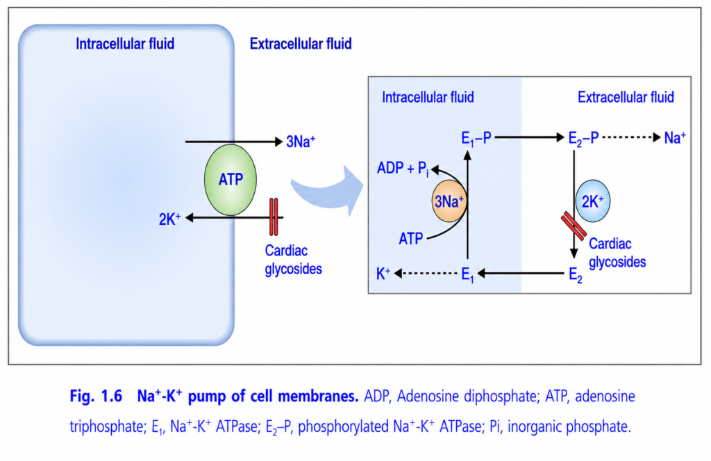
Na⁺–K⁺ Pump (Sodium–Potassium ATPase) (Figure 1.6)
This figure explains the Na⁺–K⁺ pump, one of the most important transport proteins in the cell membrane.
It continuously:
- Pumps 3 sodium (Na⁺) ions out of the cell
- Pumps 2 potassium (K⁺) ions into the cell
- Uses ATP (energy)
Without this pump, cells cannot survive.
Main Concept
3 Na⁺ go OUT + 2 K⁺ come IN + 1 ATP is used = Na⁺–K⁺ Pump
The Golden Rule
3 Na⁺  → OUT

2 K⁺   → IN

ATP → Required
This is the easiest rule to remember.
Overall View of the Figure
The figure has two parts.
Left Side
Shows the overall function of the pump.
- 3 Na⁺ leave the cell.
- 2 K⁺ enter the cell.
- ATP provides energy.
Right Side
Shows the step-by-step working mechanism of the pump.
First Understand the Cell Like a House
Imagine the cell is a house.
- Inside the house = Intracellular fluid
- Outside the house = Extracellular fluid
The Na⁺–K⁺ pump is the security guard at the door.
Its daily job is:
- Throw 3 sodium ions out.
- Allow 2 potassium ions in.
It repeats this job millions of times every second.
LEFT SIDE OF THE FIGURE
ATP
The green oval is ATP.
ATP is the battery that powers the pump.
Without ATP
❌ Pump stops.
3 Na⁺ Move Out
The arrow points outward.
The pump removes 3 sodium ions from inside the cell.
Why?
Because sodium naturally wants to enter the cell.
The pump keeps sodium low inside.
2 K⁺ Move In
The lower arrow points inward.
The pump brings 2 potassium ions into the cell.
Why?
Potassium naturally tends to leak out.
The pump replaces the lost potassium.
Simple Memory
Na⁺ = Out

K⁺ = In
RIGHT SIDE OF THE FIGURE (Mechanism)
This explains how the pump actually works.
Don’t worry about the names E₁ and E₂ at first.
Think of the pump as a revolving door that changes shape.
Step 1 – Pump Faces Inside (E₁)
The pump opens toward the inside of the cell.
It grabs:
3 sodium ions (Na⁺).
Inside Cell

Pump
  ↓
3 Na⁺ enter pump
Step 2 – ATP Gives Energy
ATP is broken down.
ATP
   ↓
ADP + Pi
This releases energy.
The phosphate (Pi) attaches to the pump.
This process is called phosphorylation.
Step 3 – Pump Changes Shape
After phosphorylation,
the pump changes its shape.
This is shown as:
E₁
   ↓
E₂-P
Now the pump faces outside.
Step 4 – Sodium is Released
The pump opens toward the outside.
The 3 sodium ions leave the pump.
Inside
   ↓
Pump
   ↓
Outside
Now sodium is outside the cell.
Step 5 – Potassium Binds
While facing outside,
the pump now binds:
2 potassium ions.
Outside

2 K⁺
   ↓
Pump
Step 6 – Pump Returns to Original Shape
The phosphate leaves the pump.
The pump changes back to its original shape.
E₂
   ↓
E₁
Now it again faces inside.
Step 7 – Potassium is Released
The pump opens toward the inside.
The 2 potassium ions are released into the cell.
The cycle is complete.
Complete Pump Cycle
Pump faces inside
        ↓
Binds 3 Na⁺
        ↓
ATP is used
        ↓
Pump changes shape
        ↓
3 Na⁺ released outside
        ↓
Binds 2 K⁺
        ↓
Pump changes back
        ↓
2 K⁺ released inside
        ↓
Cycle repeats
Why Does the Cell Need This Pump?
The Na⁺–K⁺ pump is essential because it:
1. Maintains Ion Distribution
Keeps:
- High Na⁺ outside
- High K⁺ inside
2. Maintains Resting Membrane Potential
Because 3 positive ions leave while only 2 positive ions enter, there is a net loss of one positive charge from the cell.
This makes the inside of the cell relatively more negative, helping maintain the resting membrane potential.
3. Prevents Cell Swelling
If sodium accumulated inside the cell:
- Water would follow sodium by osmosis.
- The cell would swell and could burst.
The pump removes sodium, preventing excessive water entry.
4. Provides the Sodium Gradient
Many transport systems depend on the sodium gradient created by this pump.
Examples:
- Glucose absorption in the intestine
- Amino acid transport
- Kidney tubular reabsorption
Cardiac Glycosides (Shown in Red)
The figure also shows Cardiac glycosides (e.g., digoxin, ouabain).
These drugs block the Na⁺–K⁺ pump.
What happens?
- Sodium accumulates inside the cell.
- Less calcium is removed indirectly.
- Intracellular calcium increases.
- Heart muscle contracts more strongly.
This is why digoxin can be useful in certain heart conditions.
Easy Story
Imagine a nightclub with a security guard.
The rule is:
Every time the door opens,
- 3 noisy people (Na⁺) are kicked out.
- 2 VIP guests (K⁺) are allowed in.
The guard can work only if someone gives him money (ATP).
No ATP = Guard stops working.
Easy Flow Diagram
Inside Cell
      ↓
3 Na⁺ bind pump
      ↓
ATP used
      ↓
Pump changes shape
      ↓
3 Na⁺ released outside
      ↓
2 K⁺ bind
      ↓
Pump changes back
      ↓
2 K⁺ released inside
      ↓
Repeat forever
Easy Memory Tricks
Rule 1
3 Out, 2 In
Rule 2
Na⁺ Out — K⁺ In
Rule 3
ATP Powers the Pump
Rule 4
Digoxin Blocks the Pump
Important Points from Figure 1.6
- The Na⁺–K⁺ ATPase is a primary active transport pump located in the cell membrane.
- It uses the energy from ATP hydrolysis to transport ions against their concentration gradients.
- During each cycle, the pump moves 3 Na⁺ out of the cell and 2 K⁺ into the cell.
- ATP is converted to ADP + Pi, and phosphorylation causes the pump to change shape, allowing sodium release outside the cell.
- Binding of 2 K⁺ from the extracellular fluid causes the pump to return to its original shape and release potassium inside the cell.
- Because 3 positive charges leave while 2 enter, the pump is electrogenic, contributing to the negative resting membrane potential.
- The pump helps maintain cell volume, high intracellular K⁺, low intracellular Na⁺, and the sodium gradient needed for many secondary active transport systems.
- Cardiac glycosides (e.g., digoxin) inhibit the Na⁺–K⁺ ATPase, increasing intracellular sodium and indirectly increasing intracellular calcium in cardiac muscle.
KEY CONCEPT (Figure 1.6)
The Na⁺–K⁺ pump (Na⁺–K⁺ ATPase) is an ATP-dependent membrane protein that continuously maintains the normal ionic composition of cells by transporting 3 Na⁺ out and 2 K⁺ into the cell during each cycle. ATP hydrolysis provides the energy required for the pump to change shape and move these ions against their concentration gradients. This pump is essential for maintaining the resting membrane potential, cell volume, and the sodium gradient required for many secondary active transport processes. Because it exports more positive charges than it imports, it is electrogenic. Cardiac glycosides, such as digoxin, inhibit this pump and thereby increase intracellular calcium in cardiac muscle, strengthening cardiac contraction.
Cardiac Glycosides (e.g., Ouabain and Digitalis)
- Cardiac glycosides are drugs that inhibit the Na⁺-K⁺ ATPase (Na⁺-K⁺ pump).
- Examples include ouabain and digitalis.
- These drugs cause predictable changes inside the cell:
- Intracellular Na⁺ concentration increases.
- Intracellular K⁺ concentration decreases.
- Cardiac glycosides bind to the E2~P form of the Na⁺-K⁺ ATPase.
- They bind near the K⁺ binding site on the extracellular side of the pump.
- This prevents the pump from changing from E2~P back to E1.
- As a result, the normal phosphorylation-dephosphorylation cycle stops.
- When this cycle is blocked, the entire Na⁺-K⁺ pump stops working normally.
- Therefore, the pump cannot transport Na⁺ and K⁺ properly.
KEY CONCEPT
- Cardiac glycosides (ouabain, digitalis) inhibit the Na⁺-K⁺ ATPase.
- They bind to the E2~P form near the K⁺ binding site.
- This blocks the pump from returning to the E1 state.
- As a result:
- ↑ Intracellular Na⁺
- ↓ Intracellular K⁺
- Na⁺-K⁺ pump transport stops.
Figure Fig. 1.6
Ca²⁺ ATPase (Ca²⁺ Pump)
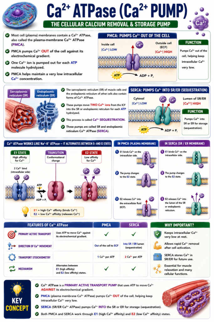
- Most cell (plasma) membranes contain a Ca²⁺ ATPase, also called the plasma-membrane Ca²⁺ ATPase (PMCA).
- PMCA pumps Ca²⁺ out of the cell against its electrochemical gradient.
- One Ca²⁺ ion is pumped out for each ATP molecule hydrolyzed.
- PMCA helps maintain a very low intracellular Ca²⁺ concentration.
- The sarcoplasmic reticulum (SR) of muscle cells and the endoplasmic reticulum of other cells also contain forms of Ca²⁺ ATPase.
- These pumps move two Ca²⁺ ions from the ICF into the SR or endoplasmic reticulum for each ATP hydrolyzed.
- This process is called Ca²⁺ sequestration.
- These pumps are called SR and endoplasmic reticulum Ca²⁺ ATPase (SERCA).
- Ca²⁺ ATPase works in a similar way to the Na⁺-K⁺ ATPase.
- It changes between E1 and E2 states.
- E1 state has a high affinity for Ca²⁺.
- E2 state has a low affinity for Ca²⁺.
- In PMCA:
- E1 binds Ca²⁺ on the intracellular side.
- The pump changes to the E2 state.
- E2 releases Ca²⁺ into the extracellular fluid (ECF).
- In SERCA:
- E1 binds Ca²⁺ on the intracellular side.
- E2 releases Ca²⁺ into the lumen of the SR or endoplasmic reticulum.
KEY CONCEPT
- Ca²⁺ ATPase is a primary active transport pump that uses ATP to move Ca²⁺ against its electrochemical gradient.
- PMCA pumps Ca²⁺ out of the cell, helping keep intracellular Ca²⁺ very low.
- SERCA pumps Ca²⁺ into the SR or endoplasmic reticulum for storage (sequestration).
- Both PMCA and SERCA work through E1 (high Ca²⁺ affinity) and E2 (low Ca²⁺ affinity) states.
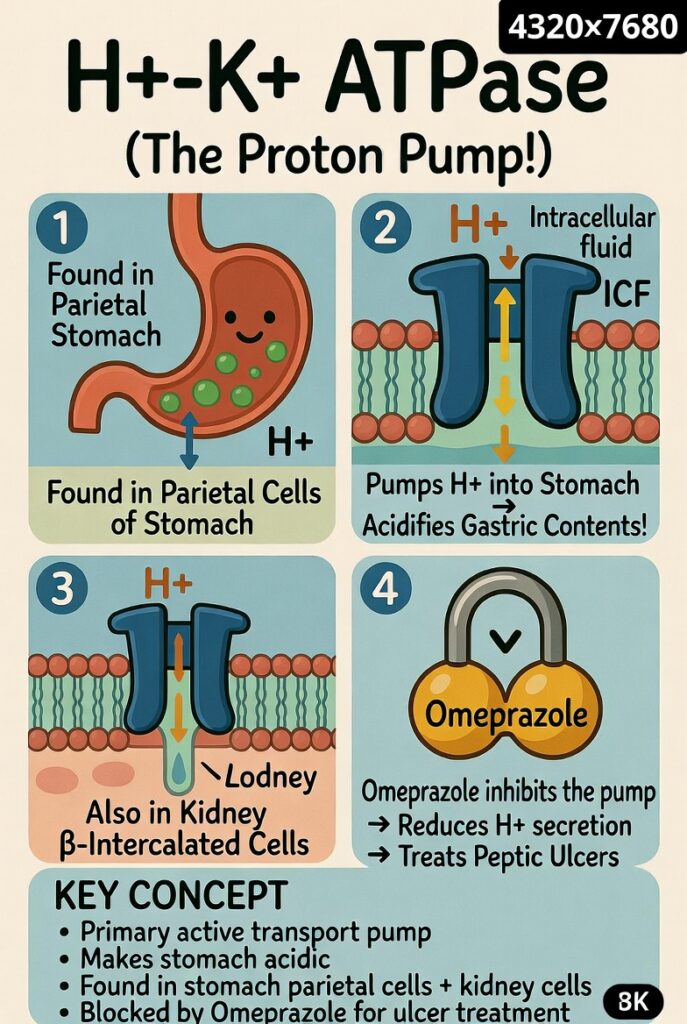
H⁺-K⁺ ATPase (H⁺-K⁺ Pump)
- H⁺-K⁺ ATPase is present in the parietal cells of the gastric mucosa.
- It is also present in the α-intercalated cells of the renal collecting duct.
- In the stomach, it pumps H⁺ from the intracellular fluid (ICF) of parietal cells into the stomach lumen.
- This process acidifies the gastric contents.
- Omeprazole inhibits the gastric H⁺-K⁺ ATPase.
- Omeprazole is used to reduce H⁺ secretion.
- It is used in the treatment of some types of peptic ulcer disease.
KEY CONCEPT
- H⁺-K⁺ ATPase is a primary active transport pump.
- It pumps H⁺ into the stomach lumen, making the stomach contents acidic.
- It is found in gastric parietal cells and renal α-intercalated cells.
- Omeprazole blocks this pump and reduces gastric acid secretion.
Secondary Active Transport
- Secondary active transport is a process in which two or more solutes are transported together.
- Usually, Na⁺ moves down its electrochemical gradient (downhill).
- The other solute moves against its electrochemical gradient (uphill).
- The downhill movement of Na⁺ provides the energy for the uphill movement of the other solute.
- ATP is not used directly in this transport.
- The energy is supplied indirectly by the Na⁺ concentration gradient across the cell membrane.
- The Na⁺-K⁺ ATPase uses ATP to create and maintain this Na⁺ gradient.
- Therefore, secondary active transport uses ATP indirectly as its energy source.
- Inhibiting the Na⁺-K⁺ ATPase (for example, with ouabain) reduces the movement of Na⁺ from the ICF to the ECF.
- As a result, intracellular Na⁺ concentration increases.
- This reduces the Na⁺ gradient across the cell membrane.
- Because the Na⁺ gradient becomes smaller, all secondary active transport processes decrease.
- This happens because their energy source (the Na⁺ gradient) is reduced.
- There are two types of secondary active transport.
- If the uphill solute moves in the same direction as Na⁺, it is called cotransport (symport).
- If the uphill solute moves in the opposite direction to Na⁺, it is called countertransport (antiport or exchange).
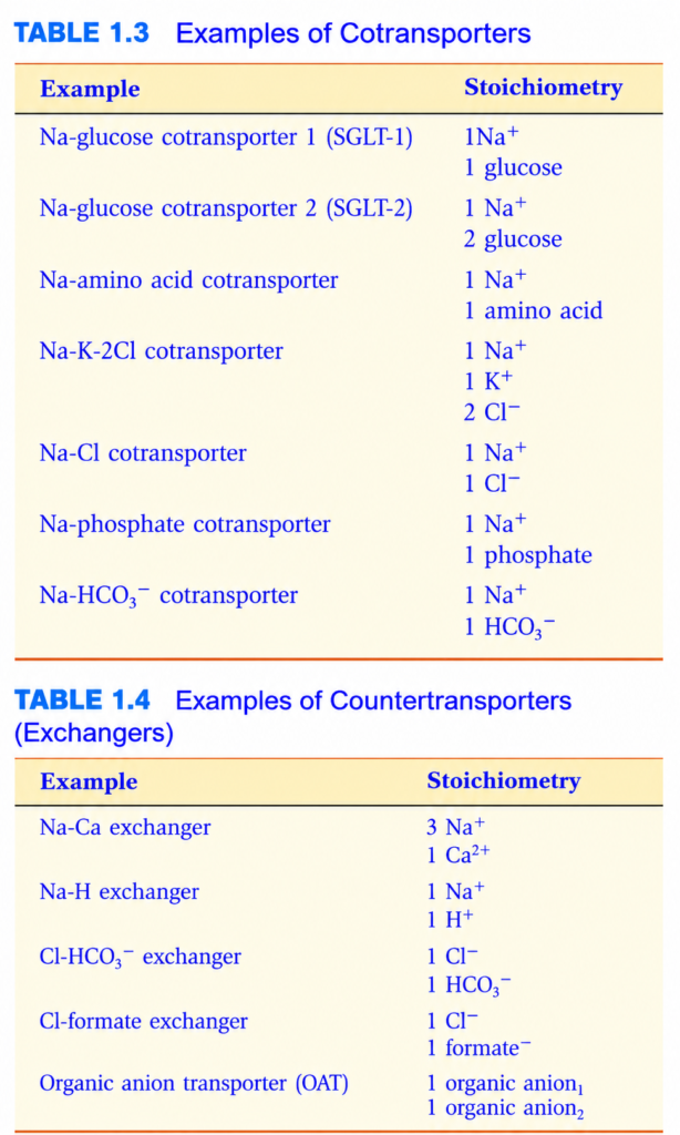
- Examples of cotransport and countertransport are shown in Tables 1.3 and 1.4.
KEY CONCEPT
- Secondary active transport uses the Na⁺ gradient as its energy source.
- Na⁺ moves downhill, while another solute moves uphill.
- ATP is used indirectly because the Na⁺-K⁺ ATPase creates the Na⁺ gradient.
- Inhibiting the Na⁺-K⁺ ATPase reduces the Na⁺ gradient and decreases all secondary active transport.
- Types:
- Cotransport (Symport): Na⁺ and the other solute move in the same direction.
- Countertransport (Antiport/Exchange): Na⁺ and the other solute move in opposite directions.
- Cotransport
- Cotransport (symport) is a type of secondary active transport.
- In cotransport, all solutes move in the same direction across the cell membrane.
- Na⁺ enters the cell through the carrier down its electrochemical gradient.
- The other solutes move into the cell together with Na⁺.
- Cotransport is important in several normal body functions, especially in the small intestine and renal tubules, where absorption occurs.
- Na⁺-glucose cotransport (SGLT) and Na⁺-amino acid cotransport are present in the luminal membrane of epithelial cells of the small intestine and renal proximal tubule.
- Another example is Na⁺-K⁺-2Cl⁻ cotransport, which is present in the luminal membrane of epithelial cells of the thick ascending limb.
- In all these examples, the Na⁺ gradient created by the Na⁺-K⁺ ATPase provides the energy to move glucose, amino acids, K⁺, or Cl⁻ against their electrochemical gradients.
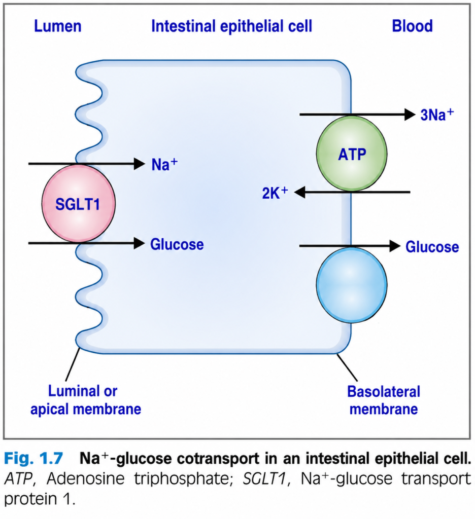
- Fig. 1.7 shows how Na⁺-glucose cotransport (SGLT1) works in intestinal epithelial cells.
- The cotransporter is present in the luminal membrane of these cells.
- It has two specific binding sites:
- One for Na⁺.
- One for glucose.
- When both Na⁺ and glucose are present in the lumen of the small intestine, both bind to the transporter.
- The cotransporter changes its shape (rotates).
- It releases both Na⁺ and glucose into the inside of the cell.
- After entering the cell:
- Na⁺ leaves the cell by the Na⁺-K⁺ ATPase.
- Glucose leaves the cell by facilitated diffusion.
- If Na⁺ or glucose is absent from the intestinal lumen, the cotransporter cannot rotate.
- Therefore, both Na⁺ and glucose are required.
- Neither Na⁺ nor glucose can be transported without the other (Box 1.1).
- The role of intestinal Na⁺-glucose cotransport can be understood in carbohydrate absorption.
- Dietary carbohydrates are digested by gastrointestinal enzymes into monosaccharides, which can be absorbed.
- Glucose is one of these monosaccharides.
- Glucose enters the intestinal epithelial cell by Na⁺-glucose cotransport through the luminal membrane.
- Glucose then leaves the cell by facilitated diffusion through the basolateral membrane.
- Na⁺-glucose cotransport is the active step.
- This active step allows glucose to be absorbed into the blood against its electrochemical gradient.
- KEY CONCEPT
- Cotransport (symport) is a type of secondary active transport.
- Na⁺ and the other solute move in the same direction into the cell.
- The Na⁺ gradient created by the Na⁺-K⁺ ATPase provides the energy.
- Important examples are:
- Na⁺-glucose cotransport (SGLT)
- Na⁺-amino acid cotransport
- Na⁺-K⁺-2Cl⁻ cotransport
- In SGLT1, both Na⁺ and glucose must bind together for transport to occur.
- During intestinal carbohydrate absorption:
Table 1.3 and Table 1.4.
BOX 1.1 Clinical Physiology: Glucosuria Due to Diabetes Mellitus
- A 14-year-old boy had frequent urination and severe thirst at his check-up.
- Urine dipstick test showed high glucose.
- Glucose tolerance test confirmed type 1 diabetes mellitus.
- He was treated with insulin injections and his urine test became normal.
- In type 1 diabetes, the body does not make enough insulin.
- Without insulin, blood glucose level rises because glucose cannot enter cells properly.
- High blood glucose leads to more glucose being filtered by the kidneys.
- Normally, the kidney filters glucose and then reabsorbs all of it in the proximal tubule.
- Reabsorption happens through Na⁺-glucose cotransporters.
- These transporters have a limited capacity (transport maximum).
- When too much glucose is filtered, the transporters become saturated.
- Extra glucose cannot be reabsorbed and spills into the urine (glucosuria).
- This causes frequent urination and thirst.
- Insulin treatment lowers blood glucose level.
- Less glucose is filtered by the kidneys.
- The Na⁺-glucose cotransporters can now reabsorb all the glucose.
- No glucose appears in the urine.
KEY CONCEPT In uncontrolled type 1 diabetes, high blood glucose overwhelms the kidney’s Na⁺-glucose cotransporters, causing glucosuria. Insulin treatment lowers blood glucose so all filtered glucose can be reabsorbed.
Countertransport
- Countertransport (antiport or exchange) is a type of secondary active transport.
- In countertransport, solutes move in opposite directions across the cell membrane.
- Na⁺ enters the cell through the carrier down its electrochemical gradient.
- The other solute moves out of the cell.
- Examples of countertransport are:
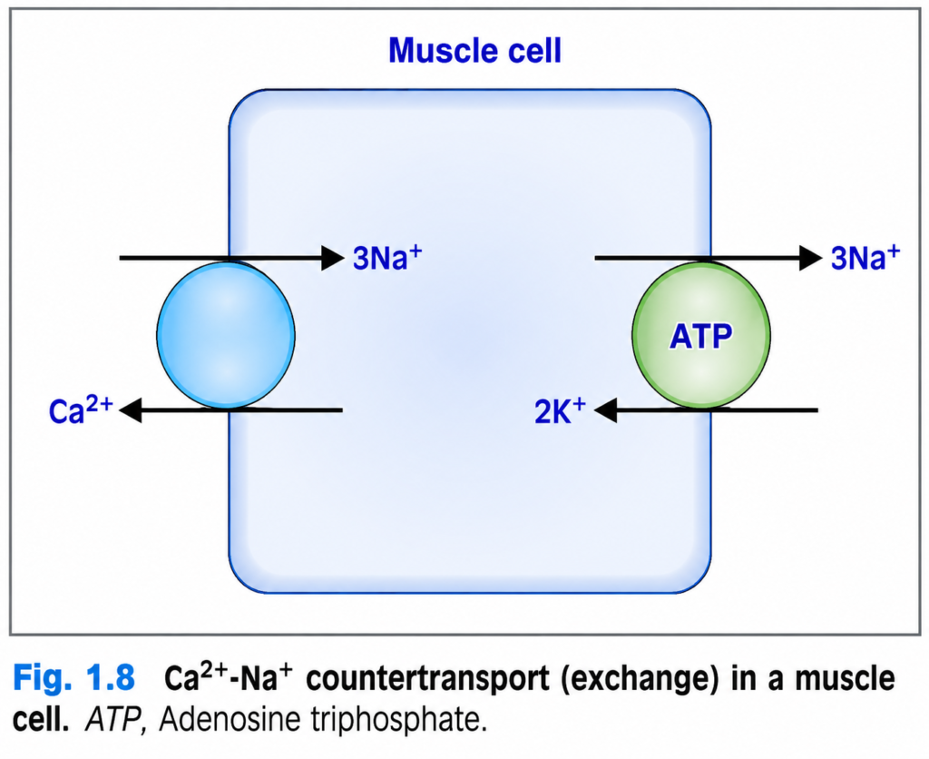
- Ca²⁺-Na⁺ exchange (Fig. 1.8)
- Na⁺-H⁺ exchange
- Like cotransport, countertransport uses the Na⁺ gradient created by the Na⁺-K⁺ ATPase as its energy source.
- Na⁺ moves downhill, while Ca²⁺ or H⁺ moves uphill against its electrochemical gradient.
- Ca²⁺-Na⁺ exchange is one of the mechanisms that helps keep intracellular Ca²⁺ concentration very low (≈10⁻⁷ molar).
- Ca²⁺ ATPase also helps maintain this low intracellular Ca²⁺ concentration.
- Because Ca²⁺ moves out of the cell against its electrochemical gradient, this process requires active transport.
- Fig. 1.8 shows Ca²⁺-Na⁺ exchange in a muscle cell membrane.
- The exchange protein has binding sites for both Ca²⁺ and Na⁺.
- It binds Ca²⁺ on the intracellular side of the membrane.
- At the same time, it binds Na⁺ on the extracellular side.
- The exchange protein then changes its shape (rotates).
- It releases Ca²⁺ outside the cell.
- It releases Na⁺ inside the cell.
- The Ca²⁺-Na⁺ exchange ratio (stoichiometry) can vary in different cell types and under different conditions.
- Usually, 3 Na⁺ ions enter the cell for every 1 Ca²⁺ ion pumped out.
- This means:
- 3 positive charges enter the cell.
- 2 positive charges leave the cell.
- Therefore, the Ca²⁺-Na⁺ exchanger is electrogenic because it creates a net movement of positive charge into the cell.
KEY CONCEPT
- Countertransport (antiport/exchange) is a type of secondary active transport.
- Na⁺ and the other solute move in opposite directions.
- The Na⁺ gradient created by the Na⁺-K⁺ ATPase provides the energy.
- Important examples are:
- Ca²⁺-Na⁺ exchange
- Na⁺-H⁺ exchange
- Ca²⁺-Na⁺ exchange helps keep intracellular Ca²⁺ concentration very low (≈10⁻⁷ molar).
- Usually, 3 Na⁺ enter for every 1 Ca²⁺ leaves, making the exchanger electrogenic.
MADE BY SELF LEARNING CEO AND FOUNDER DR SHEEN
