


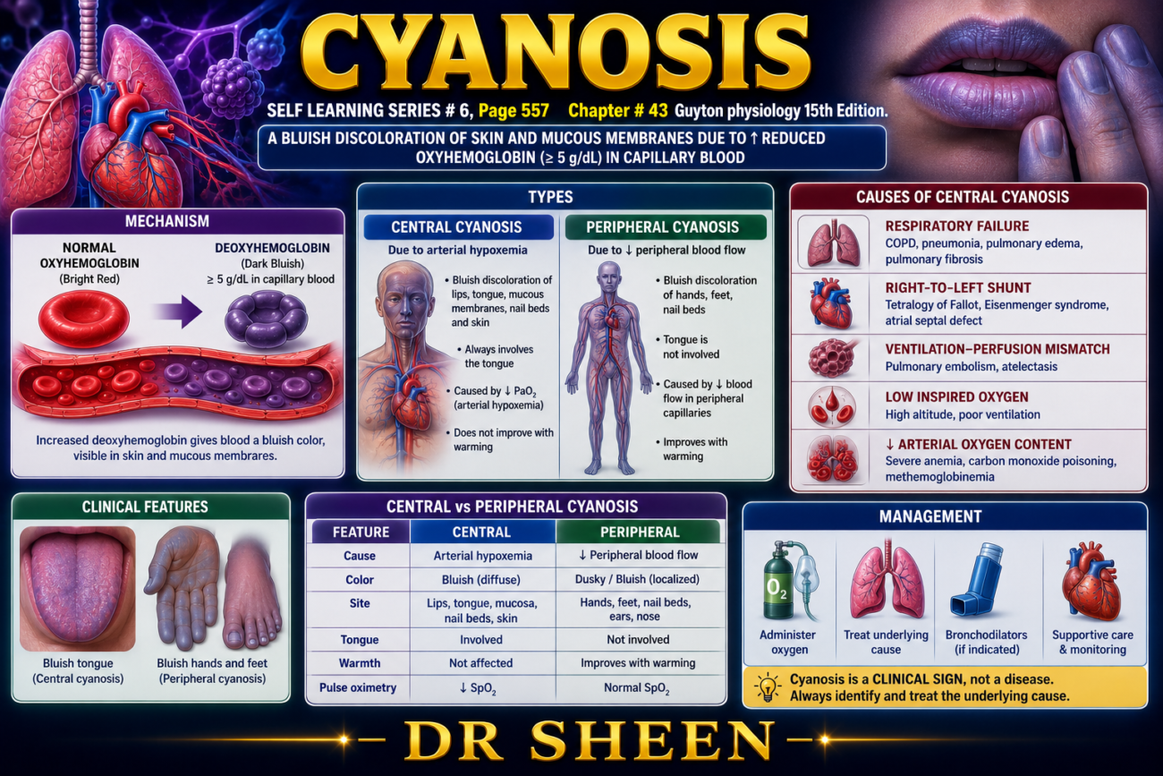
- Cyanosis means bluish discoloration of the skin.
- It is caused by an excessive amount of deoxygenated (reduced) hemoglobin in the blood vessels of the skin, especially in the capillaries.
- Deoxygenated hemoglobin has a dark blue-purple color.
- This color shows through the skin, producing the characteristic bluish appearance.
- In general, cyanosis becomes clearly visible when arterial blood contains more than 5 g of deoxygenated hemoglobin per 100 mL of blood.
Cyanosis in Anemia
- A person with anemia rarely develops cyanosis.
- This is because anemia has a low total hemoglobin level.
- Even if much of the hemoglobin loses oxygen, there is usually not enough hemoglobin present for more than 5 g/100 mL to become deoxygenated.
- Therefore, cyanosis is uncommon in anemia.
Cyanosis in Polycythemia
- A person with polycythemia vera has an abnormally high number of red blood cells.
- As a result, the total amount of hemoglobin is greatly increased.
- Even under otherwise normal conditions, more than 5 g of hemoglobin can become deoxygenated.
- Therefore, cyanosis commonly develops in polycythemia vera.
Summary
| Condition | Hemoglobin Level | Chance of Cyanosis | Reason |
|---|---|---|---|
| Normal person | Normal | Only if deoxygenated Hb > 5 g/100 mL | Sufficient reduced hemoglobin causes blue skin |
| Anemia | Low | Rare | Too little hemoglobin is available to reach 5 g/100 mL of deoxygenated hemoglobin |
| Polycythemia vera | High | Common | Excess hemoglobin allows >5 g/100 mL to become deoxygenated |
KEY CONCEPT
- Cyanosis is the bluish discoloration of the skin caused by excessive deoxygenated hemoglobin in skin capillaries.
- It becomes clinically visible when arterial blood contains more than 5 g of deoxygenated hemoglobin per 100 mL of blood.
- Anemia rarely causes cyanosis because the total hemoglobin level is too low to produce enough deoxygenated hemoglobin.
- Polycythemia vera commonly causes cyanosis because the increased hemoglobin level allows a large amount of deoxygenated hemoglobin to accumulate, even under otherwise normal conditions.
HYPERCAPNIA—EXCESS CARBON DIOXIDE IN THE BODY FLUIDS
- Hypercapnia means an excessive amount of carbon dioxide (CO₂) in the body fluids.
- At first, it may seem that every condition causing hypoxia also causes hypercapnia.
- However, this is not true.
- Hypercapnia usually occurs together with hypoxia only when the hypoxia is caused by:
- Hypoventilation
- Circulatory deficiency
1. Hypoxia Due to Low Oxygen Availability
This includes:
- Low oxygen (O₂) in the atmosphere
- Anemia (low hemoglobin)
- Poisoning of oxidative enzymes (e.g., cyanide poisoning)
- In these conditions, only oxygen supply or oxygen utilization is affected.
- Carbon dioxide (CO₂) removal remains normal.
- Therefore, hypercapnia does not occur.
2. Hypoxia Due to Poor Diffusion
- In diffusion disorders, oxygen (O₂) crosses the respiratory membrane poorly.
- However, CO₂ diffuses about 20 times faster than O₂.
- Therefore, serious hypercapnia usually does not develop.
- If CO₂ begins to increase, it strongly stimulates breathing.
- The increased ventilation removes excess CO₂.
- However, the hypoxia may still persist because oxygen diffuses much more slowly than CO₂.
3. Hypoxia Due to Hypoventilation
- In hypoventilation, too little air reaches the alveoli.
- As a result:
- Less oxygen enters the lungs.
- Less carbon dioxide leaves the lungs.
- Therefore, hypoxia and hypercapnia occur together.
4. Hypoxia Due to Circulatory Deficiency
- In circulatory deficiency, blood flow to the tissues is reduced.
- As a result:
- Less oxygen is delivered to the tissues.
- Less carbon dioxide is removed from the tissues.
- Therefore:
- Tissue hypoxia develops.
- Tissue hypercapnia also develops.
- However, blood can transport more than three times as much CO₂ as O₂.
- Therefore, the increase in tissue CO₂ is much less severe than the decrease in tissue oxygen.
Effects of Increasing PCO₂
Alveolar PCO₂ = 60–75 mm Hg
- Breathing becomes very rapid and very deep.
- Dyspnea (severe breathlessness) develops.
PCO₂ = 80–100 mm Hg
- The person becomes:
- Lethargic
- Sometimes semicomatose
PCO₂ = 120–150 mm Hg
- Anesthesia may occur.
- Death may occur if the condition is not corrected.
Vicious Cycle of Severe Hypercapnia
- At very high CO₂ levels, carbon dioxide no longer stimulates respiration.
- Instead, it depresses the respiratory center.
- This creates a vicious cycle:
- CO₂ increases
- Respiration decreases
- Even more CO₂ accumulates
- Respiration becomes weaker
- CO₂ rises further
- Respiratory failure and death may occur
Summary
| Cause of Hypoxia | Hypercapnia Present? | Reason |
|---|---|---|
| Low atmospheric O₂ | No | Only oxygen availability is reduced |
| Anemia | No | Oxygen transport is reduced, CO₂ removal remains normal |
| Oxidative enzyme poisoning (cyanide) | No | Cells cannot use O₂, but CO₂ elimination is normal |
| Diffusion impairment | Usually No | CO₂ diffuses about 20 times faster than O₂ |
| Hypoventilation | Yes | Both O₂ uptake and CO₂ removal are reduced |
| Circulatory deficiency | Yes | Reduced blood flow decreases CO₂ removal from tissues |
KEY CONCEPT
- Hypercapnia is an excessive amount of CO₂ in the body fluids.
- It usually accompanies hypoxia only in hypoventilation and circulatory deficiency.
- Hypercapnia does not occur in low atmospheric oxygen, anemia, cyanide poisoning, or most diffusion defects because CO₂ removal remains adequate or CO₂ diffuses much more rapidly than O₂.
- As PCO₂ rises, it first stimulates breathing, causing severe dyspnea.
- Very high PCO₂ (120–150 mm Hg) depresses the respiratory center, producing a vicious cycle of worsening hypercapnia, respiratory depression, and potentially respiratory death.
DYSPNEA
- Dyspnea means shortness of breath.
- It is also the mental discomfort or distress caused by the feeling that breathing is not sufficient to meet the body’s need for air.
- A common synonym for dyspnea is air hunger.
- Three main factors contribute to the sensation of dyspnea:
- Abnormal respiratory gases in the body fluids, especially:
- Hypercapnia (high CO₂) – the most important factor.
- Hypoxia (low O₂) – contributes to a much lesser extent.
- The amount of work performed by the respiratory muscles.
- The person’s mental or emotional state.
- Abnormal respiratory gases in the body fluids, especially:
1. Dyspnea Due to Abnormal Respiratory Gases
- Excess carbon dioxide (CO₂) in the body fluids is the strongest stimulus for dyspnea.
- Hypoxia (low O₂) can also cause dyspnea, but its effect is much weaker than that of hypercapnia.
2. Dyspnea Due to Increased Work of Breathing
- Sometimes, CO₂ and O₂ levels remain normal.
- However, the person must breathe forcefully to keep these gas levels normal.
- The extra effort of the respiratory muscles itself produces the sensation of dyspnea.
3. Dyspnea During Breath-Holding (Apnea)
- Most people develop severe dyspnea after only 1–2 minutes of voluntary breath-holding (apnea).
- This occurs because:
- CO₂ accumulates in the body.
- O₂ levels fall.
- However, some trained individuals can suppress the urge to breathe for more than 10 minutes, despite:
- Marked CO₂ accumulation
- Very low O₂ levels
4. Neurogenic (Emotional) Dyspnea
- Dyspnea may also occur even when breathing and blood gas levels are completely normal.
- This type is called:
- Neurogenic dyspnea
- Emotional dyspnea
- It is caused by an abnormal mental or emotional state.
Examples
- Simply thinking about breathing may make a person take deeper breaths because of a mild feeling of breathlessness.
- The sensation becomes much stronger in people who fear that they may not get enough air.
- For example:
- Entering a small room
- Entering a crowded room
- In these situations, the feeling of dyspnea is mainly psychological rather than due to a respiratory problem.
Summary
| Cause of Dyspnea | Mechanism |
|---|---|
| Hypercapnia | High CO₂ strongly stimulates the respiratory center and produces air hunger. |
| Hypoxia | Low O₂ contributes to dyspnea but is much less effective than CO₂. |
| Increased work of breathing | Forceful contraction of respiratory muscles creates the sensation of breathlessness even if blood gases are normal. |
| Breath-holding (apnea) | Rising CO₂ and falling O₂ produce severe air hunger after 1–2 minutes in most people. |
| Neurogenic (emotional) dyspnea | Psychological factors produce breathlessness despite normal respiration and blood gases. |
KEY CONCEPT
- Dyspnea is the sensation of shortness of breath or air hunger associated with difficulty satisfying the body’s need for air.
- The three major causes are abnormal respiratory gases (especially hypercapnia), increased work of breathing, and psychological factors.
- Hypercapnia is the strongest physiological stimulus for dyspnea, while hypoxia has a much smaller effect.
- Forceful breathing can produce dyspnea even when O₂ and CO₂ levels are normal.
- Neurogenic (emotional) dyspnea occurs because of psychological factors despite normal respiratory function and normal blood gas levels.
RESPIRATORY RESUSCITATION AND MECHANICAL VENTILATORS
- Respiratory resuscitation is used in patients who:
- Have respiratory arrest (stopped breathing).
- Have inadequate ventilation (cannot breathe sufficiently).
- Its purpose is to maintain adequate oxygen delivery and carbon dioxide removal until normal breathing is restored.
- During the COVID-19 pandemic, many patients with severe disease developed respiratory failure.
- These patients often required:
- Noninvasive ventilation
- Invasive mechanical ventilation through an endotracheal tube
Improvement in Respiratory Support
Manual Respiratory Resuscitator (Bag-Valve-Mask) (Fig. 43.9A)
- The simplest manual resuscitation device consists of:
- A flexible self-inflating bag
- A face mask
- A low-resistance one-way valve
- A filter
During Bag Compression
- Squeezing the bag forces air or oxygen into the patient’s lungs.
During Bag Release
- The bag automatically reinflates (self-inflates).
- During reinflation:
- Fresh air or oxygen enters the bag.
- The patient’s lungs passively deflate.
- Expired air leaves directly into the environment through the one-way valve, rather than returning to the bag.
Mechanical Ventilators (Fig. 43.9B)
- Mechanical ventilators are more advanced breathing support machines.
- They usually consist of:
- A source (tank) of oxygen or air
- A mechanism that produces intermittent positive pressure
- Some machines can also generate negative pressure
- A face mask or an endotracheal tube connector
How Mechanical Ventilators Work
- During the positive-pressure phase, the ventilator pushes air into the lungs.
- During the rest of the breathing cycle, air passively flows out of the lungs.
Modern Computer-Controlled Ventilators
- Modern ventilators are computer-controlled.
- They can be adjusted according to the patient’s respiratory needs.
- Their goal is to:
- Provide adequate ventilation.
- Reduce lung injury.
- The positive-pressure limit is usually set at:
- 12–15 cm H₂O in normal lungs.
- Higher pressures may be required in noncompliant (stiff) lungs.
- Mechanical ventilation is life-saving, but improper settings can be harmful.
- Possible complications include:
- Lung injury
- Diaphragm injury
- Impaired hemodynamics (circulation)
Effect of Mechanical Ventilation on Venous Return
- During positive-pressure ventilation, air is forced into the lungs under pressure.
- This raises the pressure inside the chest (thorax).
- The increased intrathoracic pressure reduces the pressure difference that normally helps venous blood return to the heart.
- As a result:
- Venous return decreases.
- Less blood enters the heart.
- Reduced venous return decreases cardiac output.
- If very high positive pressures are used, cardiac output may fall to dangerously low levels.
- Continuous positive pressure greater than 30 mm Hg for more than a few minutes can cause death because venous return to the heart becomes severely reduced.
Simple Flow Diagram
Respiratory Arrest / Inadequate Ventilation
                ↓
Respiratory Resuscitation Needed
                ↓
Bag-Valve-Mask or Mechanical Ventilator
                ↓
Positive Pressure Pushes Air into Lungs
                ↓
Improved O₂ Delivery + CO₂ Removal
                ↓
Supports Life Until Normal Breathing Returns
Effect of Positive-Pressure Ventilation
Positive Pressure Ventilation
            ↓
↑ Intrathoracic Pressure
            ↓
↓ Venous Return to Heart
            ↓
↓ Cardiac Filling
            ↓
↓ Cardiac Output
            ↓
Very High Pressure (>30 mm Hg)
            ↓
Severe Reduction in Venous Return
            ↓
Death (if prolonged)
KEY CONCEPT
- Respiratory resuscitation is used when patients have respiratory arrest or inadequate ventilation.
- Breathing support may be provided by a bag-valve-mask or a mechanical ventilator.
- Mechanical ventilators deliver positive-pressure breaths through a face mask or endotracheal tube, while expiration usually occurs passively.
- Modern ventilators are computer-controlled and typically use pressures of 12–15 cm H₂O in normal lungs, with higher pressures for stiff lungs.
- Positive-pressure ventilation increases intrathoracic pressure, reducing venous return and cardiac output.
- Excessive positive pressure (>30 mm Hg for several minutes) can severely impair venous return and may be fatal.
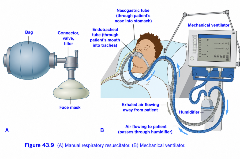
Manual Respiratory Resuscitator (Ambu Bag) and Mechanical Ventilator (Figure 43.9)
Easiest & Most Conceptual Summary
This figure explains two methods used to help a patient breathe when they cannot breathe adequately on their own.
- Manual Respiratory Resuscitator (Ambu Bag) → A person squeezes a bag to push air into the patient’s lungs.
- Mechanical Ventilator → A machine automatically pushes air into and out of the lungs.
Main Concept
If a patient cannot breathe properly, oxygen-rich air is delivered into the lungs either manually (Ambu bag) or automatically (mechanical ventilator).
Overall View of the Figure
The figure has two parts.
Part A
Manual Respiratory Resuscitator (Ambu Bag)
Human hand provides the breathing.
Part B
Mechanical Ventilator
A machine provides the breathing.
PART A – Manual Respiratory Resuscitator (Ambu Bag)
This is commonly called an Ambu Bag or Bag-Valve-Mask (BVM).
It is used during:
- CPR
- Cardiac arrest
- Emergency transport
- Before intubation
- During anesthesia if needed
Understanding Each Part
1. Bag
The large blue bag is the air reservoir.
Function
When squeezed,
it pushes air (or oxygen) into the patient’s lungs.
When released,
it automatically expands and fills with fresh air or oxygen.
Easy Concept
Think of it as a hand pump.
Squeeze →
Air goes in.
Release →
Bag refills.
2. Connector, Valve, and Filter
This small section connects the bag to the patient’s airway.
Functions
- Directs airflow toward the patient.
- Prevents exhaled air from returning to the bag.
- Filters microorganisms and secretions.
Easy Concept
It acts like a one-way traffic controller.
Air goes only toward the patient during squeezing.
3. Face Mask
The mask fits tightly over:
- Nose
- Mouth
Function
Delivers air into the airway without inserting a tube.
Easy Concept
The mask is the doorway through which air enters.
How the Ambu Bag Works
Step 1
Healthcare worker squeezes the bag.
↓
Step 2
Air travels through the valve.
↓
Step 3
Air enters the mask.
↓
Step 4
Air reaches the lungs.
↓
Step 5
The patient exhales.
↓
Step 6
The bag refills automatically.
Simple Flow
Hand squeezes bag
        ↓
Air pushed forward
        ↓
Valve
        ↓
Face mask
        ↓
Patient's lungs
PART B – Mechanical Ventilator
This machine performs breathing automatically.
It is used in:
- ICU
- Operation theatre
- Respiratory failure
- Severe pneumonia
- ARDS
- Coma
Understanding Each Part
1. Mechanical Ventilator
This is the machine itself.
Function
It automatically:
- Pushes air into the lungs.
- Allows air to come out.
- Repeats this cycle many times every minute.
Easy Concept
It is an automatic breathing machine.
2. Endotracheal Tube (ET Tube)
The figure shows a tube entering through the mouth into the trachea.
Function
Provides a direct airway to the lungs.
Why is it needed?
If the patient cannot protect or maintain the airway,
the ET tube keeps the airway open and allows the ventilator to deliver air directly to the lungs.
Easy Concept
Think of it as a special breathing pipe connecting the ventilator to the lungs.
3. Nasogastric (NG) Tube
This tube enters through the nose into the stomach.
Function
It is not part of the breathing system.
It is used to:
- Remove stomach contents.
- Prevent stomach distension.
- Provide feeding if needed.
Easy Concept
The NG tube goes to the stomach, not the lungs.
4. Humidifier
Air from the ventilator passes through the humidifier.
Function
Adds:
- Moisture
- Warmth
to the inhaled air.
Why?
Normally,
the nose warms and humidifies inspired air.
With an ET tube,
the nose is bypassed.
Therefore,
the ventilator must humidify the air.
Easy Concept
The humidifier acts like an artificial nose.
5. Air Flowing to the Patient
The blue arrows show air moving:
Ventilator
↓
Humidifier
↓
Breathing tube
↓
ET tube
↓
Lungs
6. Exhaled Air Flowing Away
The gray arrows show exhaled air leaving the lungs.
Flow
Lungs
↓
ET tube
↓
Ventilator
↓
Outside
Complete Breathing Cycle
Step 1
Ventilator pushes air.
↓
Step 2
Air passes through the humidifier.
↓
Step 3
Air travels through the breathing tube.
↓
Step 4
Air enters the ET tube.
↓
Step 5
Air reaches the lungs.
↓
Step 6
Oxygen enters the blood.
↓
Step 7
Carbon dioxide leaves the blood.
↓
Step 8
The patient exhales.
↓
Step 9
Exhaled air returns to the ventilator.
↓
The cycle repeats.
Easy Flow Diagram
Mechanical Ventilator
        ↓
Humidifier
        ↓
Breathing Tube
        ↓
Endotracheal Tube
        ↓
Lungs
        ↓
Gas Exchange
        ↓
Exhaled Air
        ↓
Back to Ventilator
Difference Between Ambu Bag and Mechanical Ventilator
| Manual Respiratory Resuscitator (Ambu Bag) | Mechanical Ventilator |
|---|---|
| Human squeezes the bag | Machine delivers breaths automatically |
| Used mainly in emergencies | Used for prolonged respiratory support |
| Simple, portable | Complex, computer-controlled |
| Temporary support | Continuous support in ICU or operating room |
Simple Real-Life Analogy
Imagine inflating a balloon.
Ambu Bag
You use your hand to squeeze air into the balloon.
This is similar to manually ventilating a patient.
Mechanical Ventilator
Now imagine a machine automatically inflating and deflating the balloon repeatedly without anyone squeezing it.
That is how a mechanical ventilator works.
Important Points from Figure 43.9
- A manual respiratory resuscitator (Ambu bag) provides manual positive-pressure ventilation by squeezing the bag.
- The bag supplies air or oxygen, the valve directs airflow toward the patient, and the face mask delivers air to the airway.
- A mechanical ventilator provides automatic positive-pressure ventilation for patients who cannot breathe adequately on their own.
- The endotracheal tube passes through the mouth into the trachea, providing a secure airway for ventilation.
- The nasogastric tube passes through the nose into the stomach and is used for gastric decompression or feeding; it is not part of the respiratory pathway.
- The humidifier warms and moistens inspired air because the upper airway is bypassed by the endotracheal tube.
- During inspiration, air flows from the ventilator → humidifier → breathing circuit → endotracheal tube → lungs.
- During expiration, air flows from the lungs back through the endotracheal tube and breathing circuit to the ventilator.
KEY CONCEPT (Figure 43.9)
Figure 43.9 illustrates two methods of assisted ventilation. The manual respiratory resuscitator (Ambu bag) requires a healthcare provider to squeeze the bag, forcing air or oxygen through a valve and face mask into the patient’s lungs. In contrast, the mechanical ventilator automatically delivers controlled breaths through an endotracheal tube. Inspired air is first humidified because the natural warming and humidifying function of the nose is bypassed. After gas exchange occurs in the lungs, carbon dioxide-rich air is exhaled back to the ventilator, and the cycle repeats. These devices are lifesaving for patients with respiratory failure or those unable to breathe effectively on their own.
Made by self learning CEO and founder Dr sheen
