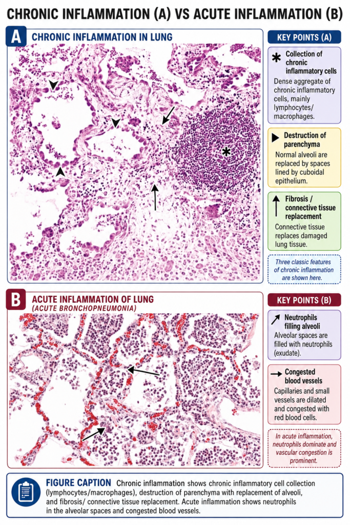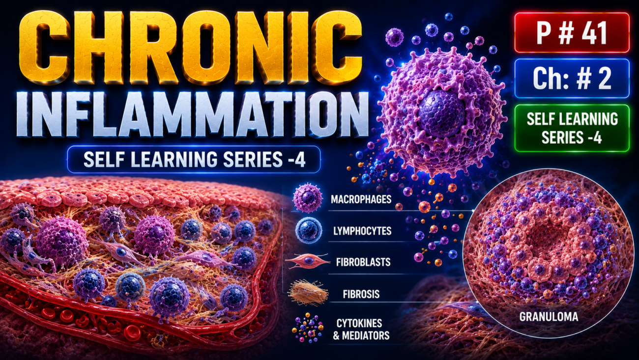
- Chronic inflammation is prolonged inflammation lasting weeks to months.
- In chronic inflammation, inflammation + tissue injury + attempts at repair occur together in different proportions.
- It may develop after acute inflammation or may start slowly as a smoldering, progressive process without a previous acute phase.
- Chronic inflammation can cause marked tissue damage and scarring, sometimes with relatively few inflammatory cells.
- Hepatic cirrhosis is an example in which extensive tissue damage and fibrosis may occur.
Causes of Chronic Inflammation
- Persistent infections: Chronic inflammation occurs when microorganisms are difficult to eliminate, such as mycobacteria, certain viruses, fungi, and parasites.
- Sometimes incompletely resolved acute inflammation → progresses to chronic inflammation.
- Example: acute bacterial lung infection → chronic lung abscess.
- Hypersensitivity diseases: Excessive or inappropriate activation of the immune system can cause persistent inflammation.
- In autoimmune diseases, the immune system reacts against self-antigens → continuous immune reaction → chronic tissue damage and inflammation.
- Examples include rheumatoid arthritis and multiple sclerosis.
- In allergic diseases, excessive immune responses against common environmental substances → chronic inflammation.
- Example: bronchial asthma.
- Repeated attacks may produce a mixture of acute and chronic inflammation, while fibrosis may dominate in late stages.
- Prolonged exposure to toxic agents: Chronic inflammation can occur after long exposure to harmful substances from outside or inside the body.
- Exogenous example: Long-term inhalation of nondegradable silica particles → chronic lung inflammation → silicosis.
- Endogenous example: Excessive production and deposition of cholesterol and other lipids in arterial walls → chronic inflammation → atherosclerosis.
- Chronic inflammation may also contribute to diseases not traditionally considered inflammatory.
- Examples include Alzheimer disease, metabolic syndrome, and associated type 2 diabetes.
KEY CONCEPT
- Chronic inflammation = prolonged inflammation + tissue injury + repair occurring together.
- Persistent infection → chronic inflammation.
- Autoimmunity/allergy → repeated immune activation → chronic inflammation.
- Long-term toxic exposure → chronic tissue injury and inflammation.
- Late chronic inflammation may lead to fibrosis and scarring.
Conceptual Examples
- Tuberculosis: Persistent mycobacteria cannot be easily eliminated → prolonged immune response → chronic inflammation.
- Rheumatoid arthritis: Immune system attacks self-antigens → repeated tissue injury → chronic inflammation.
- Silicosis: Long-term inhaled silica remains in lung tissue → persistent inflammation → lung fibrosis.
- Atherosclerosis: Cholesterol and lipids accumulate in arterial walls → persistent inflammation → chronic arterial damage.
Morphologic Features
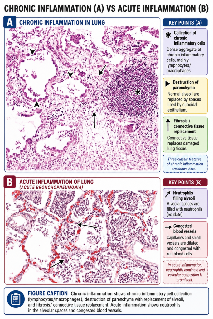
- Acute inflammation mainly shows vascular changes + edema + neutrophil infiltration.
- In contrast, chronic inflammation has three major morphologic features:
- Mononuclear cell infiltration: mainly macrophages, lymphocytes, and plasma cells (Fig. 2.14).
- Tissue destruction: caused by the persistent harmful agent or by the inflammatory cells themselves.
- Attempts at healing: damaged tissue is replaced by connective tissue.
- Healing occurs through:
- Angiogenesis → formation/proliferation of small blood vessels.
- Fibrosis → formation of fibrous connective tissue.
- Eventually → scar formation.
- Angiogenesis and fibrosis are important parts of tissue repair.
Cells and Mediators of Chronic Inflammation
- Chronic inflammation is characterized by a combination of leukocyte infiltration + tissue damage + fibrosis.
- These changes result from local activation of different cell types and production of inflammatory mediators.
Role of Macrophages
- Macrophages are the dominant cells in most chronic inflammatory reactions.
- Their major actions include:
- Destroying foreign agents and damaged tissues
- Producing cytokines and growth factors
- Activating other cells, especially T lymphocytes
- Macrophages are professional phagocytes → their main function is to ingest and destroy microbes, particulate material, and dead cells.
- They also have important roles in host defense, inflammation, and tissue repair.
- Macrophages are normally scattered throughout most connective tissues.
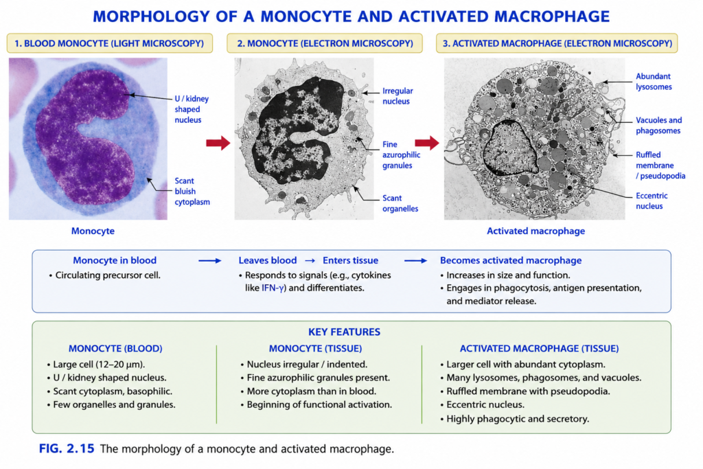
- Their circulating form in blood is called a monocyte.
- Specialized tissue macrophages include:
- Liver → Kupffer cells
- Spleen and lymph nodes → sinus histiocytes
- Central nervous system → microglial cells
- Lungs → alveolar macrophages
- Together, circulating monocytes and tissue macrophages form the mononuclear phagocyte system.
- Blood monocytes are about 10–15 μm in diameter and have a bean-shaped nucleus + finely granular cytoplasm (Fig. 2.15).
- Tissue macrophages contain abundant cytoplasm, many phagocytic vacuoles containing ingested material, and numerous lysosomes and other organelles.
KEY CONCEPT
- Chronic inflammation = mononuclear cells + tissue destruction + attempts at repair/fibrosis.
- Macrophages are the major cells of chronic inflammation.
- Monocyte in blood → macrophage in tissue.
- Macrophages → phagocytosis + cytokine/growth factor secretion + T-cell activation → inflammation and repair.
Conceptual Examples
- Persistent tissue injury: Harmful agent remains → macrophages and lymphocytes accumulate → tissue damage continues → fibrosis and scar formation.
- Liver: Tissue-resident macrophage → Kupffer cell.
- Brain: Tissue-resident macrophage → microglial cell.
- Lung: Tissue-resident macrophage → alveolar macrophage.
Role of Macrophages
- Tissue macrophages come from two main developmental sources:
- Bone marrow hematopoietic stem cells
- Embryonic yolk sac and fetal liver progenitors (Fig. 2.16)
- During inflammation → bone marrow progenitors produce monocytes → monocytes enter blood → migrate into tissues → become macrophages.
- Monocytes enter tissues using mechanisms similar to neutrophils, especially adhesion molecules and chemokines.
- Macrophages survive longer in tissues than many other leukocytes.
- Therefore, within about 48 hours after inflammation begins, macrophages often become the dominant inflammatory cells.
- Tissue-resident macrophages, such as microglia and Kupffer cells, mainly originate from the yolk sac or fetal liver during embryonic development.
- These cells enter tissues early → remain there for long periods → are maintained mainly by local proliferation of resident macrophages.
- Macrophages can be activated through two major pathways: classical and alternative activation (Fig. 2.17).
- The pathway followed depends on the type of activating signal.
- Classical macrophage activation (M1): triggered by microbial products such as endotoxin acting through TLRs and other sensors, and by IFN-γ produced during immune responses.
- M1 macrophages produce NO + ROS and increase lysosomal enzymes → stronger killing of ingested microorganisms.
- M1 macrophages also secrete cytokines that promote inflammation.
- Therefore, M1 macrophages mainly function in microbial killing and promotion of inflammation.
- They are important for eliminating infections but can also damage normal tissues.
- Alternative macrophage activation (M2): mainly stimulated by IL-4 and IL-13 produced by T lymphocytes and other cells.
- M2 macrophages are not strongly microbicidal.
- Their main role is tissue repair and control of inflammation.
- M2 macrophages release growth factors → promote angiogenesis + fibroblast activation + collagen synthesis.
- They also help suppress inflammation.
- A useful concept is:
- M1 first → destroy harmful agent and promote inflammation
- M2 later → reduce inflammation and promote tissue repair
- However, this exact sequence is not clearly established in all inflammatory reactions.
- In reality, macrophages do not exist only as pure M1 or M2 forms; many intermediate macrophage populations also exist.
KEY CONCEPT
- Bone marrow → monocyte → blood → tissue → macrophage.
- M1 macrophage → IFN-γ/microbial products → NO + ROS + lysosomal enzymes → microbial killing + inflammation.
- M2 macrophage → IL-4 + IL-13 → angiogenesis + fibroblast activation + collagen → tissue repair.
- M1 = Kill and Inflame.
- M2 = Mend and Repair.
Conceptual Examples
- Bacterial infection: Microbial products + IFN-γ → M1 activation → ROS/NO produced → microbes killed → inflammation increases.
- Healing tissue: IL-4 and IL-13 → M2 activation → fibroblasts and blood vessel growth stimulated → collagen deposition and repair.
- Persistent inflammation: Monocytes continue entering tissue → become long-lived macrophages → macrophages become the dominant cells after about 48 hours.
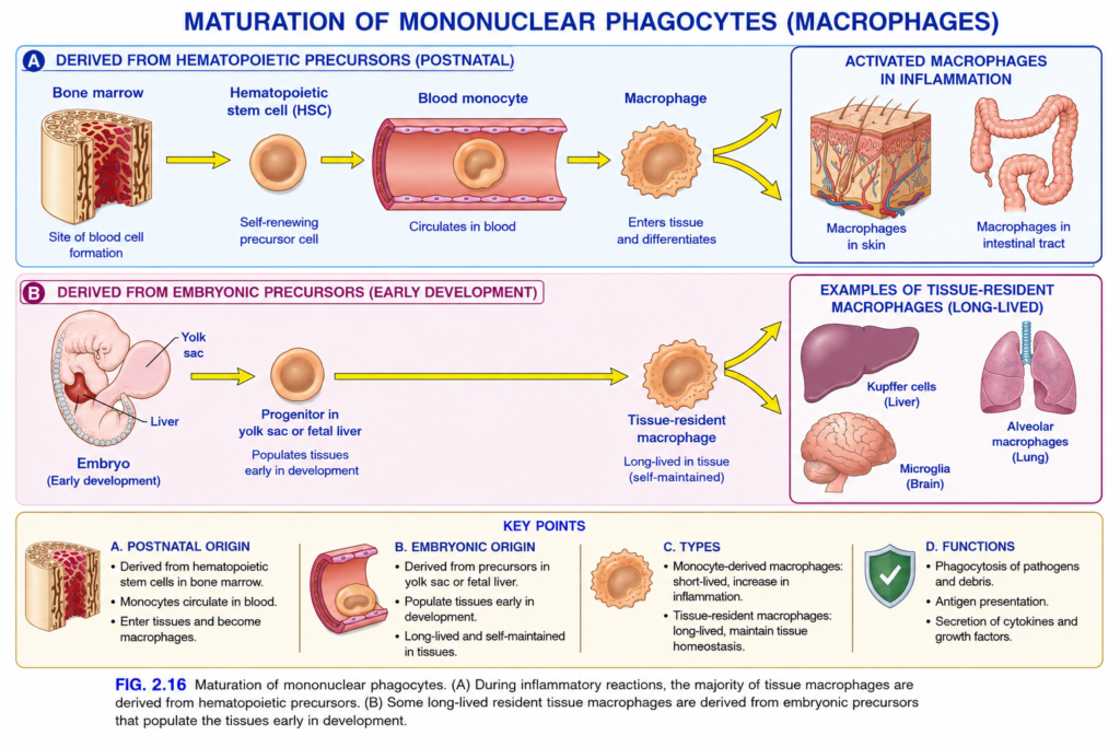
Fig. 2.16 — Maturation of Mononuclear Phagocytes (Macrophages)
This figure answers one basic question:
Where do macrophages come from?
There are 2 main routes:
Route A — After birth / during inflammation
Bone marrow → hematopoietic stem cell → blood monocyte → tissue macrophage
Route B — During embryonic development
Yolk sac / fetal liver precursor → tissue-resident macrophage
The most important difference is:
Route A passes through a blood monocyte.
Route B establishes long-lived macrophages in tissues early in life.
First: What does “mononuclear phagocyte” mean?
Break the words:
- Mono = one
- Nuclear = nucleus
- Phagocyte = cell that eats
So a mononuclear phagocyte is an immune cell with one main nucleus that can engulf microbes and dead material.
The important cells here are:
Monocytes → Macrophages
What is a macrophage?
A macrophage is a large immune cell mainly found in tissues.
Main jobs
Macrophage
→ eats microbes
→ eats dead cells/debris
→ releases cytokines
→ presents antigens to T cells
→ helps inflammation
→ helps tissue repair
Easy memory
MACRO = big
PHAGE = eater
So:
Macrophage = big eater
PANEL A — BLUE AREA
Derived from hematopoietic precursors (postnatal)
Postnatal = after birth.
This upper pathway is particularly important during inflammatory reactions.
STEP 1 — Bone marrow
The first picture on the left is:
Bone marrow
Bone marrow is the major site where blood cells are produced after birth.
The caption underneath says:
Site of blood cell formation
So the first yellow arrow means:
Bone marrow → blood-cell precursor
STEP 2 — Hematopoietic stem cell (HSC)
The orange round cell is:
Hematopoietic stem cell
Hematopoietic = blood-forming.
A hematopoietic stem cell is a parent cell capable of giving rise to all major blood-cell lineages.
For this figure, we follow only one pathway:
HSC → monocyte lineage
The figure calls it a self-renewing precursor cell.
Self-renewing means
The stem-cell population can maintain itself by making new stem cells while also producing cells that mature into blood cells.
So:
HSC
→ maintains its own population
AND
→ produces blood-cell precursors
Yellow arrow: HSC → Blood monocyte
The next yellow arrow means:
Hematopoietic stem cell
→ passes through intermediate precursor stages
→ produces a monocyte
Those intermediate stages are simply not shown.
The figure is giving you the simplified pathway.
STEP 3 — Blood monocyte
Now look at the pink/red blood vessel.
Inside it is a round orange cell labeled:
Blood monocyte
A monocyte is a white blood cell that circulates in blood.
The text underneath says:
Circulates in blood
Very easy concept
Think:
Monocyte = traveling form
It travels through the bloodstream until inflammatory signals tell it to leave.
What is the pink tube?
The pink tube represents a:
Blood vessel
- Pink lining = endothelial cells
- Orange round cell inside = monocyte
- Yellow arrow = direction of maturation/movement
STEP 4 — Monocyte leaves blood
The next yellow arrow points from:
Blood monocyte → Macrophage
During inflammation, chemokines and adhesion molecules help the monocyte:
stick to vessel wall
→ cross the endothelium
→ enter tissue
This movement out of the blood is called:
Emigration / extravasation
Very simply:
Monocyte leaves the bloodstream and enters the tissue.
STEP 5 — Monocyte becomes macrophage
Once inside tissue:
Monocyte → differentiates → macrophage
What does “differentiate” mean?
Differentiate = change into a more specialized mature cell.
So:
Blood monocyte
→ enters tissue
→ becomes larger and more specialized
→ macrophage
The irregular orange cell represents the macrophage.
Why does the macrophage look irregular?
The monocyte is shown as a fairly smooth round cell.
The macrophage has:
- more cytoplasm
- irregular edges
- more cellular machinery
because it is now specialized for:
phagocytosis + secretion + tissue defense
The yellow arrow now SPLITS
After the macrophage, the yellow arrow divides upward and downward.
This means:
Macrophages can enter and function in different tissues.
The right upper box shows:
Activated macrophages in inflammation
Examples shown:
- macrophages in skin
- macrophages in intestinal tract
What is an “activated macrophage”?
A macrophage may already be present, but during infection or tissue damage it receives activating signals.
Examples of activating signals include:
- microbial products
- inflammatory cytokines
- signals from T cells
Then:
Macrophage → activated macrophage
An activated macrophage becomes much more effective.
What does an activated macrophage do?
1. Phagocytosis
It eats:
- bacteria
- dead cells
- damaged tissue
2. Killing
It produces substances that help destroy microbes.
3. Cytokine secretion
It can release cytokines such as:
- TNF
- IL-1
- chemokines
These recruit and activate more inflammatory cells.
4. Repair
It can also release growth factors that promote healing.
So:
Activated macrophage
→ kill + clean + signal + repairComplete PANEL A pathway
Follow every yellow arrow:
Bone marrow
→ hematopoietic stem cell
→ blood monocyte
→ monocyte circulates in blood
→ monocyte enters tissue
→ macrophage
→ becomes activated during inflammation
→ participates in inflammatory defense
PANEL B — PINK AREA
Derived from embryonic precursors
Now we have a different origin.
This pathway begins during:
Early embryonic development
Look at the lower-left embryo.
Two important structures are labeled:
- Yolk sac
- Liver
Here, “liver” means the fetal liver.
STEP 1 — Yolk sac and fetal liver
Very early in development, the bone marrow is not yet functioning as the main adult blood-forming organ.
Early blood and immune-cell precursors arise in places such as:
Yolk sac
and later:
Fetal liver
These can produce macrophage precursors.
Yellow arrow → progenitor
The yellow arrow moves from the embryo toward:
Progenitor in yolk sac or fetal liver
What is a progenitor?
A progenitor cell is an immature cell that is already moving toward a specific cell lineage.
Very simply:
Stem cell
= many possible futures
Progenitor
= fewer choices; already committed toward a certain family
Here:
Embryonic progenitor
→ macrophage lineage
Important label:
“Populates tissues early in development”
This is the key concept.
These embryonic macrophage precursors move into organs while the fetus is developing.
So:
Embryonic precursor
→ migrates into developing tissue
→ becomes resident macrophage
Notice the VERY LONG yellow arrow
This long yellow arrow is important.
It goes:
Embryonic progenitor → Tissue-resident macrophage
Notice something missing:
There is NO blood monocyte stage shown.
That is intentional.
This teaches:
Not every macrophage has to arise from an adult blood monocyte.
Many long-lived tissue-resident macrophages were seeded into organs during embryonic development.
STEP 2 — Tissue-resident macrophage
The irregular orange cell is labeled:
Tissue-resident macrophage
Tissue-resident means:
A macrophage that normally lives permanently in a particular tissue.
The caption says:
Long-lived in tissue (self-maintained)What does “self-maintained” mean?
Many resident macrophage populations can maintain themselves locally.
That means:
existing resident macrophage
→ divides
→ produces more resident macrophages
They may not need constant replacement from circulating monocytes under normal conditions.
This is called:
Self-renewal / local proliferation
The yellow arrow again splits
The tissue-resident macrophage gives examples in different organs.
The figure shows three classic examples:
1. Kupffer cells — Liver
The liver is shown in the upper-left of the right box.
Its resident macrophages are called:
Kupffer cells
They help:
- remove microbes from blood
- remove cellular debris
- process material coming from the intestine
- participate in liver inflammation
Exam memory
Kupffer = Liver. Microglia — Brain
The brain is shown below.
Its specialized resident macrophage population is:
Microglia
They:
- monitor the central nervous system
- remove dead cells
- respond to infection/injury
- participate in neuroinflammation
Exam memory
Microglia = Brain
3. Alveolar macrophages — Lung
The lung is shown on the right.
These are:
Alveolar macrophages
They live around the alveoli.
Alveoli
= tiny air sacs where gas exchange occurs.
Alveolar macrophages:
- eat inhaled particles
- remove microorganisms
- clean cellular debris
Exam memory
Alveolar macrophage = Lung
NOW COMPARE PANEL A WITH PANEL B
This is the most important part of the entire figure.
| Panel A | Panel B |
|---|---|
| Postnatal route | Embryonic route |
| Begins in bone marrow | Begins in yolk sac/fetal liver |
| Hematopoietic precursor | Embryonic progenitor |
| Goes through blood monocyte | No blood-monocyte stage shown |
| Monocyte enters tissue | Cells populate tissues during development |
| Important during inflammation | Creates many long-lived resident populations |
| Recruited macrophages increase during inflammation | Often maintained locally |
KEY POINT A — Postnatal origin
The bottom box summarizes:
Bone marrow HSC
→ monocytes produced
Monocytes
→ circulate in blood
Monocytes
→ enter tissue
In tissue
→ become macrophages
So memorize:
Bone → Blood → Tissue
Bone marrow
→ monocyte in blood
→ macrophage in tissueKEY POINT B — Embryonic origin
Yolk sac / fetal liver
→ embryonic precursor
Precursor
→ enters tissues early in development
Tissue macrophage
→ remains long-lived
Resident population
→ may maintain itself by local proliferation
So:
Embryo → organ early → resident for long time
KEY POINT C — Two types emphasized in the figure
Monocyte-derived macrophages
These arise from:
blood monocytes
They increase greatly during inflammation.
Many recruited macrophages are relatively shorter-lived compared with long-lived resident populations.
Tissue-resident macrophages
These are established in particular tissues.
Many are:
- long-lived
- locally maintained
- specialized for their organ
Examples:
Liver → Kupffer cells
Brain → Microglia
Lung → Alveolar macrophages
KEY POINT D — Functions of macrophages
The bottom-right box gives 3 major functions.
1. Phagocytosis
Phagocytosis = engulfing and digesting material.
Macrophages eat:
pathogens + dead cells + debris
2. Antigen presentation
After eating a microbe:
Macrophage digests microbe
→ takes microbial antigen
→ displays it on its surface
→ presents it to T cells
So macrophages help connect:
innate immunity → adaptive immunity
3. Secretion of cytokines and growth factors
Cytokines
help regulate inflammation.
For example:
TNF / IL-1 / chemokines
→ recruit and activate inflammatory cells
Growth factors
help tissue repair.
So macrophages can participate in both:
Inflammation AND healing
What Do the COLORS Mean?
These are teaching colors, not the actual colors of the cells inside the body.
Light blue upper background
= postnatal / bone-marrow route
Light pink lower background
= embryonic route
Yellow arrows
= direction of development, migration, or differentiation
Orange round cells
= cells of the monocyte/macrophage lineage
Pink-red tube
= blood vessel
Irregular orange cell
= macrophage
Blue headings
= adult/postnatal pathway and labels
Purple/magenta heading
= embryonic pathway
Beige bottom box
= summary/key exam points
One Important Concept Students Often Mix Up
Do not memorize:
❌ “Every tissue macrophage comes from a blood monocyte.”
That is too simple and not completely correct.
Instead remember:
During inflammation
Bone marrow
→ monocytes
→ blood
→ tissue
→ macrophages
But many long-lived resident macrophages originate from:
Yolk sac/fetal liver
→ tissue during embryonic development
→ resident macrophage
Whole Figure in One Simple Story
ROUTE A — After birth
Bone marrow
↓
Hematopoietic stem cell
↓
Monocyte produced
↓
Monocyte enters blood
↓
Travels in circulation
↓
Inflammation occurs
↓
Monocyte leaves blood
↓
Enters tissue
↓
Becomes macrophage
↓
Macrophage becomes activated
↓
Kills microbes + removes debris + releases cytokines
ROUTE B — Before birth
Yolk sac / fetal liver
↓
Embryonic progenitor
↓
Moves into developing organs
↓
Becomes tissue-resident macrophage
↓
Lives there for a long time
↓
Often maintains itself locally
↓
Examples:
Liver → Kupffer cells
Brain → Microglia
Lung → Alveolar macrophages
⭐ Easiest Memory Trick
MONO = Moving in blood
MACRO = Moved into tissue
So:
Monocyte in BLOOD → Macrophage in TISSUE
For resident macrophages remember:
K-M-A
Kupffer → liver
Microglia → brain
Alveolar macrophage → lung
Key Exam Points
- Monocytes circulate in blood; macrophages mainly live in tissues.
- During inflammatory reactions, many macrophages are recruited from bone-marrow-derived monocytes.
- Monocyte enters tissue → differentiates into macrophage.
- Many long-lived tissue-resident macrophages originate from embryonic yolk sac/fetal liver precursors.
- Resident macrophages may be maintained by local self-renewal.
- Kupffer cells = liver.
- Microglia = brain.
- Alveolar macrophages = lung.
- Macrophages perform phagocytosis, antigen presentation, cytokine secretion, and tissue repair.
2-Line Exam Recall
Bone marrow → HSC → blood monocyte → tissue macrophage, especially during inflammation.
Yolk sac/fetal liver → embryonic precursor → long-lived tissue-resident macrophages such as Kupffer cells, microglia, and alveolar macrophages.
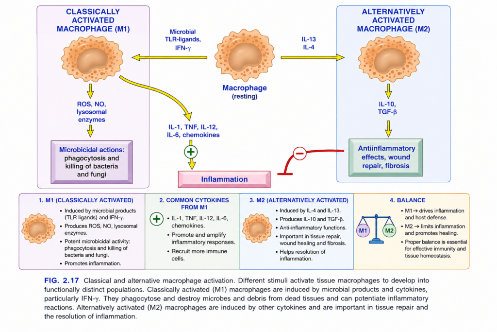
Fig. 2.17 — Classical (M1) vs Alternative (M2) Macrophage Activation
This figure is showing one very important idea:
The same macrophage can behave in two different ways depending on the signal it receives.
Think of a macrophage as having 2 major modes:
- M1 = FIGHT mode → kills microbes and increases inflammation
- M2 = FIX mode → reduces inflammation and repairs tissue
First: Start from the center
The orange cell in the middle is a:
Macrophage
At first, think of it as a macrophage waiting for instructions.
It can receive different chemical signals.
If it receives:
Microbial TLR ligands + IFN-γ
→ it becomes M1
If it receives:
IL-4 + IL-13
→ it becomes M2
So the entire figure begins with:
Macrophage
↙️ different signal different signal ↘️
M1 M2T SIDE — M1 Macrophage
The heading says:
Classically Activated Macrophage (M1)
“Classically activated” means:
A macrophage activated mainly to fight microbes and produce inflammation.
Arrow 1: Microbial TLR ligands → M1
Look at the yellow arrow pointing from the center toward the left.
Above it:
Microbial TLR-ligands, IFN-γ
These are the signals that push the macrophage toward M1 activation.hat is a TLR?
TLR = Toll-Like Receptor
TLRs are receptors present on innate immune cells such as macrophages.
Their job is to recognize common microbial molecules.
Examples:
- bacterial LPS
- bacterial cell-wall molecules
- viral nucleic acids
A microbial molecule that binds a TLR is called a:
TLR ligand
So:
Microbial molecule binds TLR on macrophage
→ macrophage detects infection
→ M1 activation
What is IFN-γ?
IFN-γ = Interferon-gamma
It is a powerful macrophage-activating cytokine.
It is produced mainly by:
- Th1 cells
- NK cells
Its message to macrophages is basically:
“Become better at killing microbes.”
So:
Microbial TLR signal + IFN-γ
→ strong classical macrophage activation
→ M1 macrophage
Why are BOTH shown together?
Because the strongest microbicidal macrophage activation occurs when it receives:
Microbial signal
+
IFN-γ
So one signal says:
“There is a microbe.”
The other says:
“Attack strongly.”
M1 now follows TWO important branches
Look at the M1 macrophage.
One yellow arrow goes downward.
Another curved yellow arrow goes toward inflammatory cytokines.
These represent two major M1 functions.
M1 Function 1 — Kill microbes
The downward arrow leads to:
ROS, NO, lysosomal enzymes
Let’s define each.
ROS
ROS = Reactive Oxygen Species
These are highly reactive oxygen-containing chemicals.
Examples include:
- superoxide
- hydrogen peroxide
They damage and kill microbes.
Very simply:
M1 macrophage → ROS → microbial damage
NO
NO = Nitric Oxide
Macrophages can produce nitric oxide using inducible nitric oxide synthase.
NO is toxic to many microbes.
So:
M1 → NO → microbial killing
Lysosomal enzymes
Macrophages contain lysosomes.
Lysosomes contain powerful digestive enzymes.
After a macrophage eats a bacterium:
Bacterium enters macrophage
→ joins lysosome
→ lysosomal enzymes digest it
Downward arrow → Microbicidal actions
The purple box says:
Microbicidal actions:
phagocytosis and killing of bacteria and fungi
Microbicidal means:
microbe-killing
So the sequence is:
M1 macrophage
→ ROS + NO + lysosomal enzymes
→ phagocytosis
→ bacterial/fungal killing
What is phagocytosis?
Phagocytosis = eating a particle or microbe
Sequence:
Macrophage recognizes microbe
→ surrounds it
→ takes it inside
→ lysosomes fuse
→ microbe is destroyed
Easy memory
M1 = Murder microbes
M1 Function 2 — Produce inflammatory cytokines
Now look at the curved yellow arrow from M1.
It leads to:
- IL-1
- TNF
- IL-12
- IL-6
- chemokines
These are inflammatory mediators produced by M1 macrophages.
IL-1 and TNF
These are major inflammatory cytokines.
They cause:
- endothelial activation
- leukocyte recruitment
- fever
- increased inflammatory responses
So:
M1 → IL-1 + TNF → inflammation ↑
IL-6
IL-6 has several systemic inflammatory effects.
A major one is:
IL-6 → liver → acute-phase proteins
For example:
CRP increases
IL-12
IL-12 helps activate:
- NK cells
- T cells
and promotes:
IFN-γ production
This can further activate macrophages.
So you can get a reinforcing loop:
M1 → IL-12 → IFN-γ ↑ → stronger macrophage activation
Chemokines
Chemokines = cytokines that attract leukocytes
Think:
Chemokines = chemical GPS signals
They tell neutrophils and other leukocytes:
“Come to this infected area.”
So:
M1 → chemokines → leukocyte recruitment → inflammation ↑
Green PLUS sign
Look at the green + before the arrow toward inflammation.
This means:
M1 promotes inflammation
So:
M1 cytokines
→ increase inflammation
That is why the green plus sign is shown.
LEFT SIDE COMPLETE PATH
Signal
Microbial TLR ligands + IFN-γ
↓
M1 macrophage
Then two major actions:
Action 1
ROS + NO + lysosomal enzymes
→ phagocytosis
→ killing bacteria and fungi
Action 2
IL-1 + TNF + IL-12 + IL-6 + chemokines
→ inflammation ↑
RIGHT SIDE — M2 Macrophage
Now look at the blue-shaded box on the right.
The heading says:
Alternatively Activated Macrophage (M2)
This macrophage has a different job.
Instead of mainly fighting microbes, M2 mainly helps:
- suppress inflammation
- repair tissue
- promote wound healing
- produce fibrosis
Arrow: IL-4 + IL-13 → M2
The yellow arrow from the central macrophage points to the right.
Above it:
IL-13
IL-4
These cytokines push the macrophage toward the M2 pathway.
So:
IL-4 + IL-13
→ alternative macrophage activation
→ M2
What are IL-4 and IL-13?
They are cytokines strongly associated with type 2 immune responses.
They can be produced by cells such as:
- Th2 cells
- mast cells
- innate lymphoid cells
Their general message is more like:
M2 → IL-10 and TGF-β
Look at the yellow downward arrow.
The M2 macrophage produces mediators including:
IL-10
IL-10 is strongly:
Anti-inflammatory
It suppresses excessive inflammatory responses.
So:
M2 → IL-10 → inflammation ↓TGF-β
TGF-β = Transforming Growth Factor-beta
It is important in:
- tissue repair
- fibroblast activation
- collagen production
- fibrosis
So:
M2 → TGF-β
→ fibroblasts activated
→ collagen deposited
→ tissue repair / fibrosis
Green box: Anti-inflammatory effects, wound repair, fibrosis
This is the main result of M2 activation.
1. Anti-inflammatory effects
M2 tries to stop excessive inflammatory damage.
Especially through:
IL-10 and TGF-β
2. Wound repair
M2 macrophages produce substances that help repair damaged tissue.
They help:
- fibroblasts
- blood vessel formation
- extracellular matrix production
So:
M2 → repair mechanisms → healing
3. Fibrosis
Fibrosis = excessive collagen/connective tissue deposition
This may be useful during wound healing.
But if too much occurs:
too much collagen → scar formation → fibrosis
So M2 is beneficial for healing, but excessive M2-type repair may contribute to pathological fibrosis.
Red inhibitory line with MINUS sign
This is extremely important.
Look at the red line coming from the M2 side toward Inflammation.
There is a white circle containing:
−
That minus sign means:
M2 inhibits inflammation
So:
M2 → IL-10 / TGF-β → inflammation ↓
Compare:
M1
green +
→ inflammation ↑
M2
red −
→ inflammation ↓
This is the central contrast of the figure.Why Does the Body Need BOTH?
Because inflammation has two phases.
Imagine bacteria enter through a wound.
Early phase
The body needs:
M1
because:
Microbes present → kill them → strong inflammation
But after the microbes have been controlled, continued strong inflammation would damage normal tissue.
Then the body needs:
M2
to say:
“The fight is finishing. Reduce inflammation and repair the damage.”
So ideally:
M1 fights → M2 fixes
Simple Real-Life Story
Imagine your skin becomes infected.
Step 1
Bacteria enter.
↓
Step 2
Microbial products activate TLRs.
↓
Step 3
IFN-γ also activates macrophages.
↓
Step 4
Macrophage becomes M1.
↓
Step 5
M1 produces:
ROS + NO + enzymes
↓
Step 6
Bacteria are killed.
At the same time:
M1 → IL-1/TNF/chemokines → inflammation ↑
↓
Step 7
After infection is controlled, repair signals increase.
↓
Step 8
IL-4 + IL-13 → M2
↓
Step 9
M2 produces:
IL-10 + TGF-β
↓
Step 10
Inflammation decreases.
↓
Step 11
Wound healing starts.
↓
Step 12
Collagen and connective tissue repair the area.
Understand the COLORS
The colors are mainly diagram teaching colors; these molecules are not literally these colors in the body.
| Figure feature | Meaning |
|---|---|
| Orange cells | Macrophages |
| Yellow arrows | Direction of activation/effect |
| White background on left | M1/classical pathway |
| Light blue background on right | M2/alternative pathway |
| Green + | Promotes inflammation |
| Red line + − | Suppresses inflammation |
| Purple boxes | Major final effects |
| Black text | Important signals/products |
M1 vs M2 — Easy Comparison
| M1 | M2 |
|---|---|
| Classical activation | Alternative activation |
| Microbial TLR ligands + IFN-γ | IL-4 + IL-13 |
| Fight microbes | Repair tissue |
| ROS | IL-10 |
| NO | TGF-β |
| Lysosomal enzymes | Growth/repair responses |
| IL-1, TNF, IL-6, IL-12 | Anti-inflammatory mediators |
| Inflammation ↑ | Inflammation ↓ |
| Microbial killing | Wound repair |
| Can cause tissue damage if excessive | Can cause fibrosis if excessive |
Very Easy Memory Trick
M1 = 1st job: Fight
Think:
M1 = Microbe killer
- microbial products
- IFN-γ
- ROS
- NO
- inflammatory cytokines
M1:
KILL + INFLAME
M2 = 2nd job: Mend
Think:
M2 = Mend tissue
- IL-4
- IL-13
- IL-10
- TGF-β
- repair
- fibrosis
M2:
CALM + REPAIR
One More Excellent Memory
M1 = FIRE 🔥
It increases inflammation and kills microbes.
M2 = FIX 🛠️
It turns inflammation down and repairs tissue.
So:
M1 FIRES — M2 FIXES
Whole Figure in One Flow
M1 pathway
Microbial TLR ligands + IFN-γ
→ macrophage becomes M1
→ ROS + NO + lysosomal enzymes
→ phagocytosis + killing of bacteria/fungi
AND
→ IL-1 + TNF + IL-12 + IL-6 + chemokines
→ inflammation ↑
M2 pathway
IL-4 + IL-13
→ macrophage becomes M2
→ IL-10 + TGF-β
→ inflammation ↓
→ wound repair
→ fibrosis
Key Exam Points
- M1 = classically activated macrophage
- M1 activated by microbial TLR ligands + IFN-γ
- M1 produces ROS, NO, lysosomal enzymes
- M1 kills bacteria and fungi
- M1 produces IL-1, TNF, IL-6, IL-12 and chemokines
- M1 promotes inflammation
- M2 = alternatively activated macrophage
- M2 activated mainly by IL-4 and IL-13
- M2 produces anti-inflammatory/repair mediators including IL-10 and TGF-β
- M2 reduces inflammation
- M2 promotes wound healing and fibrosis
2-line exam recall
M1: TLR ligands + IFN-γ → ROS/NO + inflammatory cytokines → microbial killing and inflammation.
M2: IL-4 + IL-13 → IL-10/TGF-β → anti-inflammatory effects, tissue repair and fibrosis.
Final memory: M1 = Fight; M2 = Fix.
Role of Macrophages
- Activated macrophages help eliminate microbes and other harmful agents and start tissue repair, but they can also cause much of the tissue injury in chronic inflammation.
- Important functions of macrophages include:
- Phagocytosis: ingest and remove microbes + dead tissue debris.
- Inflammation: release mediators such as TNF, IL-1, chemokines, and eicosanoids → initiate and maintain inflammatory reactions.
- Repair: initiate tissue repair + scar formation + fibrosis.
- Interaction with T lymphocytes: display antigens to T cells and respond to T-cell signals → creates a feedback loop important in cell-mediated immunity.
- After the harmful agent is removed → macrophages usually die or leave through lymphatics to lymph nodes.
- If inflammation continues → macrophages remain because of continued recruitment of blood monocytes + local macrophage proliferation.
Role of Lymphocytes
- T and B lymphocytes are activated by microbes and other environmental antigens → help amplify and maintain chronic inflammation.
- Lymphocytes mainly function in adaptive immunity, but they are also commonly present in chronic inflammation.
- When lymphocytes remain activated → inflammation tends to become persistent and severe.
- Strong chronic inflammatory reactions, especially granulomatous inflammation, depend heavily on interactions between lymphocytes and macrophages.
- In autoimmune and other hypersensitivity diseases, lymphocytes may become the dominant inflammatory cells.
- CD4+ T lymphocytes release cytokines that determine the type of inflammatory response.
- There are three important CD4+ T-cell subsets:
- Th1 cells → IFN-γ → classical (M1) macrophage activation.
- Th2 cells → IL-4 + IL-5 + IL-13 → eosinophil recruitment/activation + alternative (M2) macrophage activation.
- Th17 cells → IL-17 and other cytokines → chemokine production → mainly neutrophil recruitment.
- Th1 and Th17 cells help defend against many bacteria and viruses and also participate in chronic inflammation in diseases such as rheumatoid arthritis, psoriasis, and inflammatory bowel disease.
- Th2 cells are important in defense against helminth parasites and in allergic inflammation.
- Macrophages and T lymphocytes interact in a two-way feedback cycle.
- Macrophages → display antigen + express costimulators + produce cytokines such as IL-12 → activate T cells.
- Activated T cells → produce cytokines → recruit and activate more macrophages.
- More macrophage activation → more antigen presentation and cytokine release → chronic inflammation continues.
- Activated B lymphocytes and plasma cells are also commonly present in chronic inflammation.
- Plasma cells produce antibodies that may recognize persistent foreign antigens, self-antigens, or altered tissue components.
- However, the exact importance of these antibodies in many chronic inflammatory diseases remains unclear.
- In some chronic inflammatory conditions → lymphocytes + antigen-presenting cells + plasma cells organize into structures resembling lymph-node follicles.
- These structures are called tertiary lymphoid organs.
- They may occur in:
- Long-standing rheumatoid arthritis
- Hashimoto thyroiditis
- Some cancers
- Their exact functional importance is not established.
Other Cells in Chronic Inflammation
- Other inflammatory cells may become prominent depending on the cause of chronic inflammation.
- Eosinophils are especially abundant in IgE-mediated immune reactions and parasitic infections (Fig. 2.18).
- Eosinophils are recruited by adhesion molecules and specific chemokines such as eotaxin.
- Their granules contain major basic protein → toxic to parasites but can also damage epithelial cells.
- Therefore, eosinophils are useful against parasites but also contribute to tissue injury in allergic reactions.
- Although neutrophils are typical of acute inflammation, they may also remain abundant in some forms of chronic inflammation.
- Persistent microbes or mediators from activated macrophages and T lymphocytes can continuously recruit neutrophils.
- In chronic osteomyelitis → neutrophil-rich exudate may persist for months.
- Neutrophils also contribute to chronic lung damage caused by smoking and other irritants.
KEY CONCEPT
- Macrophages → phagocytosis + inflammatory mediators + tissue repair + T-cell activation.
- Th1 → IFN-γ → M1 macrophages.
- Th2 → IL-4/IL-5/IL-13 → eosinophils + M2 macrophages.
- Th17 → IL-17 → neutrophil recruitment.
- Macrophage ↔ T-cell activation → self-amplifying cycle → persistent chronic inflammation.
- Eosinophils → parasites + allergy; neutrophils can also persist in chronic inflammation.
Conceptual Examples
- Persistent infection: Macrophage presents antigen → T cell activated → T cell activates more macrophages → chronic inflammation continues.
- Allergy: Th2 response → IL-4/IL-5/IL-13 → eosinophils increase → allergic inflammation and tissue damage.
- Chronic bacterial bone infection: Persistent microbes → continued neutrophil recruitment → neutrophilic inflammation persists for months.
- Rheumatoid arthritis: Persistent lymphocyte activation → macrophage–T-cell interaction continues → chronic inflammation is maintained.