


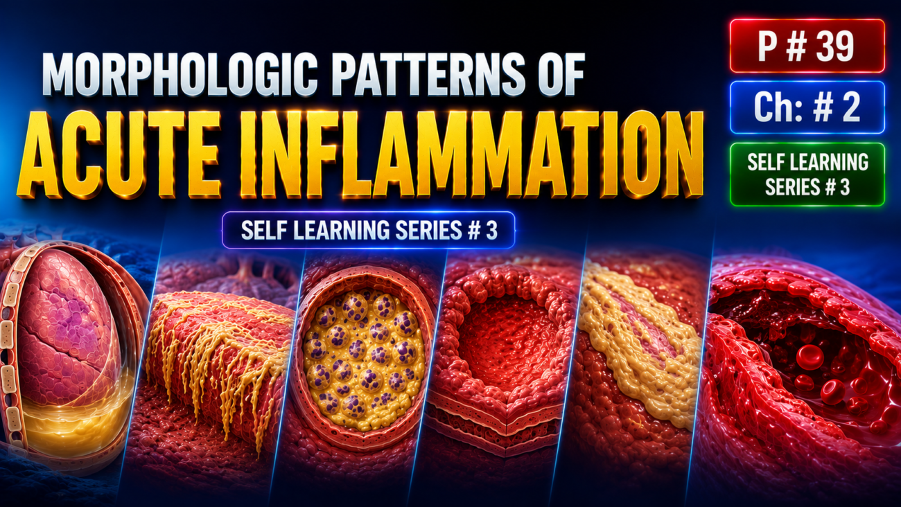
- The main morphologic features of acute inflammation are:
- Dilation of small blood vessels
- Accumulation of leukocytes and fluid in extravascular tissues
- These general features occur in most acute inflammatory reactions, but the exact appearance varies according to:
- Severity of inflammation
- Cause (etiology)
- Type and site of tissue involved
- Therefore, the gross and microscopic pattern of inflammation can provide important clues about its underlying cause.
Serous Inflammation
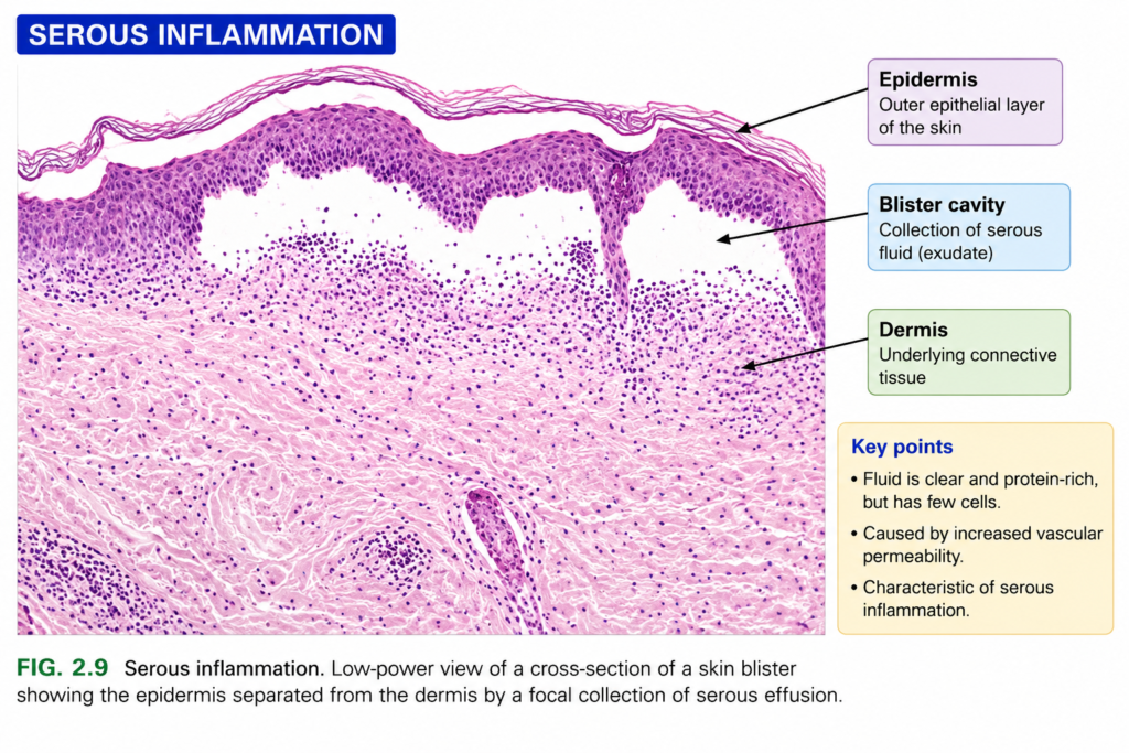
- Serous inflammation is characterized by accumulation of serum-like, protein-rich fluid (exudate) in body cavities or spaces created by tissue injury.
- It commonly occurs in cavities lined by the peritoneum, pleura, or pericardium.
- The fluid is usually sterile and contains few leukocytes.
- Large numbers of leukocytes are more typical of purulent inflammation.
- In body cavities, serous fluid may come from:
- Plasma → due to increased vascular permeability.
- Mesothelial cells → due to local irritation.
- Accumulation of fluid in a mesothelium-lined body cavity is called an effusion.
- In skin burns or some viral infections → serous fluid accumulates within or just beneath the damaged epidermis → blister formation (Fig. 2.9).
KEY CONCEPT
- Acute inflammation → dilated small vessels + leakage of fluid + leukocyte accumulation.
- Serous inflammation → mainly protein-rich fluid with few leukocytes.
- Serous fluid in a body cavity → effusion.
Conceptual Examples
- Burn: Skin injury → serous fluid accumulates beneath epidermis → blister.
- Body cavity irritation: Increased vascular permeability or mesothelial secretion → fluid collects in the cavity → effusion.
Fibrinous Inflammation
- Fibrinous inflammation is characterized by deposition of fibrin due to local activation of coagulation.
- Severe inflammation → marked increase in vascular permeability → large proteins such as fibrinogen escape from blood vessels into the exudate.
- If a procoagulant stimulus is also present → fibrinogen is converted into fibrin.
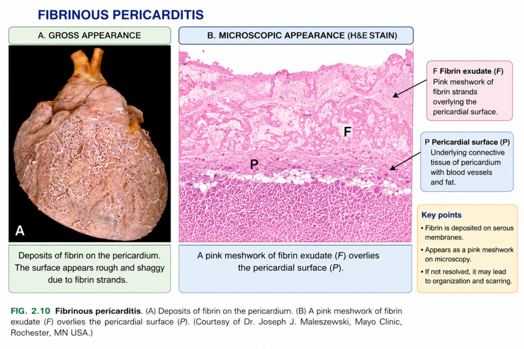
- Fibrinous exudates commonly occur on the lining of body cavities, especially the meninges, pericardium (Fig. 2.10A), and pleura.
- Histologically, fibrin appears as an eosinophilic meshwork of threads or sometimes as an amorphous coagulum (Fig. 2.10B).
- Fibrinous inflammation can have two possible outcomes:
- Resolution: Fibrin is broken down by fibrinolysis → macrophages clear the remaining material → inflammation resolves.
- Organization: If fibrin is not removed → fibroblasts and blood vessels grow into the fibrinous exudate → formation of scar tissue.
- Organization can produce harmful consequences because excessive scar formation may interfere with normal tissue function.
- For example, extensive organization of fibrin in the pericardial cavity → scar tissue forms → may obliterate the pericardial space → can result in restrictive cardiomyopathy.
KEY CONCEPT
- ↑ Vascular permeability → fibrinogen escapes → coagulation activation → fibrin deposition → fibrinous inflammation.
- Fibrinolysis + macrophage clearance → resolution.
- Persistent fibrin → organization → fibroblasts + blood vessels → scar formation.
Conceptual Examples
- Pericardial inflammation: Increased vascular permeability → fibrinogen enters pericardial cavity → fibrin forms → fibrinous pericarditis.
- Successful resolution: Fibrin → fibrinolysis → macrophage clearance → normal tissue restored.
- Failure of resolution: Persistent fibrin → organization → scar tissue → pericardial space may become obliterated → restrictive cardiomyopathy.
Purulent (Suppurative) Inflammation, Abscess
- Purulent inflammation is characterized by formation of pus.
- Pus is an exudate containing:
- Neutrophils
- Liquefied debris of necrotic cells
- Edema fluid
- The most common cause is infection by pyogenic (pus-producing) bacteria, especially bacteria such as staphylococci.
- These bacteria cause liquefactive tissue necrosis → tissue becomes liquefied → pus forms.
- Therefore, purulent inflammation is also called suppurative inflammation.
- A common example of acute suppurative inflammation is acute appendicitis.
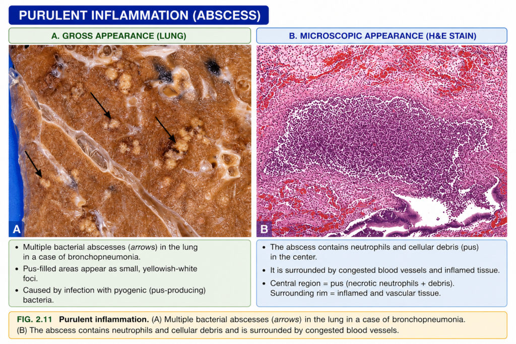
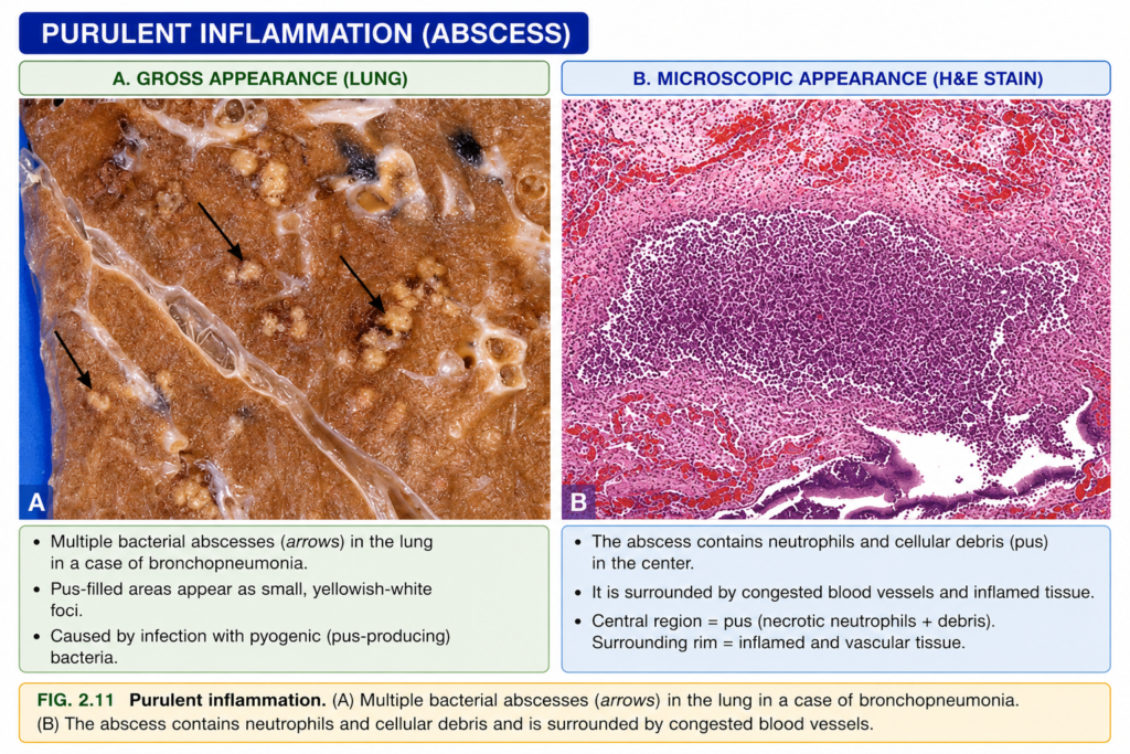
- An abscess is a localized collection of pus within a tissue, organ, or confined space.
- Abscess formation usually occurs when pyogenic bacteria are seeded into a tissue (Fig. 2.11).
- The center of an abscess contains mainly necrotic leukocytes and necrotic tissue cells.
- Around this necrotic center, there is usually a rim of preserved neutrophils.
- Outside this neutrophil-rich area, there may be vascular congestion + proliferation of parenchymal cells and fibroblasts.
- These outer changes indicate the development of chronic inflammation and tissue repair.
- With time → the abscess may become walled off → eventually replaced by connective tissue.
KEY CONCEPT
- Pyogenic bacteria → liquefactive necrosis → neutrophils + necrotic debris + edema fluid → pus.
- Localized collection of pus → abscess.
- Abscess center → necrotic cells → surrounding neutrophils → outer repair zone → connective tissue wall.
Conceptual Examples
- Acute appendicitis: Bacterial infection → neutrophil accumulation + tissue liquefaction → suppurative inflammation with pus.
- Abscess: Pyogenic bacteria enter tissue → localized pus forms → necrotic center surrounded by neutrophils.
- Healing abscess: Persistent abscess → fibroblasts and repair tissue grow around it → abscess becomes walled off by connective tissue.
Ulcers
- An ulcer is a local defect or excavation on the surface of an organ or tissue caused by shedding (sloughing) of inflamed, necrotic tissue (Fig. 2.12).
- Ulceration occurs only when tissue necrosis + inflammation are present on or close to a surface.
- Ulcers commonly occur in the mucosa of the:
- Mouth
- Stomach
- Intestines
- Genitourinary tract
- Ulcers can also occur in the skin and subcutaneous tissue of the lower limbs, especially in people with poor circulation.
- Circulatory disturbances, such as peripheral vascular disease, reduce blood supply → cause ischemic necrosis → promote ulcer formation.
- Both acute and chronic inflammation may be present in an ulcer.
- During the acute stage → large numbers of polymorphonuclear leukocytes (mainly neutrophils) infiltrate the ulcer margins + blood vessels become dilated.
- With time → the base and margins become scarred.
- Chronic inflammatory cells, including lymphocytes, plasma cells, and macrophages, then accumulate around the ulcer.
KEY CONCEPT
- Surface tissue necrosis + inflammation → dead tissue sloughs off → ulcer forms.
- Acute ulcer → neutrophils + vascular dilation.
- Chronic ulcer → lymphocytes + plasma cells + macrophages + scarring.
Conceptual Examples
- Stomach ulcer: Surface mucosal injury → necrosis + inflammation → damaged tissue is shed → ulcer forms.
- Peripheral vascular disease: Poor blood supply to lower limb → ischemic necrosis → tissue breakdown → skin ulcer.
- Long-standing ulcer: Persistent tissue injury → chronic inflammatory cells accumulate + margins and base become scarred.
OUTCOMES OF ACUTE INFLAMMATION
- The outcome of acute inflammation depends on factors such as:
- Nature and severity of injury
- Site and tissue involved
- Host response
- Acute inflammation usually ends in one of three outcomes (Fig. 2.13):
- Complete resolution: After the harmful agent is eliminated → inflammation ends → tissue returns to normal.
- Resolution usually occurs when the injury is mild or short-lived, tissue destruction is minimal, and damaged parenchymal cells can regenerate.
- During resolution → macrophages remove microbes and cellular debris + edema fluid is reabsorbed mainly through lymphatics.
- Healing by connective tissue replacement (scarring/fibrosis): Occurs when there is extensive tissue destruction, when the damaged tissue cannot regenerate, or when abundant fibrin cannot be completely removed.
- This may occur with fibrin accumulation in tissues or serous cavities such as the pleura and peritoneum.
- In these situations → connective tissue grows into the damaged area or exudate → converts it into fibrous scar tissue.
- Progression to chronic inflammation: Occurs when acute inflammation cannot completely resolve.
- This happens because the injurious agent persists or because the normal healing process is disturbed.
- Therefore → persistent acute inflammation gradually becomes chronic inflammation.
KEY CONCEPT
- Mild/short injury + regeneration possible → complete resolution.
- Severe tissue destruction / no regeneration / persistent fibrin → fibrosis and scar formation.
- Persistent injurious agent or failed healing → chronic inflammation.
Conceptual Examples
- Complete resolution: Mild infection eliminated → macrophages clean debris + edema drains → tissue returns to normal.
- Fibrosis: Extensive tissue damage → regeneration is not possible → connective tissue grows in → scar forms.
- Chronic inflammation: Injurious agent remains → acute inflammation cannot resolve → chronic inflammation develops.
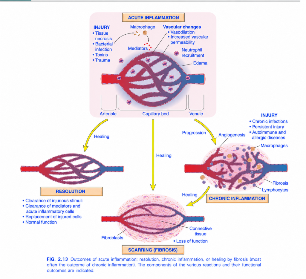
Fig. 2.13 — Outcomes of Acute Inflammation
This figure is showing one very important idea:
After acute inflammation starts, it can end in 3 main ways:
1. Resolution
2. Scarring (fibrosis)
3. Progression to chronic inflammation
I’ll explain it arrow by arrow and label by label.
1. START: INJURY
At the top-left, the figure lists causes of injury:
- Tissue necrosis
- Bacterial infection
- Toxins
- Trauma
Meaning
Any of these can damage tissue.
So:
Injury → inflammatory response starts
2. Macrophage gets activated
Near the top, you see:
Injury → Macrophage
A macrophage is an immune cell that:
- detects tissue damage
- eats microbes and dead cells
- releases inflammatory chemicals
These chemicals are called:
Mediators
Mediators = chemical substances that start and control inflammation.
Examples include:
- histamine
- prostaglandins
- cytokines
- chemokines
So:
Injury → macrophage activation → mediators released
3. Mediators cause vascular changes
The next label says:
Vascular changes
Two important vascular changes happen:
A. Vasodilation
Vasodilation = widening of blood vessels.
Result:
More blood reaches the injured area
This helps explain:
- redness
- warmth
So:
Mediators → vasodilation → increased blood flow Increased vascular permeability
Permeability = how easily fluid can leave the blood vessel.
During inflammation:
vessel wall becomes more leaky
Therefore:
fluid + proteins move from blood into tissue
This causes:
Edema
Edema = swelling caused by excess fluid in tissue.
So:
Increased permeability → fluid leaves vessel → edema
4. Neutrophil recruitment
The figure also shows:
Neutrophil recruitment
Neutrophils are the main early white blood cells in acute inflammation.
They move from blood into injured tissue.
So:
Inflammatory mediators
→ endothelial activation
→ neutrophils leave blood
→ move to injury
→ attack microbes and remove damaged material
Very simple idea
Neutrophils = first emergency soldiers of acute inflammation.
5. Arteriole → Capillary bed → Venule
At the bottom of the acute inflammation picture, you see:
- Arteriole
- Capillary bed
- Venule
Arteriole
Small artery bringing blood into the tissue.
Capillary bed
Tiny vessels where exchange happens.
Venule
Small vein carrying blood away.
In acute inflammation, vascular changes happen especially in the microcirculation.
Neutrophils usually leave the circulation mainly through:
Post-capillary venules
That is a high-yield exam point.
NOW: WHAT CAN HAPPEN AFTER ACUTE INFLAMMATION?
The yellow arrows show 3 possible directions.
OUTCOME 1 — RESOLUTION
Look at the yellow arrow going:
Acute inflammation → Healing → Resolution
What is resolution?
Resolution = tissue returns almost completely to normal.
This is the best outcome.
The figure lists four steps.
A. Clearance of injurious stimuli
The original cause is removed.
Example:
Bacteria killed
or
toxin removed
So:
Cause disappears → inflammation no longer needed
B. Clearance of mediators and acute inflammatory cells
The inflammatory chemicals stop being produced.
Neutrophils disappear, often by apoptosis and removal by macrophages.
So:
Mediators removed + neutrophils removed
C. Replacement of injured cells
Damaged cells are replaced by new cells.
This happens if the tissue has good regenerative ability.
So:
Damaged cells → regeneration → new normal cells. Normal function returns
The tissue returns to normal structure and function.
Complete sequence
Mild/short injury
→ cause removed
→ inflammatory cells cleared
→ tissue regenerates
→ resolution
When is resolution most likely?
Resolution is likely when:
- injury is mild
- injury is short-lived
- tissue destruction is small
- cells can regenerate
OUTCOME 2 — SCARRING / FIBROSIS
Look at the yellow arrow going downward:
Acute inflammation → Healing → Scarring (fibrosis)
Sometimes the tissue cannot completely return to normal.
Then the body repairs the damaged area with:
Connective tissue
This produces a:
Scar
What is fibrosis?
Fibrosis = excessive deposition of collagen and connective tissue.
The figure labels:
Fibroblasts
Fibroblasts = cells that produce collagen and extracellular matrix.
So:
Severe tissue damage
→ fibroblasts activated
→ collagen deposited
→ connective tissue forms
→ scar develops
Why does scarring happen?
Scarring happens especially when:
- tissue damage is extensive
- cells cannot regenerate well
- extracellular matrix is badly destroyed
- inflammation is severe or prolonged
Loss of function
The figure labels:
Loss of function
Scar tissue does not usually perform the original specialized function as well as normal tissue.
So:
More fibrosis → less normal tissue function
Example:
Normal heart muscle contracts.
But a fibrous scar cannot contract like heart muscle.
OUTCOME 3 — CHRONIC INFLAMMATION
Look at the yellow arrow:
Acute inflammation → Progression → Chronic inflammation
This happens when the cause of injury is not removed.
What causes chronic inflammation?
On the right side, the figure lists:
- Chronic infections
- Persistent injury
- Autoimmune diseases
- Allergic diseases
Meaning
If the damaging stimulus continues:
inflammation also continues
So:
Persistent injury → chronic inflammation
Main cells in chronic inflammation
The figure labels:
Macrophages
and
Lymphocytes
These are major cells of chronic inflammation.
Acute inflammation
Mostly:
Neutrophils
Chronic inflammation
Mostly:
Macrophages + lymphocytes
This is a very important exam difference.
Angiogenesis
The figure labels:
Angiogenesis
Angiogenesis = formation of new blood vessels.
Why does this happen?
Chronically damaged tissue needs:
- oxygen
- nutrients
- support for repair
So new blood vessels are formed.
Thus:
Chronic inflammation → angiogenesisFibrosis in chronic inflammation
The figure also shows:
Fibrosis
Chronic inflammation repeatedly causes:
damage + repair + damage + repair
This activates fibroblasts.
Then:
collagen deposition increases
So chronic inflammation often ends in:
Fibrosis / scar formation
Arrow from Chronic Inflammation → Healing → Scarring
The lower yellow arrow shows:
Chronic inflammation → healing → scarring
This means chronic inflammation very commonly heals by fibrosis.
So:
Persistent inflammation
→ ongoing tissue destruction
→ repair attempts
→ collagen deposition
→ fibrosis
The Whole Figure in One Flow
First:
Injury
→ macrophage activation
→ mediators released
→ vasodilation
→ increased permeability
→ edema
→ neutrophil recruitment
→ acute inflammation
Then there are 3 choices:
Path 1
Cause removed + tissue regenerates
→ Resolution
→ normal function
Path 2
Severe tissue destruction
→ fibroblasts
→ collagen
→ Scarring/fibrosis
→ possible loss of function
Path 3
Cause persists
→ macrophages + lymphocytes
→ angiogenesis + fibrosis
→ Chronic inflammation
→ often later heals by fibrosis
Very Easy Memory Trick
Think:
Acute inflammation has 3 endings
R — Resolution
Return to normal.
S — Scar
Replace damaged tissue with collagen.
C — Chronic
Cause continues, so inflammation continues.
So remember:
R-S-C
Resolve — Scar — ChronicAcute vs Chronic: Fast Exam Comparison
| Acute inflammation | Chronic inflammation |
|---|---|
| Short duration | Long duration |
| Mainly neutrophils | Mainly macrophages + lymphocytes |
| Edema common | Fibrosis common |
| May completely resolve | Often causes tissue destruction and fibrosis |
Key Exam Points
- Acute inflammation begins after injury
- Macrophages release inflammatory mediators
- Vasodilation → increased blood flow
- Increased permeability → edema
- Neutrophils dominate acute inflammation
- Acute inflammation can end in:
- Resolution
- Fibrosis/scarring
- Chronic inflammation
- Macrophages + lymphocytes dominate chronic inflammation
- Fibroblasts produce collagen
- Angiogenesis = formation of new blood vessels
- Chronic inflammation often ends in fibrosis
One-line recall
Acute inflammation → remove cause = resolution; severe damage = fibrosis; persistent cause = chronic inflammation.
