


Learning Objectives
After studying this topic, students will be able to:
- Describe the layers and important structural elements of the retina.
- Explain the basic structure and function of rods, cones, and the pigment layer.
- Understand the blood supply of the retina and retinal detachment.
Introduction
The retina is organized into different layers or boundaries.
Light entering the eye must pass through several retinal layers before reaching the rods and cones.
The fovea is specially designed for acute and detailed vision.
Rods and cones are the important photoreceptors of the retina.
The retina also contains a pigment layer, receives blood from the central retinal artery and choroid, and can sometimes become detached from the pigment epithelium.
The Retina Is Composed of Ten Layers or Boundaries
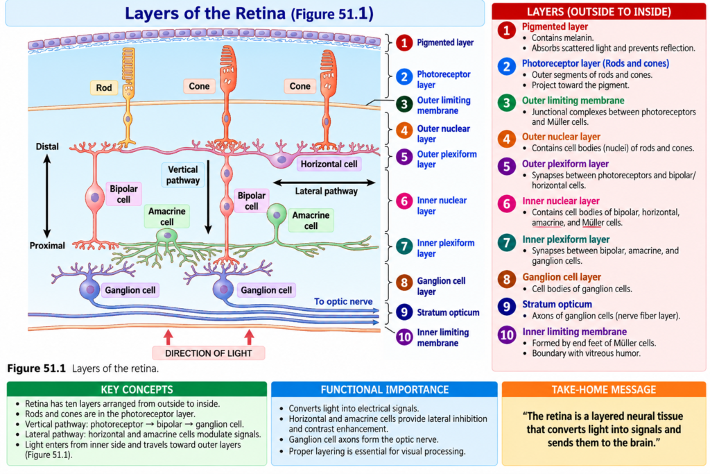
The functional components of the retina are arranged in layers from the outside toward the inside, as shown in Fig. 51.1.
The retinal layers or boundaries are:
- Pigmented layer
- Photoreceptor layer containing rods and cones
- Outer limiting membrane
- Outer nuclear layer containing the cell bodies of rods and cones
- Outer plexiform layer
- Inner nuclear layer
- Inner plexiform layer
- Ganglionic layer
- Layer of optic nerve fibers, also called stratum opticum
- Inner limiting membrane
Simple Concept
The retina is not a single sheet of cells.
It is made of several organized layers.
Each layer forms part of the retinal structure involved in vision.
Path of Light Through the Retina
After light passes through the lens system of the eye and through the vitreous humor, it enters the retina from the inside of the eye.
Light first passes through the ganglion cells.
It then passes through the:
Plexiform layers
↓
Nuclear layers
↓
Finally reaches the rods and cones
The rods and cones are located at the outer edge of the retina.
Therefore, light has to travel through several retinal layers before reaching the photoreceptors.
The distance through these layers is several hundred micrometers.
Passing through this nonhomogeneous tissue decreases some visual acuity.
However, this problem is reduced in the central foveal region because the inner retinal layers are pulled aside.
This helps decrease the loss of visual acuity.
Key Concept
Light enters the retina from the inner side but the rods and cones are located near the outer side.
Simple Flow
Light
↓
Ganglion cells
↓
Plexiform and nuclear layers
↓
Rods and cones
Example
In most of the retina, light must pass through several layers before reaching the photoreceptors.
In the central foveal region, these inner layers are moved aside, helping improve detailed vision.
Foveal Region of the Retina and Its Importance in Acute Vision
The fovea is a very small area located in the center of the retina.
Its total area is slightly more than:
1 square millimeter
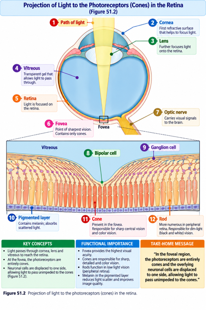
The fovea is especially capable of acute and detailed vision, as shown in Fig. 51.2.
The central part of the fovea is only:
0.3 millimeter in diameter
The central fovea is composed almost completely of cones.
These cones have a special structure that helps them detect fine details in a visual image.
The cones in the fovea have especially:
- Long bodies
- Slender bodies
They are different from the thicker cones found more peripherally in the retina.
Why Light Reaches Foveal Cones More Easily
In the foveal region, several structures are displaced to the side.
These include:
- Blood vessels
- Ganglion cells
- Inner nuclear layer
- Plexiform layers
These structures do not lie directly over the cones.
Therefore, light can pass more directly and without obstruction to the foveal cones.
Key Concept
The fovea provides detailed vision because:
Central fovea contains almost entirely cones
and
Other retinal layers are displaced away from directly over these cones
↓
Light reaches the cones with less obstruction
Example
In the central fovea, the ganglion cells, blood vessels, and other inner retinal layers are moved to one side.
Therefore, light can pass more directly to the cones.
Rods and Cones Are Essential Components of Photoreceptors
A photoreceptor may be either:
- A rod
- A cone
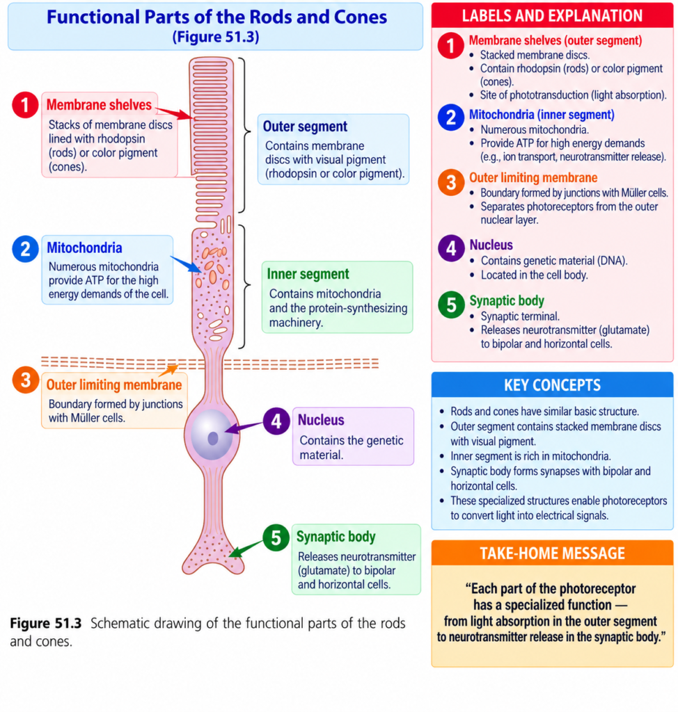
The essential components of a photoreceptor are shown in Fig. 51.3.
The outer segment of a cone is conical in shape, as shown in Fig. 51.4.
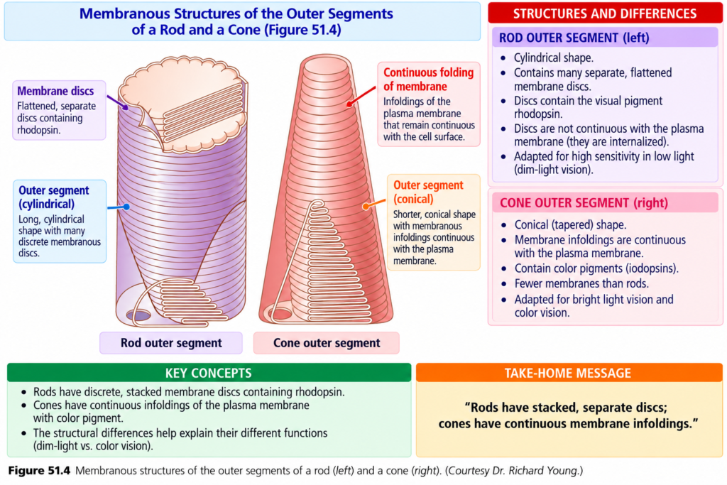
In general, rods are:
- Narrower than cones
- Longer than cones
However, this is not always the case.ze of Rods and Cones
In the peripheral parts of the retina:
Rods = 2–5 micrometers in diameter
Cones = 5–8 micrometers in diameter
In the central retina at the fovea:
- There are no rods.
- The cones are very slender.
- Foveal cones are approximately 1.5 micrometers in diameter.
Key Concept
Peripheral retina: rods and cones are present.
Central fovea: no rods are present, and the cones are especially slender.ain Functional Parts of a Rod or Cone
Each rod or cone has four major functional parts:
- Outer segment
- Inner segment
- Nucleus
- Synaptic body
Outer Segment
The light-sensitive photochemical is located in the outer segment.
In rods, this photochemical is:
Rhodopsin
In cones, the photochemicals are one of three types of:
Color pigments
The color pigments work almost like rhodopsin.
However, they differ in their spectral sensitivity.
Key Concept
Rod → rhodopsin
Cone → color pigments
Both are photosensitive chemicals located in the outer segment.
Discs in Rods and Cones
The outer segments of rods and cones contain many discs.
Each disc is an infolded shelf of the cell membrane.
Each rod or cone may contain as many as:
1000 discs
Rhodopsin and the color pigments are conjugated proteins.
They are incorporated into the membranes of these discs as transmembrane proteins.
The concentration of these photosensitive pigments is very high.
They make up approximately:
40% of the total mass of the outer segment
Key Concept
Outer segment → many discs → photosensitive pigments are present in disc membranes
Inner Segment
The inner segment contains the usual:
- Cytoplasm
- Cytoplasmic organelles
The mitochondria are especially important.
They provide energy required for the function of the photoreceptors.
Simple Concept
Outer segment = contains light-sensitive chemicals
Inner segment = contains organelles, especially mitochondria that provide energy
Synaptic Body
The synaptic body is the part of the rod or cone that connects with the next neuronal cells.
These cells include:
- Horizontal cells
- Bipolar cells
These cells form the next stages in the vision chain.
Key Concept
Rod or cone
↓
Synaptic body
↓
Horizontal and bipolar cells
↓
Next stages of the visual pathway
Pigment Layer of the Retina
The pigment layer contains the black pigment:
Melanin
Melanin prevents light from being reflected throughout the inside of the eyeball.
This is extremely important for clear vision.
The function of this black pigment is similar to the black coloring inside the bellows of a camera.
Without this pigment, light rays would reflect in many directions inside the eyeball.
This would produce diffuse lighting of the retina.
Normal sharp images require a proper contrast between:
Dark spots and light spots
Therefore, preventing unwanted light reflection helps form more precise visual images.
Importance of Melanin
The importance of melanin becomes clear in people with albinism.
Albinism is described here as a congenital absence of melanin pigment in the body.
When a person with albinism enters a bright room, light reaching the retina can be reflected in many directions inside the eyeball.
This happens because the surfaces of the retina and underlying sclera lack normal pigmentation.
Normally, one small spot of light would stimulate only a few rods or cones.
In albinism, the reflected light spreads to many areas.
As a result, many photoreceptors may become stimulated instead of only a small group.
Therefore, visual acuity in people with albinism may remain only about:
20/100 to 20/200
even with the best optical correction.
This is compared with the normal value of:
20/20
Key Concept
Normal melanin
↓
Prevents internal light reflection
↓
Maintains contrast between dark and light
↓
Helps produce a clear image
Example
In albinism:
Lack of melanin
↓
Light reflects in many directions
↓
Many receptors are stimulated
↓
Visual acuity is reduced
Vitamin A in the Pigment Layer
The pigment layer also stores large quantities of:
Vitamin A
Vitamin A moves back and forth through the cell membranes of the outer segments of rods and cones.
These outer segments are embedded in the pigment layer.
Vitamin A is an important precursor of the photosensitive chemicals of rods and cones.
Key Concept
The pigment layer has two important roles described here:
- Contains melanin to reduce light reflection.
- Stores vitamin A associated with the photosensitive chemicals of rods and cones.
Blood Supply of the Retina—The Central Retinal Artery and the Choroid
The internal layers of the retina receive their nutrient blood supply from the:
Central retinal artery
The central retinal artery enters the eyeball through the center of the optic nerve.
It then divides and supplies the entire inner retinal surface.
Therefore, the inner layers of the retina have their own blood supply.
This blood supply is independent of the other structures of the eye.
Blood Supply of the Outer Retina
The outermost layer of the retina is attached to the:
Choroid
The choroid is highly vascular.
It lies between:
Retina
and
Sclera
The outer layers of the retina depend mainly on diffusion from the choroidal blood vessels for their nutrition.
This is especially important for the:
Outer segments of rods and cones
These outer segments depend particularly on the choroid for oxygen.
Key Concept
The retina has two important nutritional sources described here:
Inner retinal layers → central retinal artery
Outer retinal layers, especially rod and cone outer segments → diffusion from choroidal blood vessels
Retinal Detachment
The neural retina can sometimes detach from the pigment epithelium.
One cause is injury to the eyeball.
Injury may allow:
- Fluid
- Blood
to collect between the neural retina and the pigment epithelium.
This can separate the two layers.
Another Cause of Retinal Detachment
Retinal detachment can also occur because of contraction of fine collagenous fibrils in the vitreous humor.
These fibrils may contract and pull parts of the retina toward the inside of the eyeball.
This pulling can cause separation of the neural retina from the pigment epithelium.
Simple Concept
Retinal detachment may occur by:
Injury
↓
Fluid or blood collects beneath neural retina
↓
Neural retina separates from pigment epithelium
or
Collagenous fibrils in vitreous contract
↓
Retina is pulled inward
↓
Retinal detachment
What Happens After Retinal Detachment
A detached retina can survive for several days.
This happens partly because nutrients can still diffuse across the gap created by the detachment.
It also happens partly because the neural retina has an independent blood supply from the retinal artery.
Therefore, if the detached retina is surgically returned to its normal relationship with the pigment epithelium, it may become functional again.
However, if it is not replaced soon, the retina can be destroyed.
Once this destruction occurs, it may no longer function even after surgical repair.
Key Concept
Early replacement of detached retina
↓
Retina may become functional again
But:
Detachment remains too long
↓
Retina is destroyed
↓
Function may not return even after repair
Clinical Note or Importance
Albinism
Melanin normally prevents excessive internal reflection of light.
When melanin is absent, light can be reflected throughout the inside of the eye and stimulate many photoreceptors.
Therefore, visual acuity in people with albinism may be only:
20/100 to 20/200
instead of normal:
20/20
Retinal Detachment
The neural retina can separate from the pigment epithelium.
It may occur because:
- Injury allows fluid or blood to collect between the layers.
- Contracting collagenous fibrils in the vitreous humor pull the retina inward.
The detached retina can remain viable for some time.
If it is surgically replaced early, it can become functional again.
If replacement is delayed too long, the retina may be permanently destroyed.
High-Yield Points
- The retina consists of ten layers or boundaries.
- Light enters the retina from its inner side.
- Light passes through ganglion cells and other retinal layers before reaching the rods and cones.
- Rods and cones lie toward the outer edge of the retina.
- The fovea is specialized for acute and detailed vision.
- The central fovea is approximately 0.3 mm in diameter.
- The central fovea is composed almost entirely of cones.
- There are no rods in the fovea.
- Foveal cones are slender and approximately 1.5 μm in diameter.
- Each rod or cone has an outer segment, inner segment, nucleus, and synaptic body.
- Rhodopsin is the photosensitive pigment of rods.
- Cones contain color pigments.
- Each rod or cone can contain as many as 1000 discs.
- Photosensitive pigments make up about 40% of the outer segment mass.
- Mitochondria in the inner segment provide energy for photoreceptor function.
- The synaptic body connects rods and cones with horizontal and bipolar cells.
- Melanin in the pigment layer prevents unwanted reflection of light.
- The pigment layer also stores vitamin A.
- Inner retinal layers are supplied by the central retinal artery.
- Outer retinal layers depend mainly on the choroid for nutrition.
- A detached retina may recover if it is surgically replaced early.
Common Student Mistakes
- Confusing the direction in which light enters the retina.
Remember:
Light enters from the inner side → rods and cones are near the outer side.
- Thinking that the fovea contains many rods.
Remember:
Central fovea contains almost entirely cones and has no rods.
- Confusing the photochemical of rods with that of cones.
Remember:
Rods → rhodopsin
Cones → color pigments
- Confusing the functions of the outer and inner segments.
Remember:
Outer segment → photosensitive chemicals
Inner segment → mitochondria and energy supply
- Forgetting the main function of melanin.
Remember:
Melanin prevents unwanted reflection of light inside the eyeball.
- Confusing the blood supply of the inner and outer retina.
Remember:
Inner retina → central retinal artery
Outer retina → mainly choroid
- Thinking that every retinal detachment is immediately irreversible.
The provided text explains that the detached retina can remain viable for days and may recover if surgically replaced early.
Quick Revision
Retina
The retina contains ten organized layers or boundaries.
Light travels:
Inside of eye
↓
Ganglion cells
↓
Plexiform and nuclear layers
↓
Rods and cones
Fovea
Fovea = area for acute and detailed vision
Central fovea:
- Diameter = 0.3 mm
- Almost entirely cones
- No rods
- Cones are slender
- Other retinal layers are displaced to the side
Rods and Cones
Main parts:
Outer segment
↓
Inner segment
↓
Nucleus
↓
Synaptic body
Outer Segment
Rods → rhodopsin
Cones → color pigments
Contains up to:
1000 discs
Inner Segment
Contains mitochondria that provide energy.
Synaptic Body
Connects to:
Horizontal cells + bipolar cells
Pigment Layer
Contains:
Melanin
↓
Prevents internal light reflection
↓
Helps maintain clear visual contrast
Also stores:
Vitamin A
Blood Supply
Inner retina → central retinal artery
Outer retina → mainly choroidal blood vessels
Retinal Detachment
Injury → fluid/blood between layers → detachment
or
Vitreous collagen fibrils contract → pull retina inward → detachment
Early surgical replacement:
May restore function
Delayed replacement:
Retina may be destroyed permanently
Conceptual Easiest Summary
The retina is made of ten organized layers or boundaries.
Although the rods and cones detect light, they are located near the outer edge of the retina.
Therefore, light entering the eye must first pass through ganglion cells, plexiform layers, and nuclear layers before reaching the rods and cones.
This passage through retinal tissue can reduce some visual acuity.
The fovea is specially adapted for detailed vision.
Its central region is very small and contains almost entirely cones.
The cones are long and slender.
Other retinal structures, including blood vessels, ganglion cells, and inner retinal layers, are moved to the side.
This allows light to reach the cones more directly.
Rods and cones are the photoreceptors of the retina.
Each has an:
Outer segment → inner segment → nucleus → synaptic body
The outer segment contains the photosensitive chemicals.
Rods contain rhodopsin, whereas cones contain color pigments.
The outer segments contain many discs, and the photosensitive pigments are incorporated into the disc membranes.
The inner segment contains mitochondria that provide energy for photoreceptor function.
The synaptic body connects the rod or cone with horizontal and bipolar cells.
The pigment layer contains melanin.
Melanin prevents excessive reflection of light within the eyeball and helps maintain the contrast needed for clear vision.
In people with albinism, the lack of melanin allows light to reflect in many directions and stimulate many receptors, reducing visual acuity.
The pigment layer also stores vitamin A, which is an important precursor of the photosensitive chemicals of rods and cones.
The inner layers of the retina receive blood from the central retinal artery.
The outer layers, especially the outer segments of rods and cones, depend mainly on the choroidal blood vessels for nutrition and oxygen.
The neural retina can sometimes separate from the pigment epithelium.
This is called retinal detachment.
It can occur when fluid or blood collects between the layers after injury or when collagenous fibrils in the vitreous pull the retina inward.
A detached retina can remain viable for several days because of diffusion across the gap and its independent retinal blood supply.
If it is surgically replaced early, it may become functional again.
If replacement is delayed too long, the retina may be destroyed and may remain nonfunctional even after surgical repair.
Final Concept
Light enters from inner retina
↓
Passes through retinal layers
↓
Reaches rods and cones
↓
Photoreceptors contain light-sensitive pigments
↓
Signals pass to horizontal and bipolar cells
At the same time:
Melanin reduces unwanted light reflection
and
Central retinal artery + choroid provide retinal nutrition
While:
Retinal detachment separates neural retina from pigment epithelium and requires early replacement to preserve function.
Reference Guyton textbook Physiology 15th Edition. page 649| Chapter 51
