


Learning Objectives
After studying this topic, students will be able to:
- Explain the rhodopsin-retinal visual cycle.
- Describe how light excites rods and causes hyperpolarization.
- Explain the role of vitamin A in rhodopsin formation.
- Understand the excitation cascade and high sensitivity of rods.
Introduction
Both rods and cones contain chemicals that break down when exposed to light.
During this process, these chemicals excite the nerve fibers that carry visual information away from the eye.
The light-sensitive chemical in rods is:
Rhodopsin
The light-sensitive chemicals in cones are called:
Cone pigments or color pigments
The composition of cone pigments is only slightly different from that of rhodopsin.
The main discussion here is about the photochemistry of rhodopsin, but the same basic principles also apply to the cone pigments.
RHODOPSIN-RETINAL VISUAL CYCLE AND EXCITATION OF THE RODS
Rhodopsin and Its Decomposition by Light Energy
The outer segment of a rod extends into the pigment layer of the retina.
About 40% of the outer segment consists of the light-sensitive pigment:
Rhodopsin
Rhodopsin is also called:
Visual purple
Rhodopsin is formed from two components:
Scotopsin + retinal
Scotopsin is a protein.
Retinal is a carotenoid pigment and is also called retinene.
The retinal present in rhodopsin is a special form called:
11-cis retinal
The 11-cis form is important because only this form can bind with scotopsin to form rhodopsin.
Simple Concept
Scotopsin + 11-cis retinal → Rhodopsin
What Happens When Light Strikes Rhodopsin?
When rhodopsin absorbs light energy, it begins to break down within a very small fraction of a second, as shown in Fig. 51.5.
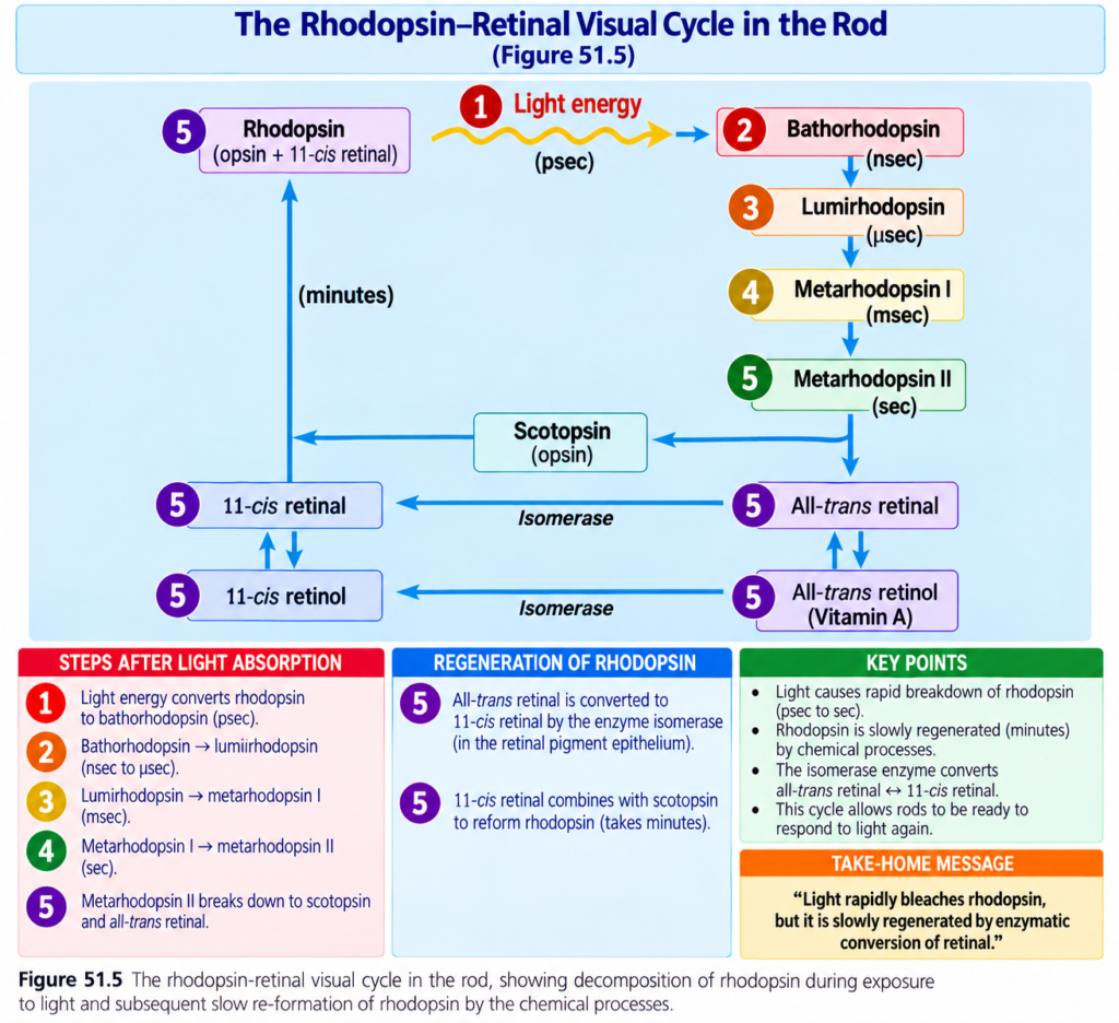
Light causes photoactivation of electrons in the retinal part of rhodopsin.
This immediately changes:
11-cis retinal
into:
All-trans retinal
The all-trans form has the same chemical structure as the cis form but has a different physical shape.
Shape Difference
11-cis retinal = angulated shape
All-trans retinal = straight shape
Because all-trans retinal now has a different three-dimensional shape, its reactive sites no longer fit properly with the reactive sites of scotopsin.
Therefore, all-trans retinal begins to separate from scotopsin.
Stages of Rhodopsin Decomposition
The first immediate product formed is:
Bathorhodopsin
Bathorhodopsin is a partially separated combination of:
All-trans retinal + scotopsin
Bathorhodopsin is extremely unstable.
It changes within nanoseconds into:
Lumirhodopsin
Lumirhodopsin then changes within microseconds into:
Metarhodopsin I
Metarhodopsin I changes in about 1 millisecond into:
Metarhodopsin II
Finally, much more slowly, over seconds, rhodopsin completely separates into:
Scotopsin + all-trans retinal
Easy Flow
Rhodopsin
↓
Light absorbed
↓
11-cis retinal changes into all-trans retinal
↓
Bathorhodopsin
↓
Lumirhodopsin
↓
Metarhodopsin I
↓
Metarhodopsin II
↓
Scotopsin + all-trans retinal
Metarhodopsin II Is the Active Form
Metarhodopsin II is also called:
Activated rhodopsin
This is the form that causes electrical changes in the rods.
The rods then transmit the visual image toward the central nervous system in the form of optic nerve action potentials.
Key Concept
The most important active product in the rhodopsin sequence is:
Metarhodopsin II = activated rhodopsin
It is this activated form that starts the electrical changes in the rod.
Re-Formation of Rhodopsin
After rhodopsin has been broken down, it must be formed again.
The first step is conversion of:
All-trans retinal
back into:
11-cis retinal
This conversion requires:
- Metabolic energy
- The enzyme retinal isomerase
Once 11-cis retinal has been formed, it automatically combines again with:
Scotopsin
This produces new:
Rhodopsin
The newly formed rhodopsin remains stable until it absorbs light again.
Easy Cycle
Light
↓
Rhodopsin breaks down
↓
All-trans retinal + scotopsin
↓
All-trans retinal is converted back to 11-cis retinal
↓
11-cis retinal + scotopsin
↓
Rhodopsin is re-formed
Key Concept
Rhodopsin can be used again because:
All-trans retinal → 11-cis retinal → combines with scotopsin → new rhodopsin
Role of Vitamin A for Formation of Rhodopsin
There is another pathway by which all-trans retinal can eventually become 11-cis retinal.
First:
All-trans retinal
is converted into:
All-trans retinol
All-trans retinol is one form of:
Vitamin A
Then:
All-trans retinol
is converted into:
11-cis retinol
This conversion occurs under the influence of the enzyme:
Isomerase
Finally:
11-cis retinol
is converted into:
11-cis retinal
The 11-cis retinal then combines with:
Scotopsin
to form:
New rhodopsin
Easy Vitamin A Pathway
All-trans retinal
↓
All-trans retinol = vitamin A
↓
11-cis retinol
↓
11-cis retinal
↓
Combines with scotopsin
↓
Rhodopsin
Where Vitamin A Is Present
Vitamin A is present in:
- The cytoplasm of the rods
- The pigment layer of the retina
Therefore, vitamin A is normally available when new retinal needs to be formed.
When there is excess retinal in the retina, some of it is converted back into:
Vitamin A
This reduces the amount of light-sensitive pigment present in the retina.
The conversion between retinal and vitamin A is especially important in the long-term adjustment of the retina to different levels of light intensity.
Key Concept
Vitamin A and retinal can be converted back and forth.
Vitamin A → retinal → rhodopsin formation
and when retinal is excessive:
Retinal → vitamin A
Night Blindness Due to Vitamin A Deficiency
Severe vitamin A deficiency can cause:
Night blindness
Without enough vitamin A, the eye cannot form normal amounts of:
- Retinal
- Rhodopsin
Therefore, the amount of rhodopsin becomes severely reduced.
The condition is called night blindness because the amount of light available at night is too small to provide adequate vision in a person with vitamin A deficiency.
Why Night Blindness Usually Takes Months to Develop
Night blindness usually does not occur immediately after vitamin A intake decreases.
A person usually has to remain on a vitamin A-deficient diet for months.
This is because large amounts of vitamin A are normally stored in the:
Liver
The stored vitamin A can be supplied to the eyes.
Once night blindness has developed, it can sometimes be reversed in less than 1 hour by intravenous injection of vitamin A.
Clinical Note or Importance
Severe vitamin A deficiency
↓
Reduced vitamin A availability
↓
Reduced retinal formation
↓
Reduced rhodopsin formation
↓
Night blindness
The condition usually develops only after months of vitamin A deficiency because vitamin A is normally stored in large quantities in the liver.
EXCITATION OF RODS WHEN RHODOPSIN IS ACTIVATED BY LIGHT
Rod Receptors Hyperpolarize in Response to Light
When a rod is exposed to light, the inside of its membrane becomes more negative.
This increase in negativity is called:
Hyperpolarization
This response is opposite to what occurs in most other sensory receptors.
Most other sensory receptors become less negative during activation.
This decreased negativity is called:
Depolarization
Key Concept
Most sensory receptors:
Activation → depolarization
Rod receptor:
Light activation → hyperpolarization
Why Does Light Cause Hyperpolarization?
When rhodopsin breaks down after exposure to light, the membrane of the rod outer segment becomes less conductive to:
Sodium ions
This reduction in sodium conductance causes the rod to become:
Hyperpolarized
Sodium and Potassium Movement in the Rod
Movement of sodium and potassium through the inner and outer segments of the rod forms a complete electrical circuit, as shown in Fig. 51.6.
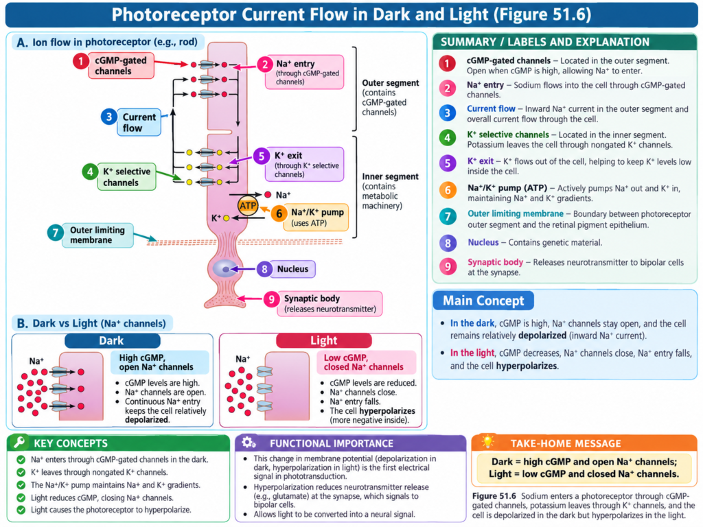
The inner segment continuously pumps:
Sodium outward from the rod
and
Potassium inward into the rod
Potassium ions then leak back out through nongated potassium channels.
These potassium channels are located in the inner segment.
The sodium-potassium pump creates a negative electrical potential inside the cell.
What Happens in Darkness?
The outer segment behaves differently from the inner segment.
The outer segment contains the photoreceptor discs.
In darkness, its membrane is leaky to:
Sodium ions
Sodium enters through:
cGMP-gated channels
In darkness, the level of:
cGMP
is high.
The high cGMP level keeps the sodium channels open.
Therefore, positively charged sodium ions continually move back into the rod.
These positive sodium ions neutralize much of the negative charge inside the cell.
As a result, in normal darkness, when the rod is not excited, its membrane potential is approximately:
−40 mV
This is less negative than the usual:
−70 to −80 mV
found in most other sensory receptors.
Easy Dark-State Concept
Darkness
↓
High cGMP
↓
cGMP-gated Na⁺ channels open
↓
Na⁺ enters the rod
↓
Inside becomes less negative
↓
Membrane potential ≈ −40 mV
What Happens When Light Reaches the Rod?
When rhodopsin in the rod outer segment absorbs light, it becomes activated and starts to break down.
The cGMP-gated sodium channels then close.
The reduction in sodium movement into the rod occurs through the process shown in Fig. 51.7.
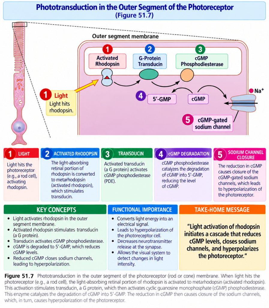
Easy Sequence
Light is absorbed by rhodopsin
↓
Electrons in retinal are photoactivated
↓
Rhodopsin becomes activated
↓
Activated rhodopsin stimulates the G protein:
Transducin
↓
Transducin activates:
cGMP phosphodiesterase
↓
Phosphodiesterase breaks:
cGMP → 5′-GMP
↓
cGMP concentration falls
↓
cGMP-gated sodium channels close
↓
Less Na⁺ enters the rod
↓
Na⁺ continues to be pumped outward from the inner segment
↓
More Na⁺ leaves than enters
↓
Inside becomes more negative
↓
Hyperpolarization
Effect of Increasing Light Intensity
Sodium ions carry positive charge.
When less sodium enters the rod while sodium continues to be pumped outward, positive charge is lost from inside the rod.
Therefore, the inside of the rod becomes increasingly negative.
The more light energy that strikes the rod:
The greater the membrane negativity
Therefore:
More light → greater hyperpolarization
At maximum light intensity, the rod membrane potential approaches:
−70 to −80 mV
This is near the equilibrium potential for potassium ions across the membrane.
Key Concept
Dark → Na⁺ channels open → about −40 mV
Light → Na⁺ channels close → increased negativity → hyperpolarization
At very high light intensity:
Membrane potential approaches −70 to −80 mV
Duration of the Receptor Potential, and Logarithmic Relation of the Receptor Potential to Light Intensity
When a sudden pulse of light reaches the retina, rods develop a temporary hyperpolarization.
This electrical change is called the:
Receptor potential
The rod receptor potential reaches its peak in about:
0.3 second
It lasts for:
More than 1 second
In cones, this change occurs approximately:
4 times faster than in rods
Persistence of a Visual Image
A visual image may strike the rods for only:
One-millionth of a second
Yet it can sometimes produce the sensation of seeing that image for:
Longer than 1 second
Key Concept
A very brief light stimulus can produce a rod response that lasts much longer than the actual light exposure.
Relation Between Receptor Potential and Light Intensity
The receptor potential is approximately proportional to the:
Logarithm of light intensity
This relationship is important because it allows the eye to distinguish between light intensities over a range that is many thousands of times greater than would otherwise be possible.
Simple Concept
As light intensity changes greatly, the receptor potential does not need to change by the same enormous amount.
This logarithmic relationship allows the eye to discriminate a very wide range of light intensities.
Mechanism Whereby Rhodopsin Decomposition Decreases Membrane Sodium Conductance—The Excitation “Cascade”
Under optimal conditions, a single:
Photon of light
can produce a receptor potential of approximately:
1 mV
in a rod.
A photon is the smallest possible quantal unit of light energy.
Only about:
30 photons
are required to produce half-saturation of a rod.
This very high sensitivity occurs because rods have a powerful chemical excitation cascade.
This cascade amplifies the effect of light by approximately:
One million times
Excitation Cascade in the Easiest Sequence
Photon Activates Rhodopsin
A photon activates an electron in the:
11-cis retinal
part of rhodopsin.
This leads to formation of:
Metarhodopsin II
Metarhodopsin II is the active form of rhodopsin.
Activated Rhodopsin Activates Transducin
Activated rhodopsin behaves as an enzyme.
It activates many molecules of:
Transducin
Transducin is normally present in an inactive form in the:
- Disc membranes
- Cell membrane of the rod
Transducin Activates Phosphodiesterase
Activated transducin activates many molecules of:
Phosphodiesterase
Phosphodiesterase Destroys cGMP
Activated phosphodiesterase rapidly hydrolyzes many molecules of:
cGMP
Before light stimulation, cGMP is attached to sodium channel proteins and helps keep these sodium channels:
Open
When phosphodiesterase destroys cGMP:
cGMP decreases
↓
Sodium channels close
Several hundred sodium channels can close for each originally activated rhodopsin molecule.
Because sodium had been moving very rapidly through these channels, their closure can block the entry of:
More than 1 million sodium ions
before the channels open again.
This reduction in sodium movement is what excites the rod.
The Cascade Is Switched Off
Within approximately:
1 second
another enzyme called:
Rhodopsin kinase
inactivates the activated rhodopsin, or metarhodopsin II.
Rhodopsin kinase is always present in the rod.
When activated rhodopsin is inactivated, the entire cascade reverses toward its normal state.
The sodium channels then become:
Open again
Complete Excitation Cascade
Photon
↓
Activates 11-cis retinal in rhodopsin
↓
Metarhodopsin II
↓
Activates many molecules of transducin
↓
Transducin activates many molecules of phosphodiesterase
↓
Phosphodiesterase destroys many molecules of cGMP
↓
cGMP decreases
↓
Many Na⁺ channels close
↓
More than 1 million Na⁺ ions may be prevented from entering
↓
Inside of rod becomes more negative
↓
Rod is hyperpolarized
↓
Rhodopsin kinase inactivates metarhodopsin II
↓
Cascade reverses
↓
Na⁺ channels reopen
Why Rods Are Extremely Sensitive to Light
The excitation cascade greatly amplifies the effect of light.
A single photon can ultimately affect the movement of millions of sodium ions.
This explains the extreme sensitivity of rods under:
Dark conditions
Key Concept
A tiny light stimulus becomes a much larger electrical effect because each stage activates many molecules in the next stage.
One photon
↓
Activated rhodopsin
↓
Many transducins
↓
Many phosphodiesterase molecules
↓
Large cGMP reduction
↓
Hundreds of Na⁺ channels close
↓
Movement of millions of Na⁺ ions is affected
Sensitivity of Cones Compared With Rods
Cones are approximately:
30 to 300 times less sensitive than rods
Despite being less sensitive, cones still have enough sensitivity to provide:
Color vision
at light intensities greater than extremely dim twilight.
Key Concept
Rods = much more sensitive to light
Cones = 30–300 times less sensitive
But cones can still provide color vision when the light intensity is above extremely dim twilight.
Key Concepts With Examples
Rhodopsin Formation
Scotopsin + 11-cis retinal → rhodopsin
Only the 11-cis form of retinal can bind properly with scotopsin.
Rhodopsin Breakdown
Light
↓
11-cis retinal → all-trans retinal
↓
Rhodopsin passes through several intermediate forms
↓
Metarhodopsin II
↓
Electrical changes in rod
Rhodopsin Re-Formation
All-trans retinal
↓
11-cis retinal
↓
Combines with scotopsin
↓
Rhodopsin
Vitamin A Example
Vitamin A can participate in the pathway:
All-trans retinal
↓
All-trans retinol
↓
11-cis retinol
↓
11-cis retinal
↓
New rhodopsin
Dark Versus Light Example
In Darkness
High cGMP
↓
Na⁺ channels remain open
↓
Na⁺ enters
↓
Rod membrane potential about −40 mV
In Light
Activated rhodopsin
↓
Transducin
↓
Phosphodiesterase
↓
cGMP decreases
↓
Na⁺ channels close
↓
Less Na⁺ enters
↓
Membrane becomes more negative
↓
Hyperpolarization
Amplification Example
Under optimal conditions:
1 photon → about 1 mV receptor potential
About:
30 photons → half-saturation of the rod
A single activated rhodopsin can eventually cause closure of several hundred sodium channels and prevent the movement of more than 1 million sodium ions.
Clinical Note or Importance
Vitamin A Deficiency and Night Blindness
Severe vitamin A deficiency reduces the formation of:
Retinal and rhodopsin
As a result, a person may develop:
Night blindness
The condition usually requires months of vitamin A deficiency because vitamin A is normally stored in large quantities in the liver.
Once night blindness has developed, it can sometimes be reversed in less than 1 hour by intravenous vitamin A.
High-Yield Points
- Rods contain rhodopsin.
- Cones contain cone pigments or color pigments.
- Rhodopsin consists of scotopsin + 11-cis retinal.
- Only 11-cis retinal can bind with scotopsin to form rhodopsin.
- Light converts 11-cis retinal into all-trans retinal.
- Metarhodopsin II is the activated form of rhodopsin.
- Metarhodopsin II causes the electrical changes in rods.
- Rhodopsin can be re-formed by converting all-trans retinal back into 11-cis retinal.
- Retinal isomerase helps this conversion.
- Vitamin A participates in the formation of retinal and therefore rhodopsin.
- Severe vitamin A deficiency can cause night blindness.
- Rods respond to light by hyperpolarization, not depolarization.
- In darkness, cGMP is high and cGMP-gated Na⁺ channels are open.
- In darkness, the rod membrane potential is approximately −40 mV.
- Light activates rhodopsin.
- Activated rhodopsin activates transducin.
- Transducin activates phosphodiesterase.
- Phosphodiesterase breaks down cGMP to 5′-GMP.
- Reduced cGMP closes sodium channels.
- Closure of sodium channels causes hyperpolarization.
- At maximum light intensity, rod membrane potential approaches −70 to −80 mV.
- Rod receptor potential peaks in about 0.3 second and lasts more than 1 second.
- Cones respond approximately four times faster than rods.
- A single photon can cause approximately a 1-mV receptor potential in a rod.
- About 30 photons can produce half-saturation of a rod.
- The rod excitation cascade can amplify the effect of light about one million times.
- Cones are approximately 30–300 times less sensitive than rods.
Common Student Mistakes
- Confusing the forms of retinal.
Remember:
11-cis retinal = binds with scotopsin
All-trans retinal = produced after light activation
- Thinking metarhodopsin I is the active form.
Remember:
Metarhodopsin II = activated rhodopsin
- Thinking rods depolarize when exposed to light.
Remember:
Light causes rods to hyperpolarize.
- Thinking sodium channels open when light strikes the rod.
Remember:
Darkness → Na⁺ channels open
Light → Na⁺ channels close
- Confusing the relationship between cGMP and sodium channels.
Remember:
High cGMP → sodium channels open
Low cGMP → sodium channels close
- Confusing the sequence of the excitation cascade.
Remember:
Rhodopsin → transducin → phosphodiesterase → ↓cGMP → Na⁺ channels close
- Forgetting why vitamin A deficiency causes night blindness.
Remember:
↓ Vitamin A → ↓ retinal → ↓ rhodopsin → poor vision in low light
- Thinking cones are more sensitive than rods.
Remember:
Cones are 30–300 times less sensitive than rods.
Quick Revision
Rhodopsin
Scotopsin + 11-cis retinal
↓
Rhodopsin
Light Activation
Light
↓
11-cis retinal → all-trans retinal
↓
Bathorhodopsin
↓
Lumirhodopsin
↓
Metarhodopsin I
↓
Metarhodopsin II
↓
Scotopsin + all-trans retinal
Active Form
Metarhodopsin II = activated rhodopsin
Rhodopsin Re-Formation
All-trans retinal
↓
11-cis retinal
↓
- Scotopsin
↓
Rhodopsin
Vitamin A Route
All-trans retinal
↓
All-trans retinol
↓
11-cis retinol
↓
11-cis retinal
↓
- Scotopsin
↓
Rhodopsin
Night Blindness
Vitamin A deficiency
↓
↓ Retinal
↓
↓ Rhodopsin
↓
Night blindness
Rod in Darkness
High cGMP
↓
Na⁺ channels open
↓
Na⁺ enters
↓
Membrane potential ≈ −40 mV
Rod in Light
Light
↓
Activated rhodopsin
↓
Transducin
↓
Phosphodiesterase
↓
cGMP decreases
↓
Na⁺ channels close
↓
Na⁺ entry decreases
↓
Inside becomes more negative
↓
Hyperpolarization
Maximum Light
Membrane potential approaches:
−70 to −80 mV
Rod Response
Peak:
About 0.3 second
Duration:
More than 1 second
Cone response:
About 4 times faster
Rod Sensitivity
1 photon → about 1 mV receptor potential
30 photons → half-saturation
Cascade amplification:
About one millionfold
Cones:
30–300 times less sensitive than rods
Conceptual Easiest Summary
Both rods and cones contain chemicals that react to light.
The main light-sensitive chemical in rods is rhodopsin, whereas cones contain color pigments.
Rhodopsin consists of:
Scotopsin + 11-cis retinal
The 11-cis form is important because it can fit with scotopsin and form rhodopsin.
When light is absorbed by rhodopsin, the 11-cis retinal rapidly changes into:
All-trans retinal
Its shape changes from an angulated form into a straight form.
Because the new shape no longer fits scotopsin correctly, retinal starts to separate from scotopsin.
Rhodopsin then passes rapidly through:
Bathorhodopsin → lumirhodopsin → metarhodopsin I → metarhodopsin II
Metarhodopsin II is the activated form of rhodopsin.
It produces the electrical changes that excite the rod.
Finally, rhodopsin separates completely into:
Scotopsin + all-trans retinal
To make rhodopsin again, all-trans retinal must be converted back into:
11-cis retinal
This requires metabolic energy and retinal isomerase.
The 11-cis retinal then automatically combines with scotopsin to produce new rhodopsin.
Vitamin A also participates in this cycle.
All-trans retinal can be converted into:
All-trans retinol
which is a form of vitamin A.
It can then become:
11-cis retinol → 11-cis retinal
The 11-cis retinal again combines with scotopsin to form rhodopsin.
Vitamin A is stored in the rods, pigment layer of the retina, and in large quantities in the liver.
If vitamin A deficiency becomes severe, less retinal and rhodopsin can be produced.
This can cause night blindness.
The rod behaves differently from most sensory receptors when it is stimulated.
Light causes the rod to become:
Hyperpolarized
rather than depolarized.
In darkness, cGMP levels are high.
High cGMP keeps sodium channels open.
Positive sodium ions continuously enter the rod, making its membrane potential only about:
−40 mV
When light activates rhodopsin, activated rhodopsin stimulates:
Transducin
Transducin activates:
Phosphodiesterase
Phosphodiesterase breaks down:
cGMP
As cGMP decreases, the cGMP-gated sodium channels close.
Sodium continues to be pumped outward, but much less sodium can enter.
Therefore, positive charge is lost from inside the rod.
The inside becomes more negative.
This is:
Hyperpolarization
The stronger the light, the greater the hyperpolarization.
At maximum light intensity, the membrane potential approaches:
−70 to −80 mV
The receptor potential of rods reaches its peak in about:
0.3 second
and can last:
More than 1 second
Cones respond about four times faster.
The rod system is extremely sensitive because it has a powerful chemical amplification cascade.
A single photon can activate rhodopsin.
Activated rhodopsin activates many transducin molecules.
These activate many phosphodiesterase molecules.
Phosphodiesterase destroys many cGMP molecules.
This causes hundreds of sodium channels to close and can prevent the movement of more than a million sodium ions.
Therefore, a very small amount of light can produce a significant response in a rod.
Within about 1 second, rhodopsin kinase inactivates metarhodopsin II.
The cascade then reverses and the sodium channels reopen.
Rods are therefore extremely sensitive in darkness.
Cones are approximately 30–300 times less sensitive than rods, but their sensitivity is still sufficient for color vision at light intensities above extremely dim twilight.
Final Concept
In darkness:
High cGMP
↓
Na⁺ channels open
↓
Na⁺ enters
↓
Rod ≈ −40 mV
When light arrives:
Light
↓
Rhodopsin activated
↓
Metarhodopsin II
↓
Transducin
↓
Phosphodiesterase
↓
cGMP decreases
↓
Na⁺ channels close
↓
Na⁺ entry decreases
↓
Rod becomes more negative
↓
Hyperpolarization
↓
Visual signal is produced
For rhodopsin recovery:
All-trans retinal
↓
11-cis retinal
↓
- Scotopsin
↓
New rhodopsin
And:
Vitamin A helps provide the retinal needed for this cycle.
Reference Guyton Textbook Physiology 15th Edition
