


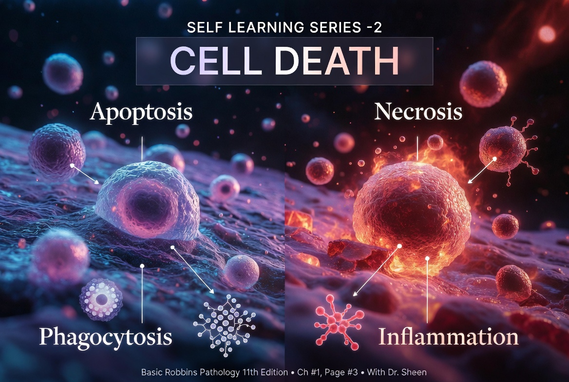
- When cells are injured, they can die by different mechanisms.
- The type of cell death depends on:
- The nature of the injury.
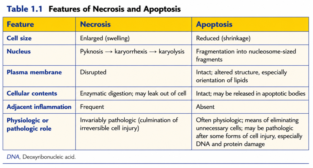
- The severity of the injury (Table 1.1).
- Necrosis
- Necrosis occurs after severe cell injury.
- It is caused by:
- Loss of oxygen
- Loss of nutrient supply
- Toxins
- This type of cell death is rapid and uncontrolled.
- It is also called “accidental” cell death.
- The structural (morphologic) appearance of accidental cell death is called necrosis.
- The word necrosis comes from the Greek word “necros,” meaning death.
- Necrosis is the main pathway of cell death in many common injuries, including:
- Ischemia
- Exposure to toxins
- Infections
- Trauma
- Necrosis is the final result of severe cell damage that cannot be repaired.
- It occurs when the injury is beyond the cell’s ability to survive or recover.
- Necrosis is not considered to be controlled by specific cellular signals or biochemical regulatory mechanisms.
KEY CONCEPT
- Cells die by different mechanisms depending on the type and severity of injury (Table 1.1).
- Necrosis is a rapid, uncontrolled (“accidental”) form of cell death.
- Common causes of necrosis are:
- Loss of oxygen
- Loss of nutrients
- Toxins
- Ischemia
- Infections
- Trauma
- Necrosis occurs when cell injury is too severe to be repaired, leading to irreversible cell death.
- Apoptosis
- Apoptosis removes cells without causing a reaction from the body (Table 1.1).
- It occurs by activating specific molecular pathways inside the cell.
- This type of cell death is called apoptosis.
- Apoptosis depends on specific genes and biochemical pathways.
- It is tightly controlled.
- Once apoptosis begins, it cannot be reversed.
- Therefore, apoptosis is called regulated cell death.
- The discovery of apoptosis showed that:
- Cell death can be an intentional and highly controlled process.
- Apoptosis helps to:
- Remove cells with serious internal abnormalities.
- Remove dead cell fragments without causing inflammation.
- This is a clean form of cell suicide.
- Apoptosis occurs when:
- DNA is damaged beyond repair.
- Proteins are damaged beyond repair.
- The cell does not receive essential survival signals.
- Unlike necrosis:
- Apoptosis does not always indicate disease.
- It also occurs in healthy tissues.
- In healthy tissues, apoptosis:
- Removes unnecessary cells during development.
- Maintains a constant number of cells.
- Physiologic apoptosis is also called programmed cell death.
- Loss of cell function occurs before cell death.
- Structural (morphologic) changes appear after loss of function and viability (Fig. 1.5).
- Example:
- Heart muscle (myocardial) cells stop contracting 1–2 minutes after ischemia begins.
- These cells usually die after 20–30 minutes of ischemia.
- Electron microscopy shows signs of cell death 2–3 hours later.
- Light microscopy shows these changes 6–12 hours later.
KEY CONCEPT
- Apoptosis is a regulated, controlled form of cell death (Table 1.1).
- It occurs through specific genes and biochemical pathways.
- Apoptosis:
- Removes damaged or unnecessary cells.
- Does not cause inflammation.
- Is also called programmed cell death in normal tissues.
- Necrosis is always associated with pathologic injury, whereas apoptosis can occur in both normal and diseased tissues.
- Cell function is lost before the cell dies, and morphologic changes appear later (Fig. 1.5).
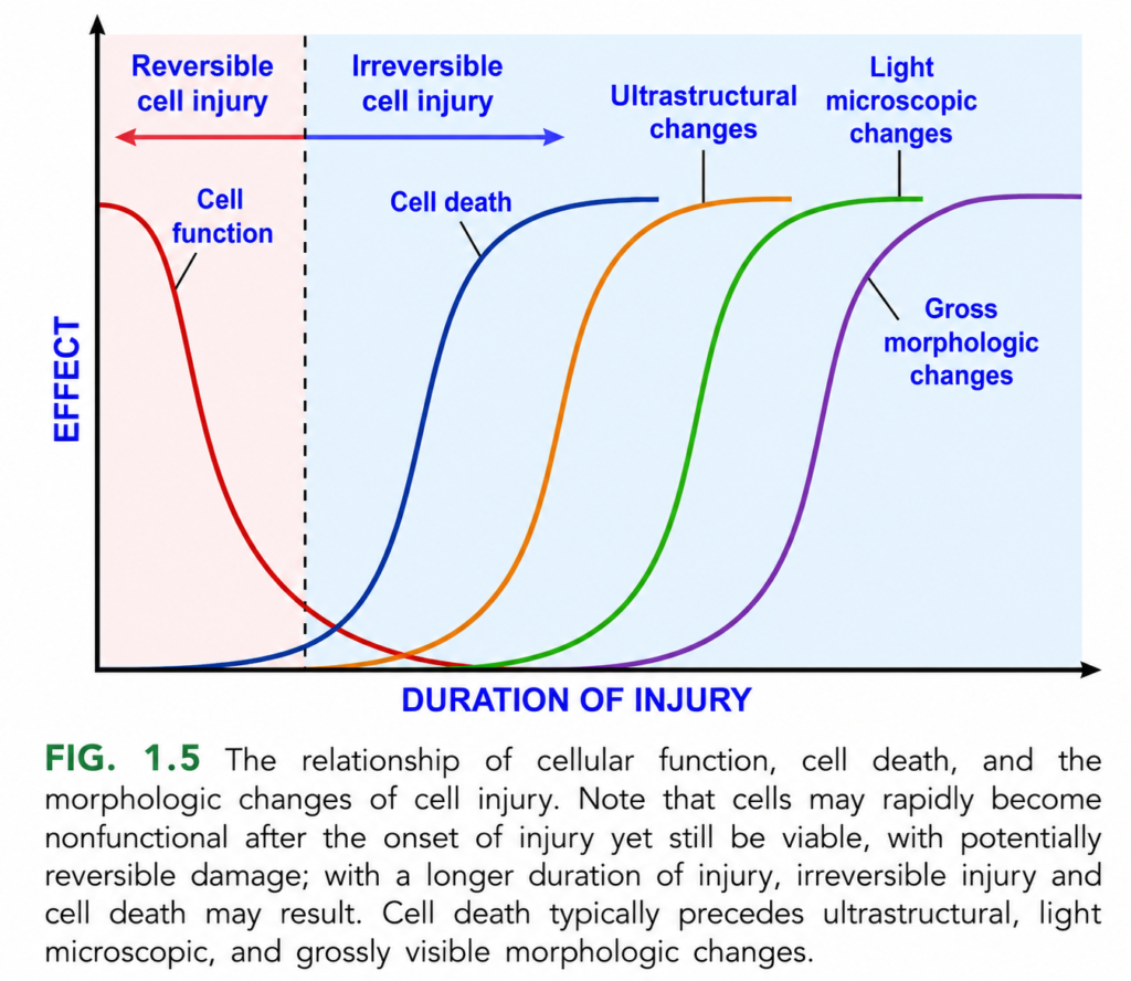
Relationship Between Cell Function, Cell Death, and Morphologic Changes (Figure 1.5)
This figure explains what happens to a cell after injury over time.
The most important concept is:
A cell loses its function first, dies later, and only after death do visible structural changes appear.
This is one of the most important pathology concepts because loss of function occurs before we can see cell damage under the microscope.ain Concept
Cell injury → Loss of function → Irreversible injury → Cell death → Ultrastructural changes → Light microscopic changes → Gross morphologic changes
Understanding the Axes
X-Axis = Duration of Injury
This shows how long the injury continues.
Moving from left to right means:
- More time passes.
- The injury becomes more severe.
Y-Axis = Effect
This shows how much effect the injury has on the cell.
Higher on the graph means:
- Greater cellular damage or visible changes.
Stage 1: Reversible Cell Injury (Left Side)
The pink shaded area represents reversible injury.
At this stage:
- The cell is injured.
- The cell is still alive.
- Recovery is possible if the harmful stimulus is removed.
Examples:
- Mild hypoxia
- Short-term ischemia
- Mild toxin exposure
Red Curve = Cell Function
The red curve represents cell function.
Notice that it falls very early.
Meaning
The first thing a cell loses is its function.
Even though the cell is still alive,
it cannot work normally.
Examples
A heart muscle cell may stop contracting.
A kidney cell may stop transporting ions.
A liver cell may stop producing proteins.
Important Point
The cell is alive but not functioning properly.
Why Does Function Fall First?
Because injury causes:
- ATP depletion
- Failure of ion pumps
- Reduced protein synthesis
- Disturbed metabolism
These changes occur before the cell dies.Can the Cell Recover Here?
Yes.
If oxygen returns or the damaging stimulus is removed,
the cell can return to normal.
That is why this phase is called reversible injury.
Stage 2: Irreversible Cell Injury
The dotted vertical line marks the point of no return.
Beyond this point,
the injury becomes irreversible.
Meaning
Even if the cause of injury is removed now,
the cell cannot recover.
Blue Curve = Cell Death
The blue curve represents cell death.
Notice:
It rises after the red curve has already fallen.
Meaning
The cell first loses function,
then it dies.
Very Important Concept
Loss of function occurs BEFORE cell death.
What Happens at Cell Death?
Once the cell dies:
- Membrane integrity is lost.
- ATP production stops permanently.
- The nucleus breaks down.
- Organelles are destroyed.
- Intracellular enzymes leak out.
At this stage,
necrosis or apoptosis may occur, depending on the type of injury.
Orange Curve = Ultrastructural Changes
These are the earliest visible structural changes after cell death.
They are seen only with an electron microscope.
Examples:
- Mitochondrial swelling
- Membrane damage
- Ribosome detachment
- ER swelling
- Myelin figures
Important Point
These changes are not visible under a light microscope.
Green Curve = Light Microscopic Changes
These changes appear later.
They can be seen using a light microscope.
Examples:
- Cell swelling
- Increased eosinophilia
- Nuclear pyknosis
- Karyorrhexis
- Karyolysis
- Necrosis
Important Point
By the time these changes appear,
the cell is already dead.
Purple Curve = Gross Morphologic Changes
These are the last changes to appear.
They are visible to the naked eye (gross examination).
Examples:
- Pale infarct
- Large necrotic area
- Tissue discoloration
- Organ swelling
- Tissue softening
Important Point
Gross changes require many dead cells, not just one.
Timeline of Events
Cell Injury
      ↓
Loss of Cell Function
      ↓
Reversible Injury
      ↓
Point of No Return
      ↓
Irreversible Injury
      ↓
Cell Death
      ↓
Ultrastructural Changes
(Electron microscope)
      ↓
Light Microscopic Changes
(Light microscope)
      ↓
Gross Morphologic Changes
(Naked eye)
Easy Memory Rule
Function Dies First
The cell stops working before it dies.
Cell Dies Second
The cell becomes irreversibly injured.
Microscope Finds Damage Third
Electron microscope detects injury first.
Light microscope detects injury later.
Gross Changes Come Last
Visible tissue damage appears last.
Simple Clinical Example: Myocardial Infarction (Heart Attack)
Immediately after blocked blood flow
- Heart muscle stops contracting.
- Function is lost.
Cell is still alive.
If blood flow is restored quickly
- The cell recovers.
- No permanent damage.
If ischemia continues
- Cell dies.
After cell death
Electron microscope detects:
- Swollen mitochondria
- Membrane disruption
Later
Light microscope shows:
- Necrosis
- Nuclear changes
- Eosinophilic cytoplasm
Finally
The infarct becomes visible to the naked eye.
Why Is This Figure Clinically Important?
This figure explains why:
- A patient may lose organ function before tissue damage is visible under a microscope.
- Early restoration of blood flow can save cells during the reversible injury stage.
- By the time obvious microscopic or gross changes appear, many cells have already died.
Important Points from Figure 1.5
- Cell function declines first after injury and may be lost while the cell is still alive.
- During reversible cell injury, recovery is possible if the damaging stimulus is removed.
- The dotted vertical line represents the transition to irreversible cell injury, after which recovery is no longer possible.
- Cell death occurs before structural changes become visible.
- Ultrastructural changes are the earliest detectable morphologic changes and require an electron microscope.
- Light microscopic changes appear later and indicate that the cell is already dead.
- Gross morphologic changes are the last to develop and are visible to the naked eye.
- The sequence is always:
Loss of function → Cell death → Ultrastructural changes → Light microscopic changes → Gross morphologic changes.
KEY CONCEPT (Figure 1.5)
This figure demonstrates the time course of cell injury. Following injury, cell function is impaired first, but the cell may still be alive, making the damage reversible. If the injury persists beyond the point of no return, the cell undergoes irreversible injury and dies. Importantly, cell death occurs before any visible structural changes. Morphologic changes appear in a predictable order: ultrastructural changes (seen only by electron microscopy), followed by light microscopic changes, and finally gross morphologic changes visible to the naked eye. Thus, functional impairment precedes cell death, and cell death precedes visible morphologic evidence of injury.
Necrosis
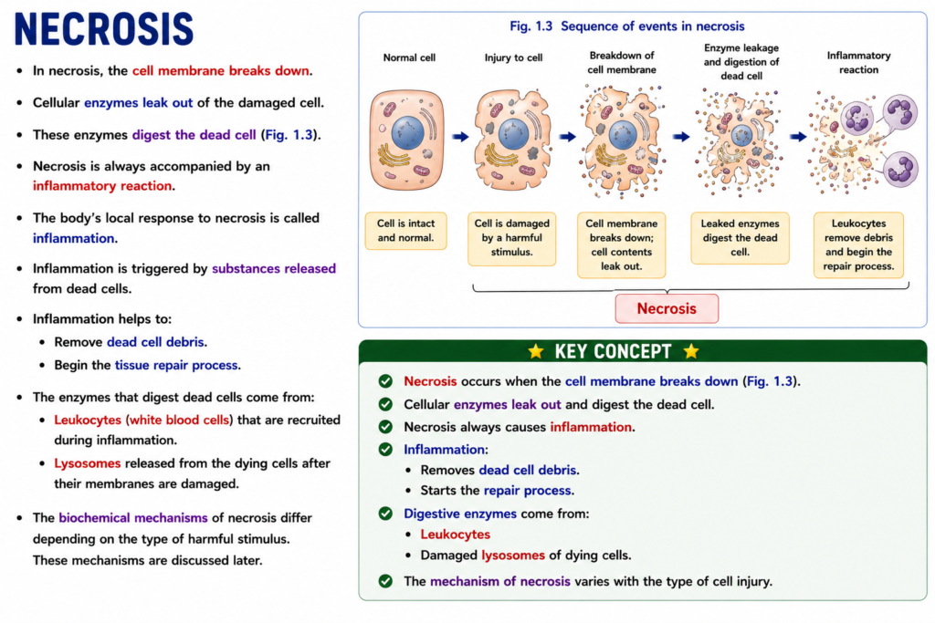
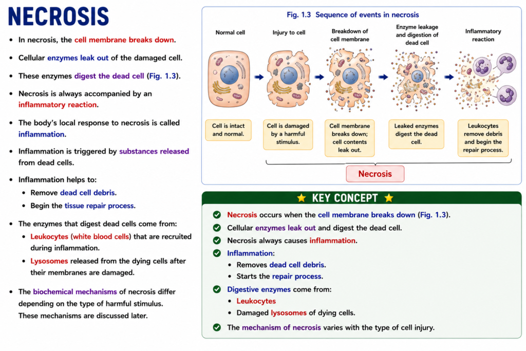
- In necrosis, the cell membrane breaks down.
- Cellular enzymes leak out of the damaged cell.
- These enzymes digest the dead cell (Fig. 1.3).
- Necrosis is always accompanied by an inflammatory reaction.
- The body’s local response to necrosis is called inflammation.
- Inflammation is triggered by substances released from dead cells.
- Inflammation helps to:
- Remove dead cell debris.
- Begin the tissue repair process.
- The enzymes that digest dead cells come from:
- Leukocytes (white blood cells) that are recruited during inflammation.
- Lysosomes released from the dying cells after their membranes are damaged.
- The biochemical mechanisms of necrosis differ depending on the type of harmful stimulus.
- These mechanisms are discussed later.
KEY CONCEPT
- Necrosis occurs when the cell membrane breaks down (Fig. 1.3).
- Cellular enzymes leak out and digest the dead cell.
- Necrosis always causes inflammation.
- Inflammation:
- Removes dead cell debris.
- Starts the repair process.
- Digestive enzymes come from:
- Leukocytes
- Damaged lysosomes of dying cells.
- The mechanism of necrosis varies with the type of cell injury.
MORPHOLOGY
- Cytoplasmic changes
- The cytoplasm becomes more eosinophilic (red-staining).
- This happens because:
- Eosin binds more strongly to denatured cytoplasmic proteins.
- Cytoplasmic RNA is lost, reducing basophilic (blue) staining.
- Compared with normal cells, necrotic cells may appear:
- Glassy
- Necrosis causes structural changes in both the cytoplasm and the nucleus of injured cells (Figs. 1.3 and 1.4C).
- Homogeneous (uniform)
- This appearance is mainly due to the loss of glycogen particles.
- When enzymes digest the cell organelles:
- The cytoplasm develops vacuoles.
- It appears “moth-eaten.”
- Electron microscopy (eFig. 1.1) shows:
- Breaks in the plasma membrane and organelle membranes.
- Marked swelling (dilation) of mitochondria.
- Large amorphous densities inside mitochondria.
- Disruption of lysosomes.
- Intracytoplasmic myelin figures.
- These changes are more prominent in necrotic cells than in cells with reversible injury.
- Nuclear changes
- Nuclear changes occur because DNA and chromatin break down.
- Three characteristic patterns are seen:
- Pyknosis
- The nucleus shrinks.
- DNA becomes condensed.
- The nucleus becomes dark and deeply stained.
- Karyorrhexis
- The pyknotic nucleus breaks into fragments.
- Karyolysis
- The nucleus gradually disappears.
- DNA is digested by DNase.
- Basophilic staining fades.
- Pyknosis
- Within 1–2 days, the nucleus of a dead cell may disappear completely.
- Fates of necrotic cells
- Dead cells may remain in the tissue for some time.
- They may be digested by enzymes and disappear.
- Dead cells may be replaced by myelin figures.
- Myelin figures are:
- Engulfed (phagocytosed) by other cells, or
- Broken down into fatty acids.
- Fatty acids bind with calcium salts.
- This can cause dystrophic calcification in dead cells.
KEY CONCEPT
- Necrosis produces changes in the cytoplasm and nucleus (Figs. 1.3 and 1.4C).
- Cytoplasmic changes include:
- Increased eosinophilic (red) staining.
- Glassy appearance.
- Vacuolation.
- “Moth-eaten” cytoplasm.
- Membrane damage.
- Mitochondrial swelling.
- Lysosomal disruption.
- Myelin figures (eFig. 1.1).
- Nuclear changes occur in the following sequence:
- Pyknosis → Nuclear shrinkage.
- Karyorrhexis → Nuclear fragmentation.
- Karyolysis → Nuclear dissolution.
- Necrotic cells are eventually:
- Digested and removed, or
- Undergo dystrophic calcification after calcium deposition.
Morphologic Patterns of Tissue Necrosis
- Severe injury can cause the death of many cells or even all cells in a tissue or an entire organ.
- This commonly occurs in:
- Severe ischemia
- Infections
- Inflammatory reactions
- Tissue necrosis appears in different morphologic patterns.
- Each pattern can provide clues about the underlying cause of the injury.
- The names of these necrosis patterns describe their appearance only.
- They do not explain the actual mechanism of cell death.
- These terms are widely used.
- Pathologists and clinicians understand their meanings and use them in diagnosis.
- Most types of necrosis have distinctive gross (visible) appearances.
- Fibrinoid necrosis is an exception.
- It can be identified only by microscopic examination.
KEY CONCEPT
- Severe injury may destroy many cells or an entire tissue/organ.
- Common causes include:
- Severe ischemia
- Infections
- Inflammatory reactions
- Tissue necrosis occurs in different morphologic patterns.
- These patterns help identify the possible cause of injury.
- The pattern names describe appearance, not mechanism.
- Most necrosis types are visible grossly, while fibrinoid necrosis is seen only under the microscope.
MORPHOLOGY
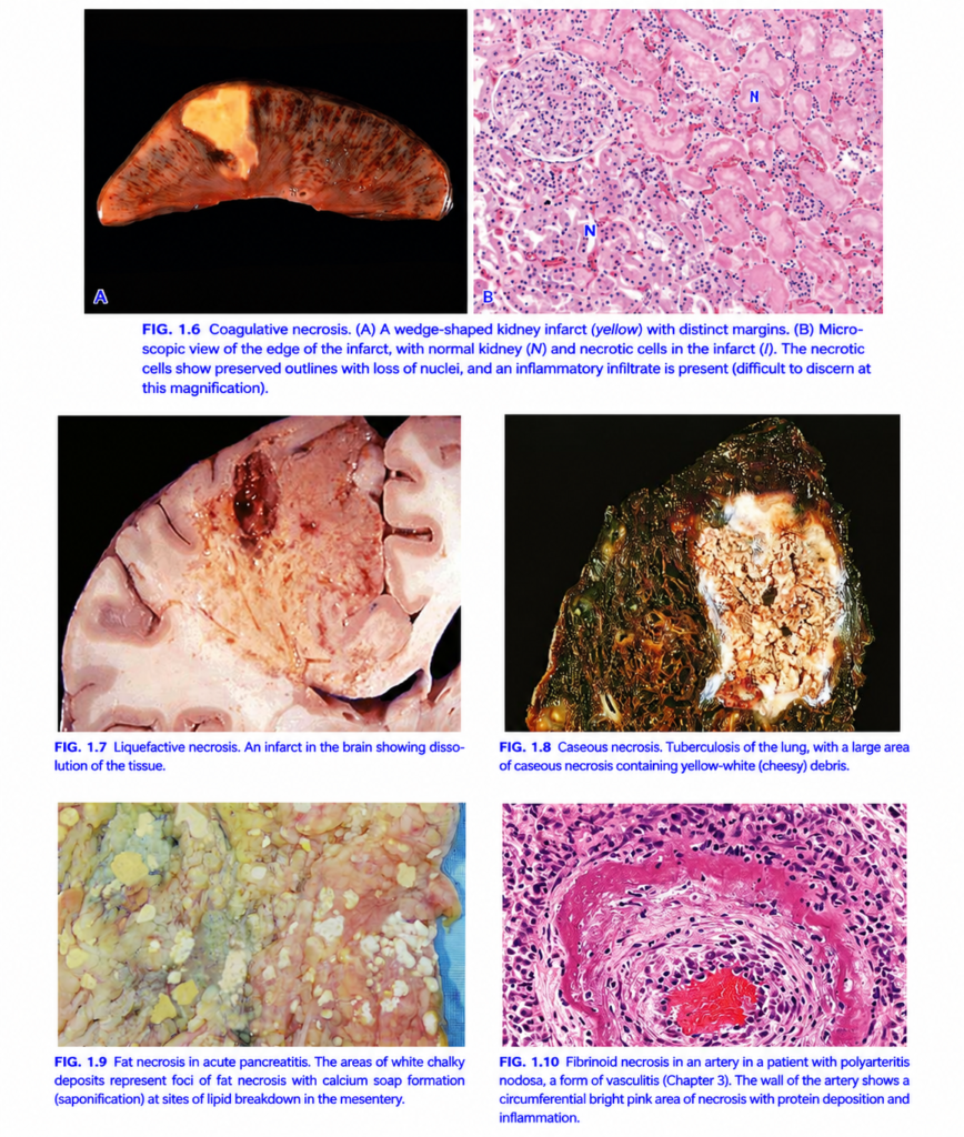
- Coagulative necrosis (Fig. 1.6)
- The basic tissue structure is preserved for several days after injury.
- The affected tissue becomes firm.
- Injury denatures:
- Structural proteins
- Cellular enzymes
- Because enzymes are inactivated, dead cells are not digested immediately.
- Dead cells remain as eosinophilic, anucleate cells for days or weeks.
- Later, lysosomal enzymes from recruited leukocytes digest the dead cells.
- Cell debris is removed by phagocytosis.
- It is the typical pattern of infarction (ischemia) in all solid organs except the brain.
- Liquefactive necrosis (Fig. 1.7)
- Commonly occurs in:
- Bacterial infections
- Occasionally fungal infections
- Microbes attract inflammatory cells.
- Leukocyte enzymes digest (liquefy) the tissue.
- Hypoxic injury in the central nervous system (brain) also causes liquefactive necrosis.
- Dead tissue is completely digested into a thick liquid (viscous).
- Phagocytes eventually remove the liquid material.
- In acute bacterial infection:
- The creamy yellow material is called pus.
- A localized collection of pus is called an abscess.
- Commonly occurs in:
- Gangrenous necrosis
- Gangrene is not a separate type of cell death.
- It is a commonly used clinical term.
- It usually affects a limb, especially the lower leg.
- It develops after loss of blood supply, producing coagulative necrosis.
- If bacterial infection is added:
- Liquefactive changes also develop.
- This is called wet gangrene.
- Caseous necrosis (Fig. 1.8)
- Most commonly seen in tuberculosis.
- Caseous means “cheese-like.”
- The necrotic area appears soft, friable, and yellow-white.
- Microscopically:
- It consists of amorphous, granular, pink cellular debris.
- Normal tissue architecture is completely lost.
- Individual cell outlines cannot be identified.
- It is commonly surrounded by:
- Macrophages
- Other inflammatory cells
- This forms a granuloma.
- Fat necrosis (Fig. 1.9)
- Refers to localized destruction of fat tissue.
- Common causes:
- Abdominal trauma
- Acute pancreatitis
- Pancreatic enzymes leak out and digest surrounding fat.
- Released fatty acids combine with calcium.
- This forms chalky white deposits.
- Microscopically:
- Necrotic fat cells appear as shadowy outlines.
- Calcium deposits and inflammation surround the dead fat cells.
- Fibrinoid necrosis (Fig. 1.10)
- A special type of necrosis seen only by light microscopy.
- Commonly occurs in:
- Immune reactions
- Severe hypertension
- Immune complexes and plasma proteins are deposited in blood vessel walls.
- The vessel wall appears bright pink and amorphous on H&E stain.
- This appearance is called fibrinoid (fibrin-like).
- It is commonly seen in:
- Vasculitis
- Rejected transplanted organs
KEY CONCEPT
- Coagulative necrosis (Fig. 1.6) → Tissue architecture preserved; typical of ischemic infarction in solid organs (except brain).
- Liquefactive necrosis (Fig. 1.7) → Tissue becomes liquid; seen in brain infarction and bacterial/fungal infections; forms pus and abscesses.
- Gangrenous necrosis → Clinical term for ischemic limb necrosis; bacterial infection causes wet gangrene.
- Caseous necrosis (Fig. 1.8) → Cheese-like appearance; characteristic of tuberculosis; associated with granuloma.
- Fat necrosis (Fig. 1.9) → Fat destruction due to pancreatitis or trauma; produces chalky calcium deposits.
- Fibrinoid necrosis (Fig. 1.10) → Bright pink vessel wall change seen in immune-mediated vascular injury and severe hypertension.
Leakage of Intracellular Proteins Through Damaged Cell Membranes
- When cells undergo necrosis, the cell membrane is damaged.
- Because of this damage, proteins inside the cell leak into the blood.
- Different tissues contain their own specific proteins.
- Therefore, the leaked proteins help identify which tissue has been damaged.
- Blood or serum tests can detect these leaked proteins.
- These proteins are useful clinical markers of tissue necrosis.
- Cardiac muscle
- Contains a unique form of the contractile protein troponin.
- Damage to heart muscle causes troponin to leak into the blood.
- Increased blood troponin indicates cardiac muscle injury.
- Hepatic bile duct epithelium
- Contains a heat-resistant (temperature-resistant) isoform of alkaline phosphatase (ALP).
- Damage to bile duct cells releases alkaline phosphatase into the blood.
- Hepatocytes (liver cells)
- Contain transaminases.
- Liver cell injury causes transaminases to leak into the blood.
- Measurement of these proteins in blood helps doctors:
- Detect tissue damage.
- Identify the affected organ.
KEY CONCEPT
- Necrosis damages the cell membrane, allowing intracellular proteins to leak into the blood.
- These leaked proteins serve as tissue-specific markers of cell injury.
- Examples:
- Troponin → Cardiac muscle injury.
- Alkaline phosphatase (ALP) → Bile duct epithelial injury.
- Transaminases → Liver cell (hepatocyte) injury.
- Blood measurement of these proteins helps diagnose organ-specific tissue necrosis.
Apoptosis
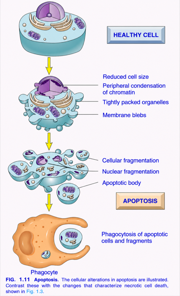
- Apoptosis is a regulated form of cell death.
- During apoptosis:
- The cell activates its own enzymes.
- These enzymes break down:
- Nuclear DNA
- Nuclear proteins
- Cytoplasmic proteins (Fig. 1.11)
- As the cell breaks down:
- Small fragments separate from the cell.
- This gives the appearance of “falling off,” which is the meaning of the word apoptosis.
- The plasma membrane remains intact during apoptosis.
- However, the plasma membrane changes in a way that allows the cell fragments to be recognized.
- These membrane-bound fragments are called apoptotic bodies.
- Macrophages quickly recognize and engulf (phagocytose) the apoptotic bodies.
- Unlike necrosis (Table 1.1):
- Cell contents do not leak out.
- Apoptotic cells are removed before their contents are released.
- Therefore, apoptosis does not cause an inflammatory reaction.
KEY CONCEPT
- Apoptosis is a regulated, programmed form of cell death.
- Cell enzymes digest:
- DNA
- Nuclear proteins
- Cytoplasmic proteins (Fig. 1.11)
- The cell breaks into apoptotic bodies.
- The plasma membrane remains intact.
- Macrophages rapidly remove apoptotic bodies.
- No leakage of cell contents occurs, so apoptosis does not produce inflammation, unlike necrosis (Table 1.1).
Causes of Apoptosis
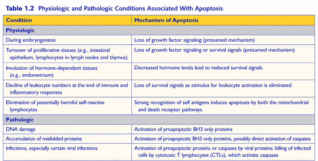
- Apoptosis occurs in many normal (physiologic) and disease (pathologic) conditions (Table 1.2).
- It helps remove:
- Cells that may be harmful.
- Cells that are no longer needed.
- It also removes cells that are damaged beyond repair, especially when DNA or proteins are severely damaged.
Physiologic apoptosis
- During normal development, some cells naturally die.
- These cells are replaced by new cells.
- In mature tissues:
- Some tissues continuously grow and lose cells.
- This happens especially in:
- Highly proliferative tissues.
- Hormone-responsive tissues.
- The balance between cell growth and cell death depends on:
- Growth factors.
- Survival signals.
- In these situations:
- Cell death always occurs by apoptosis.
- Unwanted cells are removed without causing inflammation.
- In the immune system, apoptosis removes:
- Excess leukocytes after an immune response.
- B lymphocytes in germinal centers that fail to produce high-affinity antibodies.
- Self-reactive lymphocytes that recognize the body’s own antigens and could cause autoimmune disease if they survive (Chapter 5).
Apoptosis in pathologic conditions
- Apoptosis removes cells with irreparable damage.
- One important cause is severe DNA damage, such as after:
- Radiation exposure.
- Cytotoxic drugs.
- Accumulation of misfolded proteins also triggers apoptosis.
- The mechanism and role of this process are discussed later in relation to endoplasmic reticulum (ER) stress.
- Some infectious agents, especially certain viruses, also induce apoptosis in infected cells.
KEY CONCEPT
- Apoptosis occurs in both physiologic and pathologic conditions (Table 1.2).
- Physiologic apoptosis:
- Removes unnecessary cells during development.
- Maintains normal cell numbers.
- Removes excess immune cells.
- Eliminates self-reactive lymphocytes.
- Occurs without inflammation.
- Pathologic apoptosis:
- Removes cells with severe DNA damage.
- Is triggered by misfolded proteins.
- Can be induced by certain viral infections.
- Apoptosis protects the body by removing damaged or potentially harmful cells.
Mechanisms of Apoptosis
- Apoptosis is controlled by biochemical pathways.
- These pathways maintain the balance between:
- Cell survival signals
- Cell death signals
- The final step is the activation of enzymes called caspases.
- Caspases are enzymes that:
- Break down proteins.
- Cut proteins after aspartic acid residues.
- Are cysteine proteases.
- Two major pathways activate caspases (Fig. 1.12):
- Mitochondrial (intrinsic) pathway
- Death receptor pathway
- Although these pathways can interact:
- They are usually activated under different conditions.
- They use different molecules.
- They perform different roles in normal body function and disease.
- Mitochondrial (intrinsic) pathway
- This is the main pathway responsible for apoptosis in most normal and disease conditions.
- Mitochondria contain proteins that can trigger apoptosis.
- One important protein is cytochrome c.
- When the mitochondrial membrane becomes permeable:
- Cytochrome c leaks into the cytoplasm.
- This activates caspases.
- The cell undergoes apoptosis.
- Mitochondrial membrane permeability is controlled by the BCL-2 family of proteins.
- The BCL-2 family contains more than 20 proteins.
- BCL-2 is the prototype member of this family.
- In healthy cells:
- BCL-2 and BCL-XL are produced in response to:
- Growth factors.
- Other survival signals.
- These proteins are antiapoptotic.
- They keep the mitochondrial membrane intact.
- They prevent apoptosis.
- BCL-2 and BCL-XL are produced in response to:
- BCL-2 and BCL-XL keep two proapoptotic proteins under control:
- BAX
- BAK
- When cells:
- Lose growth factors.
- Lose survival signals.
- Have DNA damage.
- Accumulate excessive misfolded proteins.
- The most important sensor proteins are BH3-only proteins.
- BH3-only proteins:
- Shift the balance toward BAX and BAK.
- Allow BAX and BAK to join together (dimerize).
- They insert into the mitochondrial membrane.
- They form channels (pores) in the membrane.
- Through these channels:
- Cytochrome c
- Other mitochondrial proteins
- At the same time:
- Levels of BCL-2 and BCL-XL decrease.
- This further increases mitochondrial membrane permeability.
- Once cytochrome c enters the cytoplasm:
- It combines with specific cofactors.
- It activates caspase-9.
- Caspase-9 starts the caspase cascade.
- The caspase cascade leads to apoptotic cell death.
KEY CONCEPT
- Apoptosis is controlled by biochemical pathways that regulate cell survival and cell death signals.
- Caspases are the key enzymes that execute apoptosis (Fig. 1.12).
- Two pathways activate caspases:
- Mitochondrial (intrinsic) pathway
- Death receptor pathway
- The mitochondrial pathway is the most common pathway of apoptosis.
- Cytochrome c released from mitochondria activates caspase-9, initiating the caspase cascade.
- BCL-2 and BCL-XL prevent apoptosis by protecting the mitochondrial membrane.
- BAX and BAK promote apoptosis by forming pores in the mitochondrial membrane.
- BH3-only proteins activate BAX and BAK when cells experience severe stress, DNA damage, loss of survival signals, or accumulation of misfolded proteins.
- Death receptor (extrinsic) pathway
- Many cells have death receptors on their surface.
- These receptors can trigger apoptosis.
- Most death receptors belong to the tumor necrosis factor (TNF) receptor family.
- These receptors contain a death domain in their cytoplasmic region.
- The death domain helps the receptor interact with proteins involved in apoptosis.
- The main death receptors are:
- Type I TNF receptor
- Fas (CD95)
- Fas ligand (FasL) is a membrane protein.
- It is mainly present on activated T lymphocytes.
- When activated T cells recognize a target cell that expresses Fas:
- FasL binds to Fas receptors.
- Fas receptors become linked together (cross-linked).
- Adaptor proteins attach to the death domain (Fig. 1.12).
- These adaptor proteins:
- Recruit caspase-8.
- Activate caspase-8.
- Activated caspase-8 then activates other downstream caspases.
- This leads to apoptotic cell death.
- The death receptor pathway helps:
- Eliminate self-reactive lymphocytes.
- Kill target cells by cytotoxic T lymphocytes that express FasL.
- Terminal phase of apoptosis
- Activated caspase-8 and caspase-9 enter a common final pathway.
- They activate additional caspases.
- These caspases activate enzymes that:
- Break down cellular proteins.
- Digest the nucleus.
- The final result is fragmentation of the cell, which is the hallmark of apoptosis.
- Clearance of apoptotic cells
- Apoptotic cells produce “eat-me” signals.
- These signals attract phagocytes.
- In normal cells:
- Phosphatidylserine is located on the inner layer of the plasma membrane.
- During apoptosis:
- Phosphatidylserine moves to the outer surface of the membrane.
- Tissue macrophages recognize this signal.
- Apoptotic cells also release soluble factors.
- These factors recruit phagocytes.
- Macrophages bind to and engulf apoptotic cells.
- This removal is rapid and efficient.
- Dead cells disappear without leaving debris.
- No inflammation occurs.
- Other pathways of cell death
- Besides necrosis and apoptosis, other forms of cell death include:
- Necroptosis
- Triggered by the cytokine tumor necrosis factor (TNF).
- Shows features of both necrosis and apoptosis.
- Pyroptosis
- Triggered by activation of inflammasomes (Chapter 5).
- Causes release of interleukin-1 (IL-1).
- IL-1 produces:
- Inflammation
- Fever
- Ferroptosis
- Depends on the level of cellular iron.
- Necroptosis
- The normal and disease roles of these newer forms of cell death are still being investigated.
KEY CONCEPT
- The death receptor (extrinsic) pathway uses death receptors such as TNF receptor and Fas (CD95) to initiate apoptosis (Fig. 1.12).
- FasL from activated T lymphocytes binds Fas, activating caspase-8.
- Caspase-8 and caspase-9 activate a common caspase cascade, leading to:
- Protein breakdown
- Nuclear degradation
- Cell fragmentation
- Apoptotic cells expose phosphatidylserine as an “eat-me” signal.
- Macrophages rapidly remove apoptotic bodies without causing inflammation.
- Other forms of cell death include:
- Necroptosis → Features of necrosis and apoptosis.
- Pyroptosis → Inflammasome-mediated; releases IL-1, causing inflammation and fever.
- Ferroptosis → Iron-dependent cell death.
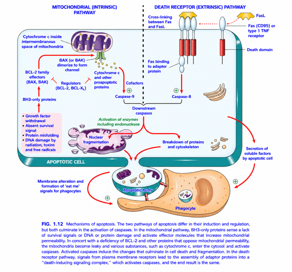
Mechanisms of Apoptosis (Figure 1.12)
This figure explains how cells perform programmed (controlled) cell death, called apoptosis.
Unlike necrosis, apoptosis is an organized, energy-dependent process that removes damaged or unwanted cells without causing inflammation.
Main Concept
There are two pathways that start apoptosis:
- Intrinsic (Mitochondrial) Pathway → Triggered by damage inside the cell.
- Extrinsic (Death Receptor) Pathway → Triggered by signals outside the cell.
Although they begin differently, both pathways activate caspases, which execute apoptosis.
Intrinsic Pathway + Extrinsic Pathway → Caspase Activation → Apoptosis
Overall Flow of the Figure
                 APOPTOSIS

        ┌─────────────────────┐
        │                     │
Intrinsic Pathway      Extrinsic Pathway
(Mitochondria)        (Death Receptors)
        │                     │
        ▼                     ▼
   Caspase-9            Caspase-8
        │                     │
        └──────────┬──────────┘
                   ▼
          Executioner Caspases
                   ▼
      DNA Fragmentation
      Cytoskeleton Breakdown
      Cell Shrinkage
      Apoptotic Bodies
                   ▼
        Phagocytosis (No Inflammation)
PART 1 – Intrinsic (Mitochondrial) Pathway
This pathway is activated when the cell detects internal damage.
Step 1. Internal Cell Stress
The figure lists several triggers:
- Growth factor withdrawal
- Loss of survival signals
- Protein misfolding
- DNA damage
- Radiation
- Toxins
- Free radicals
Concept
The cell asks,
“Am I too damaged to survive?”
If the answer is yes, apoptosis begins.
Step 2. BH3-Only Proteins Become Active
These proteins are the damage sensors.
Function
They detect cellular stress and activate the apoptotic pathway.
Step 3. BCL-2 Family Proteins
The figure shows two groups.
A. Pro-apoptotic Proteins
These promote apoptosis.
Examples:
- BAX
- BAK
Function
They form pores (channels) in the outer mitochondrial membrane.B. Anti-apoptotic Proteins
These prevent apoptosis.
Examples:
- BCL-2
- BCL-xL
Function
They keep the mitochondrial membrane intact and prevent cytochrome c release.
Easy Memory
BAX & BAK = Break the membrane
BCL-2 & BCL-xL = Block apoptosis
Step 4. Cytochrome c Release
Normally,
cytochrome c remains inside the mitochondria.
When BAX and BAK create channels,
cytochrome c escapes into the cytoplasm.
Meaning
This is the point of commitment to apoptosis in the intrinsic pathway.
Step 5. Caspase-9 Activation
Released cytochrome c combines with cofactors to activate caspase-9.
Caspase-9
This is the initiator caspase of the intrinsic pathway.
PART 2 – Extrinsic (Death Receptor) Pathway
This pathway begins with signals coming from outside the cell.
Step 1. Death Receptors
The figure shows:
- Fas (CD95) receptor
- Type I TNF receptor
These receptors are present on the cell membrane.
Step 2. Fas Ligand (FasL)
A neighboring immune cell presents Fas ligand (FasL).
When FasL binds Fas receptor, the receptors cluster together.
Concept
It is like pressing the self-destruct button on the cell.
Step 3. Death Domain
Inside the receptor is a death domain.
Function
It recruits adaptor proteins that form the death-inducing signaling complex (DISC).
Step 4. Caspase-8 Activation
The adaptor proteins activate caspase-8.
Caspase-8
This is the initiator caspase of the extrinsic pathway.
Both Pathways Meet Here
Whether apoptosis begins through:
- mitochondria
- death receptors
both pathways activate downstream (executioner) caspases.
Examples:
- Caspase-3
- Caspase-6
- Caspase-7
These enzymes actually dismantle the cell.
What Do Executioner Caspases Do?
1. Nuclear Fragmentation
Executioner caspases activate endonucleases.
These enzymes cut DNA into small fragments.
Result
The nucleus breaks into pieces.
2. Breakdown of Proteins and Cytoskeleton
Caspases digest:
- Cytoskeletal proteins
- Structural proteins
- Nuclear proteins
Result
The cell loses its shape and shrinks.
3. Cell Shrinkage
Unlike necrosis,
the cell becomes smaller, not swollen.
4. Formation of Apoptotic Bodies
The dying cell breaks into small, membrane-bound fragments.
These are called apoptotic bodies.
Each contains:
- Cytoplasm
- Organelles
- Nuclear fragments
5. “Eat-Me” Signals
The membrane changes and displays “eat-me” signals (such as exposed phosphatidylserine).
These signals attract phagocytes.
6. Phagocytosis
Macrophages (phagocytes) rapidly engulf apoptotic bodies.
Result
Dead cells are removed cleanly.
There is no leakage of cellular contents.
7. Soluble Factors
The apoptotic cell also releases soluble factors that help attract phagocytes and promote efficient clearance.
Why Is There No Inflammation?
Because:
- Cell membrane remains intact.
- Cellular contents do not spill out.
- Apoptotic bodies are rapidly engulfed.
Therefore
Apoptosis does not cause inflammation.
Complete Sequence of Apoptosis
Intrinsic Pathway
Cell stress
      ↓
BH3 proteins
      ↓
BAX / BAK activation
      ↓
Cytochrome c release
      ↓
Caspase-9
      ↓
Executioner caspases
      ↓
Apoptosis
Extrinsic Pathway
FasL binds Fas receptor
        ↓
Death domain activated
        ↓
Adaptor proteins
        ↓
Caspase-8
        ↓
Executioner caspases
        ↓
Apoptosis
Final Common Pathway
Executioner caspases
        ↓
DNA fragmentation
        ↓
Protein breakdown
        ↓
Cell shrinkage
        ↓
Apoptotic bodies
        ↓
Phagocytosis
        ↓
No inflammation
Apoptosis vs Necrosis
| Feature | Apoptosis | Necrosis |
|---|---|---|
| Type of death | Programmed | Accidental/pathologic |
| Energy | ATP-dependent | Usually ATP depletion |
| Cell size | Shrinks | Swells |
| Membrane | Intact until apoptotic bodies form | Ruptures |
| DNA | Fragmented in an orderly way | Random degradation |
| Cell contents | Remain enclosed | Leak out |
| Inflammation | Absent | Present |
| Outcome | Phagocytosis of apoptotic bodies | Tissue injury and inflammation |
Easy Memory Tricks
Intrinsic Pathway
BAX & BAK = Break the mitochondria
BCL-2 = Blocks apoptosis
Cytochrome c = Caspase-9Extrinsic Pathway
Fas → Caspase-8
Final Pathway
Caspases → Cell Cutters
They cut:
- DNA
- Cytoskeleton
- Structural proteins
Result:
Apoptotic bodies → Phagocytosis → No inflammation
Important Points from Figure 1.12
- Apoptosis occurs through two initiation pathways: the intrinsic (mitochondrial) pathway and the extrinsic (death receptor) pathway.
- The intrinsic pathway is triggered by DNA damage, growth factor withdrawal, protein misfolding, toxins, radiation, and free radicals.
- BH3-only proteins activate BAX and BAK, while BCL-2 and BCL-xL oppose apoptosis by preserving mitochondrial integrity.
- BAX and BAK form channels in the mitochondrial membrane, allowing cytochrome c to escape and activate caspase-9.
- The extrinsic pathway begins when Fas ligand (FasL) binds the Fas (CD95) receptor or when related death receptors are activated, leading to activation of caspase-8.
- Caspase-9 (intrinsic) and caspase-8 (extrinsic) activate executioner caspases, which digest cellular proteins and DNA.
- Executioner caspases cause DNA fragmentation, cytoskeletal breakdown, cell shrinkage, and formation of apoptotic bodies.
- The apoptotic cell displays “eat-me” signals, allowing phagocytes to remove apoptotic bodies rapidly.
- Because the plasma membrane remains largely intact and cellular contents are not released, apoptosis does not provoke inflammation.
KEY CONCEPT (Figure 1.12)
Apoptosis is a regulated, ATP-dependent form of cell death that removes damaged or unnecessary cells without causing inflammation. It can begin through the intrinsic (mitochondrial) pathway, in which BAX and BAK promote cytochrome c release and activation of caspase-9, or through the extrinsic (death receptor) pathway, in which Fas/FasL or related death receptors activate caspase-8. Both pathways converge on executioner caspases, which fragment DNA, degrade cytoskeletal proteins, and package the dying cell into apoptotic bodies. These apoptotic bodies are rapidly engulfed by phagocytes, allowing the cell to be removed without leakage of contents or inflammation.
MADE BY SELF LEARNING DR SHEEN CEO AND FOUNDER
