


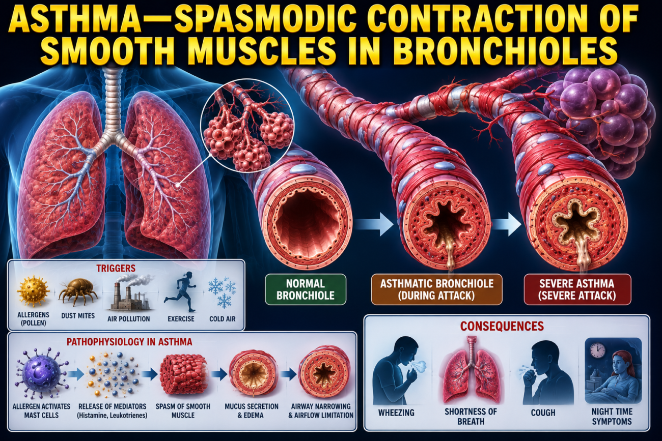
- Asthma is a disease in which the smooth muscles of the bronchioles contract suddenly (spasm).
- This partially narrows the bronchioles, making breathing very difficult.
- Asthma is becoming more common.
- It affects about 7%–8% of people in the United States.
- The World Health Organization (WHO) estimates that more than 262 million people worldwide have asthma.
- The usual cause of asthma is excessive contraction (hypersensitivity) of the bronchiolar smooth muscle in response to foreign substances in the air.
- In about 70% of patients younger than 30 years, asthma is caused by allergic hypersensitivity, especially to plant pollens.
- In older people, asthma is usually caused by hypersensitivity to non-allergic airborne irritants, such as smog.
- People with allergic asthma produce unusually large amounts of immunoglobulin E (IgE) antibodies.
- These IgE antibodies attach to mast cells present in the lung interstitium near the bronchioles and small bronchi.
- When the person inhales an allergen (such as pollen) to which they are sensitive:
- The allergen binds to IgE antibodies on the mast cells.
- This activates the mast cells.
- Activated mast cells release several substances, including:
- Histamine
- Slow-reacting substance of anaphylaxis (a mixture of leukotrienes)
- Eosinophilic chemotactic factor
- Bradykinin
- The combined effects of these substances, especially the slow-reacting substance of anaphylaxis, cause:
- Edema (swelling) of the walls of the small bronchioles
- Secretion of thick mucus into the bronchioles
- Spasm of the bronchiolar smooth muscle
- These changes greatly increase airway resistance.
- During expiration, the bronchioles become narrower than during inspiration because they collapse more easily during expiratory effort.
- Since the bronchioles are already partially blocked in asthma, this additional narrowing causes severe airway obstruction during expiration.
- Therefore, a person with asthma can usually inhale fairly well but has great difficulty exhaling.
- Clinical findings include:
- Greatly reduced maximum expiratory flow rate
- Reduced timed expiratory volume
- These changes produce dyspnea (air hunger).
- During an acute asthma attack, the functional residual capacity (FRC) and residual volume (RV) increase because air cannot be expelled completely from the lungs.
- After many years of asthma, the chest becomes permanently enlarged, producing a barrel chest.
- In long-standing asthma, the functional residual capacity and residual volume remain permanently increased.
KEY CONCEPT
- Asthma is caused by spasm of bronchiolar smooth muscle, leading to partial airway obstruction and difficult breathing.
- The most common cause is hypersensitivity of the bronchioles to allergens or airborne irritants.
- In allergic asthma, IgE antibodies activate mast cells, which release histamine, leukotrienes, eosinophilic chemotactic factor, and bradykinin.
- These mediators cause bronchiolar edema, thick mucus secretion, and smooth muscle spasm, greatly increasing airway resistance.
- Airway obstruction is more severe during expiration, making exhalation more difficult than inhalation.
- Clinical findings include reduced expiratory flow rate, reduced timed expiratory volume, and dyspnea.
- Acute asthma increases FRC and RV, while long-standing asthma may cause a permanent barrel chest with persistently increased FRC and RV.
TUBERCULOSIS
- In tuberculosis (TB), tubercle bacilli infect the lungs and produce a characteristic tissue reaction.
- This tissue reaction includes:
- Invasion of the infected area by macrophages.
- Formation of a fibrous wall around the infected area, creating a tubercle.
- The fibrous wall helps prevent the spread of tubercle bacilli within the lungs.
- Therefore, walling off the infection is a protective mechanism that limits the extension of tuberculosis.
- If tuberculosis is not treated, the walling-off process fails in about 3% of affected people.
- When this happens, tubercle bacilli spread throughout the lungs.
- The infection may destroy large areas of lung tissue.
- This destruction can lead to the formation of large abscess cavities in the lungs.
- In the late stages of tuberculosis, the lungs contain many areas of fibrosis (scar tissue).
- There is also a reduction in the amount of functional lung tissue.
- These changes produce the following effects:
- The respiratory muscles must work harder to ventilate the lungs.
- Vital capacity and breathing capacity decrease.
- The total surface area of the respiratory membrane decreases.
- The respiratory membrane becomes thicker.
- Pulmonary diffusing capacity progressively decreases.
- The ventilation-perfusion (V/Q) ratio becomes abnormal.
- Diffusion of both O₂ and CO₂ across the lungs decreases further.
KEY CONCEPT
- Tuberculosis is caused by tubercle bacilli, which produce macrophage infiltration and fibrous walling-off of the infected area to form a tubercle.
- The fibrous wall limits the spread of infection within the lungs.
- If untreated, walling off fails in about 3% of patients, allowing widespread spread of bacilli, severe lung destruction, and formation of large abscess cavities.
- Late-stage tuberculosis causes extensive fibrosis and loss of functional lung tissue.
- These changes increase the work of breathing, reduce vital capacity, decrease respiratory membrane surface area, increase membrane thickness, reduce pulmonary diffusing capacity, produce an abnormal ventilation-perfusion ratio, and impair O₂ and CO₂ diffusion.
MADE BY SELF LEARNING CEO AND FOUNDER DR SHEEN
