


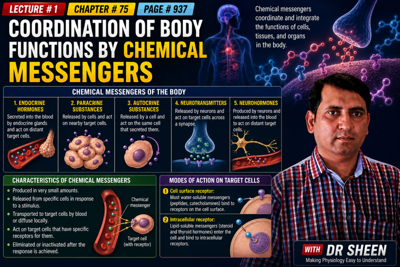
- Many activities of the body’s cells, tissues, and organs are coordinated by different types of chemical messengers.
- 1. Neurotransmitters
- Neurotransmitters are released from the axon terminals of neurons.
- They enter the synaptic junctions.
- They act locally to control the functions of nerve cells.
- 2. Endocrine hormones
- Endocrine hormones are released by glands or specialized cells.
- They enter the circulating blood.
- Blood carries them to target cells at another location in the body.
- They then change the function of those target cells.
- 3. Neuroendocrine hormones
- Neuroendocrine hormones are secreted by neurons.
- They enter the circulating blood.
- Blood carries them to target cells at another location in the body.
- They then influence the function of those target cells.
- 4. Paracrines
- Paracrines are secreted by cells into the extracellular fluid.
- They act on nearby neighboring target cells.
- The neighboring target cells are different from the cells that produced the paracrines.
- 5. Autocrines
- Autocrines are secreted by cells into the extracellular fluid.
- They act on the same cells that produced them.
- Therefore, a cell can release a chemical messenger that affects itself.
- 6. Cytokines
- Cytokines are peptides secreted by cells into the extracellular fluid.
- They can act as autocrines, paracrines, or endocrine hormones.
- Examples include interleukins and other lymphokines.
- These substances are secreted by helper cells.
- They act on other cells of the immune system (see Chapter 35).
- Cytokine hormones, such as leptin, are produced by adipocytes.
- These cytokine hormones are sometimes called adipokines.
- The next few chapters mainly discuss the endocrine and neuroendocrine hormone systems.
- However, many chemical messenger systems interact with each other to help maintain homeostasis.
- For example, the adrenal medullae and pituitary gland mainly secrete their hormones in response to neural stimuli.
- Neuroendocrine cells are located in the hypothalamus.
- These cells have axons that end in the posterior pituitary gland and median eminence.
- These cells secrete several neurohormones.
- These neurohormones include antidiuretic hormone, oxytocin, and hypophysiotropic hormones.
- These hormones control the secretion of anterior pituitary hormones.
- Endocrine hormones are transported by the circulatory system to cells throughout the body.
- In some cases, they also reach the nervous system.
- These hormones bind to specific receptors on their target cells.
- After binding to receptors, they start many cellular reactions.
- Some endocrine hormones affect many different types of cells in the body.
- For example, growth hormone from the anterior pituitary causes growth in most parts of the body.
- Thyroxine from the thyroid gland increases the rate of many chemical reactions in almost all cells of the body.
- Other hormones mainly affect specific target tissues.
- This happens because these tissues have many receptors for that particular hormone.
- For example, adrenocorticotropic hormone (ACTH) from the anterior pituitary specifically stimulates the adrenal cortex.
- ACTH causes the adrenal cortex to secrete adrenocortical hormones.
- Ovarian hormones mainly affect the female sex organs and the secondary sexual characteristics of the female body.
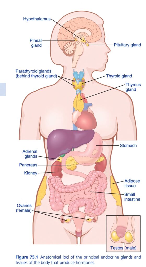
- Fig. 75.1 shows the anatomical locations of the major endocrine glands and endocrine tissues of the body.
- The placenta is not included in this figure.
- The placenta is another source of sex hormones in women who become pregnant.
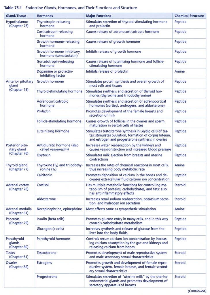
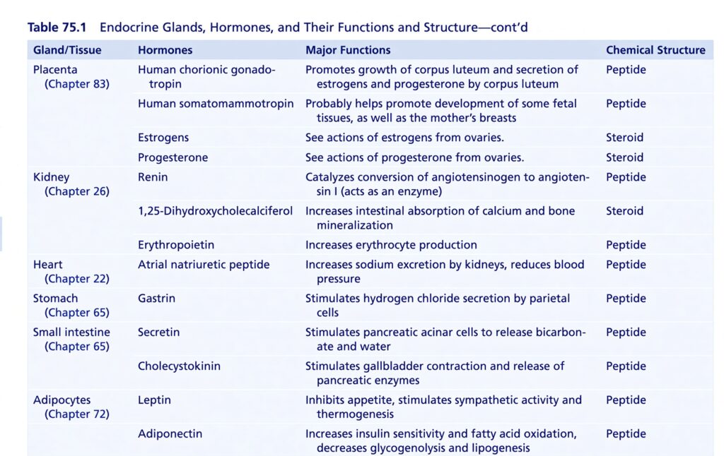
- Table 75.1 gives an overview of the different hormone systems and their major actions.
- The many hormone systems have an important role in regulating almost all body functions.
- These functions include metabolism, growth and development, water and electrolyte balance, reproduction, and behavior.
- For example, without growth hormone, a person would have very short stature.
- Without thyroxine and triiodothyronine from the thyroid gland, almost all chemical reactions in the body would become slow.
- Because of this, the person would also become sluggish.
- Without insulin from the pancreas, the body’s cells could use only a small amount of food carbohydrates for energy.
- Without sex hormones, sexual development and sexual functions would be absent.
KEY CONCEPT
- Neurotransmitter → neuron → nearby synaptic junction
- Endocrine hormone → blood → distant target cell
- Neuroendocrine hormone → neuron → blood → distant target cell
- Paracrine → nearby different cell
- Autocrine → same cell that released it
- Cytokine → can act as autocrine, paracrine, or endocrine
- Hormones work by binding to receptors on target cells.
- Hormones can affect many tissues or mainly specific tissues, depending on where their receptors are present.
- Hormone systems together regulate metabolism, growth, development, water/electrolyte balance, reproduction, and behavior.
CONCEPTUAL EXAMPLES
- Neurotransmitter: neuron → synaptic junction → nearby nerve cell.
- Endocrine hormone: gland → blood → distant target cell.
- Neuroendocrine hormone: neuron → blood → distant target cell.
- Paracrine: cell → nearby different cell.
- Autocrine: cell → chemical messenger → same cell.
- Growth hormone: anterior pituitary → blood → many body tissues → growth.
- ACTH: anterior pituitary → blood → adrenal cortex → adrenal hormones.
- Insulin: pancreas → body cells → helps cells use food carbohydrates for energy.
CHEMICAL STRUCTURE AND SYNTHESIS OF HORMONES
- There are three general classes of hormones.
- 1. Proteins and polypeptides
- This group includes protein and polypeptide hormones.
- They are secreted by the anterior and posterior pituitary glands.
- They are also secreted by the pancreas, such as insulin and glucagon.
- They are also secreted by the parathyroid gland, such as parathyroid hormone.
- Many other hormones also belong to this group (see Table 75.1).
- 2. Steroids
- Steroid hormones are secreted by the adrenal cortex.
- Examples are cortisol and aldosterone.
- Steroid hormones are also secreted by the ovaries.
- Examples are estrogen and progesterone.
- The testes secrete the steroid hormone testosterone.
- The placenta secretes estrogen and progesterone.
- 3. Derivatives of the amino acid tyrosine
- These hormones are made from the amino acid tyrosine.
- The thyroid gland secretes thyroxine and triiodothyronine.
- The adrenal medullae secrete epinephrine and norepinephrine.
- There are no known polysaccharide hormones or nucleic acid hormones.
KEY CONCEPT
- Hormones are mainly divided into 3 classes:
- Proteins/polypeptides → insulin, glucagon, parathyroid hormone, pituitary hormones.
- Steroids → cortisol, aldosterone, estrogen, progesterone, testosterone.
- Tyrosine derivatives → thyroxine, triiodothyronine, epinephrine, norepinephrine.
- Easy memory:
- Protein → Pituitary + Pancreas + Parathyroid
- Steroid → Adrenal cortex + Ovary + Testis + Placenta
- Tyrosine → Thyroid + Adrenal medulla
CONCEPTUAL EXAMPLES
- Insulin → protein/polypeptide hormone
- Cortisol → steroid hormone
- Testosterone → steroid hormone
- Thyroxine → tyrosine derivative
- Epinephrine → tyrosine derivative
- No known hormone is a polysaccharide or nucleic acid.
Polypeptide and Protein Hormones Are Stored in Secretory Vesicles Until Needed
- Many hormones in the body are polypeptides and proteins.
- These hormones can be very different in size.
- Some are very small peptides containing only 3 amino acids.
- Example: thyrotropin-releasing hormone (TRH).
- Some are much larger proteins containing almost 200 amino acids.
- Examples: growth hormone and prolactin.
- In general:
- 100 or more amino acids → protein
- Fewer than 100 amino acids → peptide
- Protein and peptide hormones are made in the rough endoplasmic reticulum (RER) of endocrine cells.
- They are synthesized in the same basic way as most other proteins (Fig. 75.2).
- These hormones are usually first made as larger, inactive proteins called preprohormones.
- Inside the endoplasmic reticulum, preprohormones are cut into smaller molecules called prohormones.
- Prohormones are then transferred to the Golgi apparatus.
- The Golgi apparatus packages them into secretory vesicles.
- Inside these vesicles, enzymes cut the prohormones into:
- Smaller active hormones
- Inactive fragments
- The secretory vesicles are stored inside the cytoplasm.
- Many vesicles remain attached near the cell membrane until the hormone is needed.
- When the hormone needs to be released, the secretory vesicles fuse with the cell membrane.
- Their contents are then released outside the cell by exocytosis.
- The hormones and inactive fragments may be released into:
- Interstitial fluid, or
- Directly into the bloodstream.
- In many cases, exocytosis is triggered by an increase in cytosolic calcium (Ca²⁺).
- This increase in calcium can occur because the plasma membrane becomes depolarized.
- In other cases, stimulation of a receptor on the surface of the endocrine cell increases cyclic adenosine monophosphate (cAMP).
- Increased cAMP activates protein kinases.
- These protein kinases then initiate hormone secretion.
- Peptide hormones are water soluble.
- Because they are water soluble, they can easily enter the circulatory system.
- The blood then transports them to their target tissues.
KEY CONCEPT
Easy sequence:
Rough ER → Preprohormone → Prohormone → Golgi apparatus → Secretory vesicle → Active hormone → Exocytosis → Blood → Target tissue
- RER: makes the hormone.
- Preprohormone: large, inactive starting form.
- Prohormone: smaller inactive form.
- Golgi: packages it.
- Secretory vesicle: stores it.
- Enzymes: convert prohormone → active hormone.
- Ca²⁺ or cAMP/protein kinases: trigger secretion.
- Exocytosis: releases the hormone.
- Water soluble: allows easy transport in blood.
CONCEPTUAL EXAMPLES
- TRH: very small peptide → only 3 amino acids.
- Growth hormone: large protein → almost 200 amino acids.
- Hormone production: RER → preprohormone → prohormone → Golgi → secretory vesicle.
- Hormone release: vesicle fuses with cell membrane → exocytosis → hormone enters blood.
- One common trigger: membrane depolarization → ↑ Ca²⁺ → exocytosis → hormone secretion.
- Another trigger: receptor stimulation → ↑ cAMP → protein kinase activation → hormone secretion.
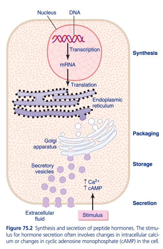
Figure 75.2 — ELI5: Synthesis and Secretion of Peptide Hormones
🧠 First, the BIG IDEA
Think of a peptide hormone-producing cell like a factory:
DNA = instruction book
⬇️
mRNA = copied instruction
⬇️
Endoplasmic reticulum = production line
⬇️
Golgi apparatus = packaging department
⬇️
Secretory vesicles = storage boxes
⬇️
Stimulus → Ca²⁺/cAMP increase = “SEND IT!” signal
⬇️
Hormone released into extracellular fluid
So the entire figure is basically:
MAKE → PACKAGE → STORE → RELEASE
1. 🧬 Nucleus — where the instructions are kept
At the top of the cell is the nucleus.
Inside the nucleus:
DNA
- DNA contains the instructions for making the peptide hormone.
- Think of DNA as the original recipe book.
But DNA itself does not leave the nucleus.
2. ✍️ Transcription — DNA → mRNA
The figure shows:
DNA → Transcription → mRNA
What does transcription mean?
It means:
A copy of the DNA instructions is made.
That copy is called mRNA.
ELI5:
Imagine DNA is a recipe book locked inside an office.
You cannot take the whole recipe book to the kitchen.
So you make a copy of the recipe.
That copy = mRNA.
3. 🚚 mRNA moves toward the ER
The mRNA then moves from the nucleus toward the endoplasmic reticulum (ER).
The ER is shown as the folded purple structures covered with black dots.
The black dots represent ribosomes.
Remember:
mRNA = instructions
Ribosomes = machines that read the instructions
4. 🏭 Translation — mRNA → peptide hormone
The figure then shows:
mRNA → Translation
What is translation?
Translation means:
Ribosomes read the mRNA instructions and build the peptide/protein.
ELI5:
Think of:
- mRNA = recipe
- Ribosome = chef
- Amino acids = ingredients
- Peptide hormone = finished product
So:
mRNA instructions → ribosome → peptide/protein
5. 🟣 Endoplasmic reticulum — production line
The endoplasmic reticulum (ER) is the large folded structure in the middle of the cell.
It has many black dots on it.
Those black dots are ribosomes.
Its main job in this figure:
The peptide hormone is synthesized here.
So think:
ER = hormone manufacturing/production area
6. 📦 Golgi apparatus — packaging department
After being produced, the hormone moves toward the Golgi apparatus.
The Golgi is the stack of flattened curved structures near the bottom-middle.
Think of Golgi as:
The packaging department of the cell.
The hormone is prepared and packaged into secretory vesicles.
7. 🟣 Secretory vesicles — storage boxes
The small purple circles are:
Secretory vesicles
They contain the peptide hormone.
Their job is essentially:
Store the hormone until the cell receives the signal to release it.
So:
Golgi → secretory vesicles → storage
8. 🚨 Stimulus — the “RELEASE!” signal
At the bottom, the figure shows:
Stimulus
This is the signal telling the cell:
“Release the stored hormone!”
The figure specifically shows that the stimulus often involves:
↑ Ca²⁺
and/or
↑ cAMP
So remember:
Stimulus → ↑ intracellular Ca²⁺ or ↑ cAMP → hormone secretion
9. ⚡ Ca²⁺ and cAMP — intracellular signals
The figure shows:
↑ Ca²⁺
↑ cAMP
These are signals inside the cell.
They help convert the external stimulus into the action of releasing the stored hormone.
ELI5:
Imagine the secretory vesicles are boxes waiting inside a warehouse.
The stimulus is the manager saying:
“Send the boxes out!”
Ca²⁺ and cAMP help transmit that command inside the cell.
10. 🚪 Secretion — hormone leaves the cell
The secretory vesicles move toward the cell membrane.
They release their contents outside the cell.
The outside is labeled:
Extracellular fluid
Therefore:
Secretory vesicle → release hormone → extracellular fluid
This is called:
SECRETION
🔥 Understand the whole figure as ONE STORY
Imagine a hormone factory:
STEP 1 — 🧬 INSTRUCTIONS
DNA in nucleus
DNA contains the hormone-making instructions.
⬇️
STEP 2 — ✍️ COPY
Transcription
DNA instructions are copied into mRNA.
⬇️
STEP 3 — 🏭 MAKE
Translation at ribosomes on ER
mRNA is read and the peptide hormone is synthesized.
⬇️
STEP 4 — 📦 PACKAGE
Golgi apparatus
The hormone is processed/packaged.
⬇️
STEP 5 — 🗃️ STORE
Secretory vesicles
Hormone is stored inside vesicles.
⬇️
STEP 6 — 🚨 SIGNAL
Stimulus
A stimulus causes:
↑ intracellular Ca²⁺ and/or ↑ cAMP
⬇️
STEP 7 — 🚪 RELEASE
Secretion
Secretory vesicles release the hormone into the extracellular fluid.
🧠 The easiest memory chain
DNA → mRNA → ER → Golgi → Vesicle → Stimulus → Secretion
Or even easier:
MAKE → PACKAGE → STORE → RELEASE
| Part | Think of it as | Main idea |
|---|---|---|
| Nucleus/DNA | 📖 Recipe book | Instructions |
| Transcription | ✍️ Copying recipe | DNA → mRNA |
| mRNA | 📄 Recipe copy | Carries instructions |
| Ribosome/ER | 🏭 Factory | Makes peptide |
| Translation | 👨🍳 Cooking | mRNA → peptide |
| Golgi | 📦 Packaging department | Packages product |
| Secretory vesicles | 🗃️ Storage boxes | Store hormone |
| Ca²⁺/cAMP | ⚡ Internal signal | Helps trigger release |
| Stimulus | 🚨 Order | Tells cell to release |
| Extracellular fluid | 🌎 Outside | Hormone is released here |
⭐ Figure 75.2 — EXAM-READY CONCEPT
Peptide hormone synthesis and secretion:
DNA
↓ Transcription
mRNA
↓ Translation
Peptide hormone synthesized
↓
Endoplasmic reticulum
↓
Golgi apparatus
↓
Secretory vesicles
↓ Storage
Stimulus
↓
↑ intracellular Ca²⁺ / ↑ cAMP
↓
Secretion into extracellular fluid
🔑 One-line concept:
Peptide hormones are synthesized from DNA instructions through mRNA and ribosomes, packaged by the Golgi, stored in secretory vesicles, and released when a stimulus increases intracellular Ca²⁺ or cAMP.
Steroid Hormones Are Usually Synthesized From Cholesterol and Are Not Stored
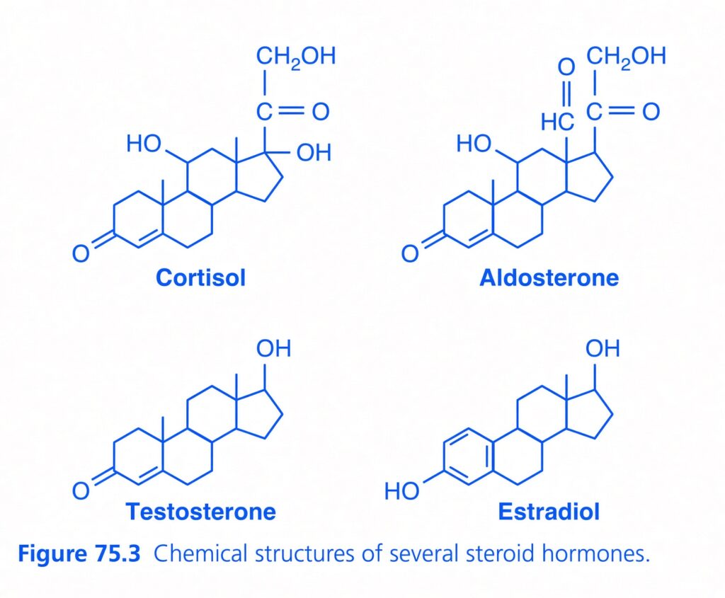
- Steroid hormones have a chemical structure that is similar to cholesterol.
- Most steroid hormones are made from cholesterol.
- They are lipid soluble.
- Their basic structure contains:
- 3 cyclohexyl rings
- 1 cyclopentyl ring
- These rings are joined together into one structure (Fig. 75.3).
- Steroid-producing endocrine cells usually have very little stored steroid hormone.
- However, these cells contain large amounts of cholesterol esters stored in cytoplasmic vacuoles.
- When a stimulus occurs, these cholesterol stores can be rapidly mobilized.
- The cholesterol is then used to make steroid hormones.
- Much of the cholesterol used by these cells comes from the plasma.
- Steroid-producing cells can also make cholesterol themselves.
- Because steroid hormones are highly lipid soluble, they can easily pass through the cell membrane after they are synthesized.
- They then enter the interstitial fluid and finally the blood.
Amine Hormones Are Derived From Tyrosine
- There are two groups of hormones derived from tyrosine:
- Thyroid hormones
- Adrenal medullary hormones
- These hormones are formed by the action of enzymes inside specific parts of the glandular cells.
- Thyroid hormones are synthesized and stored in the thyroid gland.
- They are incorporated into a large protein called thyroglobulin.
- Thyroglobulin is stored in large follicles inside the thyroid gland.
- When thyroid hormones need to be released, the hormones are split away from thyroglobulin.
- The free thyroid hormones are then released into the bloodstream.
- After entering the blood, most thyroid hormones attach to plasma proteins.
- One important plasma protein is thyroxine-binding globulin.
- This protein slowly releases the thyroid hormones to the target tissues.
- Epinephrine and norepinephrine are formed in the adrenal medulla.
- The adrenal medulla normally secretes about 4 times more epinephrine than norepinephrine.
- Catecholamines are taken into preformed vesicles.
- They are stored in these vesicles until they are needed.
- Like protein hormones stored in secretory granules, catecholamines are released from adrenal medullary cells by exocytosis.
- After catecholamines enter the circulation, they can be present in the plasma:
- In free form, or
- Attached/conjugated with other substances.
KEY CONCEPT
- Steroid hormones → cholesterol → synthesized when needed → not stored as hormones → diffuse through cell membrane
- Thyroid hormones → tyrosine → stored in thyroglobulin → released → blood → target tissues
- Epinephrine/norepinephrine → tyrosine → stored in vesicles → exocytosis → blood
CONCEPTUAL EXAMPLES
- Cortisol: cholesterol → steroid hormone → made when needed → diffuses out of the cell.
- Aldosterone: cholesterol → steroid hormone → little hormone storage.
- Thyroxine: tyrosine → stored in thyroglobulin → released into blood.
- Epinephrine: tyrosine → stored in vesicles → exocytosis → blood.
- Steroid vs peptide: steroid diffuses out after synthesis; peptide hormone is stored in secretory vesicles.
HORMONE SECRETION, TRANSPORT, AND CLEARANCE FROM THE BLOOD
- Hormone secretion after a stimulus and duration of action of different hormones
- Different hormones have different speeds of secretion and effects.
- Some hormones, such as norepinephrine and epinephrine, are secreted within seconds after the gland is stimulated.
- Their full effects may develop within another few seconds to minutes.
- Other hormones, such as thyroxine and growth hormone, may require months to produce their full effects.
- Therefore, every hormone has its own characteristic:
- Onset of action → how quickly its effect begins.
- Duration of action → how long its effect lasts.
- These differences are suited to the specific control function performed by each hormone.
- Concentrations of hormones in the circulating blood and hormonal secretion rates
- The amount of hormone needed to control most metabolic and endocrine functions is extremely small.
- Hormone concentrations in blood can be as low as 1 picogram per milliliter (pg/mL).
- 1 picogram = one-millionth of one-millionth of a gram.
- Hormone concentrations can increase up to only a few micrograms per milliliter (µg/mL).
- 1 microgram = one-millionth of a gram.
- The amount of hormone secreted by glands is also extremely small.
- Hormonal secretion rates are usually measured in micrograms or milligrams per day.
- Even these very tiny amounts of hormones can produce powerful effects.
- This is possible because target tissues have highly specialized mechanisms that allow small amounts of hormones to strongly control physiological functions.
KEY CONCEPT
- Different hormones = different speed + different duration
- Epinephrine/norepinephrine → very fast → seconds to minutes
- Thyroxine/growth hormone → slow full effect → may take months
- Only tiny amounts of hormones are present in blood.
- Despite these tiny amounts, hormones can produce powerful physiological effects because target tissues are highly sensitive to them.
CONCEPTUAL EXAMPLES
- Epinephrine: stimulus → secretion within seconds → effect develops within seconds to minutes.
- Thyroxine: hormone action develops much more slowly → full effects may require months.
- Hormone concentration: even 1 pg/mL can be physiologically important.
- Easy idea: “Tiny amount, powerful effect.”
FEEDBACK CONTROL OF HORMONE SECRETION
- Negative Feedback Mechanisms Control Activity of Hormone Systems
- The blood levels of many hormones change in response to different stimuli during the day.
- Even though hormone levels change, all studied hormones appear to be closely controlled.
- In most cases, this control occurs through negative feedback mechanisms (described in Chapter 1).
- Negative feedback helps maintain the proper level of hormone activity at the target tissue.
- A stimulus first causes the hormone to be released.
- The effects or products produced by the hormone then tend to reduce further hormone release.
- In simple words, the hormone or one of its products tells the system to slow down.
- This prevents:
- Too much hormone secretion
- Too much activity of the target tissue
- Feedback control can occur at different stages.
- It can control gene transcription and translation involved in hormone synthesis.
- It can also control the steps involved in processing hormones.
- It can also control the release of stored hormones.
- Sometimes the controlled variable is not the amount of hormone made or secreted.
- Instead, the controlled variable may be the activity level of the target tissue.
- Therefore, feedback signals become strong enough to slow hormone synthesis and secretion only when target tissue activity reaches an appropriate level.
- Surges of Hormones Can Occur With Positive Feedback
- In a few situations, positive feedback occurs.
- In positive feedback, the biological action of a hormone causes more secretion of the same hormone.
- An example is the LH surge before ovulation.
- Estrogen stimulates the anterior pituitary to release more LH.
- The increased LH acts on the ovaries.
- LH stimulates the ovaries to produce more estrogen.
- More estrogen then causes even more LH secretion.
- Therefore:
- Estrogen ↑ → LH ↑
- LH ↑ → Estrogen ↑
- This continues until LH reaches an appropriate concentration.
- Then normal negative feedback takes over and controls hormone secretion.
- Cyclical Variations Occur in Hormone Release
- Hormone secretion also changes in repeating cycles.
- These cycles occur in addition to negative and positive feedback.
- Hormone release can be influenced by:
- Seasonal changes
- Stages of development and aging
- Daily (diurnal) cycle
- Sleep
- For example, growth hormone secretion increases greatly during the early part of sleep.
- Growth hormone secretion then decreases during later stages of sleep.
- In many cases, these cyclical changes occur because of changes in the activity of neural pathways that control hormone release.
- Endocrine signaling also shows oscillations, partly because of circadian clocks.
- The suprachiasmatic nucleus (SCN) of the hypothalamus acts as the body’s “master clock.”
- The SCN controls rhythmic patterns of biological clocks in many parts of the body, including:
- Neuroendocrine cells
- Endocrine glands
- There is also evidence that some endocrine tissues have their own local clocks.
- Examples include the adrenal gland and pancreas.
- These local clocks can cause cyclical changes in how sensitive these tissues are to different signals.
- Cyclical changes in tissue responsiveness and changes in hormone concentrations help the body prepare for and adapt to changing demands during the day.
- These changes include transitions from:
- Restful sleep
- Eating meals
- High mental activity
- High physical activity
- Rhythmic changes in female sex hormones also occur over an average 28-day cycle.
- These hormonal changes are essential for reproduction, as discussed in Chapter 82.
KEY CONCEPT
- Negative feedback = hormone effect reduces further hormone secretion.
- Main purpose → prevent oversecretion and overactivity.
- Positive feedback = hormone effect increases further hormone secretion.
- Example → estrogen ↑ → LH ↑ → estrogen ↑ → LH surge before ovulation.
- Hormone secretion can also follow cycles/rhythms.
- These rhythms are influenced by season, development, aging, daily cycle, and sleep.
- SCN of hypothalamus = master circadian clock controlling rhythmic hormone activity.
CONCEPTUAL EXAMPLES
- Negative feedback:
Hormone ↑ → target tissue effect ↑ → feedback signal ↑ → hormone secretion ↓. - Positive feedback:
Estrogen ↑ → LH ↑ → estrogen ↑ → more LH ↑ → LH surge. - Sleep rhythm:
Early sleep → growth hormone secretion ↑ → later sleep → growth hormone secretion ↓. - Daily rhythm:
The body’s hormone activity changes according to the circadian clock. - Easy memory:
Negative = “STOP/slow down.”
Positive = “MORE/more.”
TRANSPORT OF HORMONES IN THE BLOOD
- Water-soluble hormones, such as peptides and catecholamines, dissolve directly in the plasma.
- They are transported by the blood from their site of synthesis to their target tissues.
- At the target tissues, they move out of the capillaries.
- They enter the interstitial fluid.
- They then reach the target cells.
- Steroid and thyroid hormones are different.
- They are mainly transported in the blood bound to plasma proteins.
- Usually, less than 10% of steroid and thyroid hormones in plasma are present freely dissolved.
- For example, more than 99% of thyroxine in the blood is bound to plasma proteins.
- Hormones that are bound to plasma proteins cannot easily pass through the capillary walls to reach target cells.
- Therefore, protein-bound hormones are generally biologically inactive until they separate from the plasma proteins.
- The large amount of hormone bound to plasma proteins acts as a reservoir.
- This reservoir can replace free hormones when they:
- Bind to target receptors, or
- Are removed from the circulation.
- Binding to plasma proteins also greatly slows hormone clearance from plasma.
- Clearance of Hormones From the Blood
- The concentration of a hormone in the blood can increase or decrease because of two main factors:
- 1. Rate of hormone secretion into the blood
- 2. Rate of hormone removal from the blood
- The rate of hormone removal is called the metabolic clearance rate.
- It is usually expressed as the milliliters of plasma cleared of the hormone per minute.
- The concentration of a hormone in the blood can increase or decrease because of two main factors:
- To calculate metabolic clearance rate, two things are measured:
- Rate at which the hormone disappears from plasma.
- Plasma concentration of the hormone.
- The formula is:
Metabolic clearance rate = Rate of disappearance of hormone from plasma ÷ Plasma concentration of hormone
- For example, if the disappearance rate is measured in nanograms/minute and the plasma concentration is measured in nanograms/mL, the clearance will be expressed as mL/minute.
- The usual method for measuring clearance is as follows:
- A purified sample of the hormone is attached to a radioactive substance.
- The radioactive hormone is then infused into the bloodstream at a constant rate.
- Infusion continues until the radioactive hormone concentration in plasma becomes steady.
- At this steady level, the rate at which the radioactive hormone disappears from plasma equals the rate at which it is infused.
- This gives the rate of disappearance.
- At the same time, the radioactive hormone concentration in plasma is measured using a radioactive counting method.
- The measured values are then placed into the formula to calculate the metabolic clearance rate.
- Hormones can be cleared from plasma in several ways:
- 1. Metabolic destruction by tissues
- 2. Binding with tissues
- 3. Excretion by the liver into bile
- 4. Excretion by the kidneys into urine
- If the metabolic clearance rate of a hormone decreases, the hormone may accumulate in the blood.
- This can cause an excessively high hormone concentration.
- For example, excessive steroid hormones can accumulate when the liver is diseased.
- This occurs because steroid hormones are mainly conjugated in the liver and then cleared into the bile.
- Some hormones are destroyed directly at their target cells.
- Enzymatic processes can cause endocytosis of the hormone-receptor complex from the cell membrane.
- The hormone is then metabolized inside the cell.
- The receptors are usually recycled back to the cell membrane.
- Most peptide hormones and catecholamines are water soluble.
- Therefore, they circulate freely in the blood.
- They are usually broken down by enzymes in the blood and tissues.
- They are also rapidly excreted by the kidneys and liver.
- Therefore, they remain in the blood for only a short time.
- For example, the half-life of angiotensin II in the blood is less than 1 minute.
- Catecholamines have an even shorter half-life of only 10–15 seconds.
- Hormones bound to plasma proteins are removed from the blood much more slowly.
- Therefore, they can remain in the circulation for hours or even days.
- The half-life of adrenal steroids is about 20–100 minutes.
- The half-life of protein-bound thyroid hormones can be as long as 1–6 days.
KEY CONCEPT
Easy transport idea
Water-soluble hormones → dissolve in plasma → travel freely → rapid clearance → short action
Steroid/thyroid hormones → bind plasma proteins → slower clearance → longer stay in blood
- Free hormone = biologically available
- Protein-bound hormone = mainly inactive reservoir
- Plasma proteins protect hormones from rapid clearance and provide a reservoir of hormone.
Clearance idea
Blood hormone level depends on:
Secretion into blood ↔ Removal from blood
- More secretion → hormone concentration ↑
- More clearance → hormone concentration ↓
- Less clearance → hormone concentration ↑
Formula
Metabolic clearance rate = Rate of disappearance ÷ Plasma concentration
Example:
If hormone disappearance = 100 ng/min
and plasma concentration = 10 ng/mL
Clearance = 100 ÷ 10 = 10 mL/min
So, 10 mL of plasma is effectively cleared of the hormone each minute.
CONCEPTUAL EXAMPLES
- Peptide hormone: travels freely in plasma → reaches target cell → rapidly broken down → short half-life.
- Catecholamine: freely dissolved → very rapid clearance → half-life about 10–15 seconds.
- Thyroxine: >99% protein-bound → slowly released from protein → slow clearance → half-life 1–6 days.
- Steroid hormone: mainly protein-bound → protein acts as a reservoir → slower clearance.
- Liver disease: steroid clearance ↓ → steroid concentration in blood ↑.
- Simple memory:
FREE = FAST OUT
BOUND = SLOW OUT
MADE BY SELF LEARNING CEO AND FOUNDER DR SHEEN
