


- The first step of hormone action is binding of the hormone to a specific receptor on or inside the target cell.
- A cell without the correct receptor does not respond to that hormone.
- Hormone receptors may be located on the cell membrane, in the cytoplasm, or in the nucleus.
- Hormone-receptor binding usually starts a cascade of cellular reactions, where each step amplifies the next.
- Therefore, even a small amount of hormone can produce a large effect.
- Hormone receptors are large proteins.
- A target cell may have about 2,000–100,000 receptors.
- Usually, each receptor is highly specific for one hormone.
- Therefore, a tissue responds to a hormone only when it has the specific receptor for that hormone.
- The main receptor locations are:
- Cell membrane: mainly for protein, peptide, and catecholamine hormones.
- Cytoplasm: mainly for steroid hormones.
- Nucleus: for thyroid hormones, whose receptors are associated with chromosomes.
- The Number and Sensitivity of Hormone Receptors Are Regulated
- The number of active receptors in a target cell can change from day to day or even minute to minute.
- Receptors may be inactivated or destroyed, while others may be reactivated or newly produced.
- Increased hormone concentration and increased receptor binding can sometimes cause the number of active receptors to decrease.
- This is called down-regulation.
- Down-regulation can occur through:
- Inactivation of receptor molecules.
- Inactivation of intracellular signaling molecules.
- Temporary movement of receptors inside the cell, away from the hormone.
- Destruction of internalized receptors by lysosomes.
- Decreased production of new receptors.
- Result: down-regulation → fewer active receptors → decreased tissue responsiveness to the hormone.
- Some hormones cause up-regulation of receptors and intracellular signaling proteins.
- In up-regulation, the hormone causes the target cell to produce more receptors or signaling molecules, or makes more receptors available to interact with the hormone.
- Result: up-regulation → more receptor availability → increased tissue sensitivity to the hormone.
KEY CONCEPT
Hormone → specific receptor → cellular signaling cascade → biological effect
- No receptor → no response
- Membrane receptor → protein/peptide/catecholamine hormones
- Cytoplasmic receptor → steroid hormones
- Nuclear receptor → thyroid hormones
- Down-regulation → receptors ↓ → sensitivity ↓
- Up-regulation → receptors ↑ → sensitivity ↑
- Small hormone concentration can produce a large effect because the receptor-linked signaling cascade amplifies the response.
CONCEPTUAL EXAMPLES
- Protein/peptide hormone: hormone → membrane receptor → cellular signaling → response.
- Steroid hormone: hormone → cytoplasmic receptor → response.
- Thyroid hormone: hormone → nuclear receptor → response.
- High hormone exposure: receptor number ↓ → down-regulation → target becomes less sensitive.
- Up-regulation: receptor number/availability ↑ → target becomes more sensitive.
INTRACELLULAR SIGNALING AFTER HORMONE RECEPTOR ACTIVATION
- A hormone usually affects its target tissue by first forming a hormone–receptor complex.
- When the hormone binds, the receptor’s function changes.
- The activated receptor then starts the cellular processes that produce the hormone’s effects.
- Ion Channel–Linked Receptors
- Almost all neurotransmitters, such as acetylcholine and norepinephrine, bind to receptors on the postsynaptic membrane.
- Binding usually changes the receptor structure and causes an ion channel to open or close.
- These channels may control the movement of:
- Na⁺
- K⁺
- Ca²⁺
- Other ions.
- The resulting change in ion movement produces the effects in the postsynaptic cell.
- Some hormones can also act through ion channel-linked receptors.
- However, many hormones affect ion channels indirectly through G protein-coupled receptors or enzyme-linked receptors.
- G Protein–Coupled Hormone Receptors
- Many hormones activate receptors that indirectly control target proteins such as enzymes and ion channels.
- They do this through G proteins located in the cell membrane (Fig. 75.4).
- Known G protein-coupled receptors have 7 transmembrane segments that pass in and out of the cell membrane.
- The inside part of the receptor is connected to a G protein.
- The G protein has 3 subunits: α, β, and γ.
- When a hormone binds to the outside of the receptor, the receptor changes shape.
- This activates the G protein and produces intracellular signals that can:
- Open or close ion channels
- Change enzyme activity
- Activate gene transcription
- G proteins are named because they bind guanine nucleotides.
- In the inactive state, the α, β, and γ subunits are together.
- The α subunit contains GDP.
- When the hormone activates the receptor:
- The G protein attaches to the activated receptor.
- GDP is exchanged for GTP.
- The α subunit separates from the βγ complex.
- The activated α subunit interacts with other intracellular signaling proteins.
- These proteins can change the activity of ion channels or enzymes such as adenylyl cyclase and phospholipase C.
- These changes then alter cell function.
- The signal stops after the hormone is removed.
- The α subunit converts its bound GTP → GDP and becomes inactive.
- The α subunit then joins the β and γ subunits again.
- The inactive αβγ G protein complex is re-formed.
- Some hormones use inhibitory Gi proteins.
- Other hormones use stimulatory Gs proteins.
- Therefore, depending on the type of G protein involved, a hormone can increase or decrease intracellular enzyme activity.
- This G protein system allows different hormones to produce many different responses in different target tissues.
KEY CONCEPT
Hormone → receptor → intracellular signal → cell response
G protein pathway
Hormone binds receptor → receptor changes shape → GDP → GTP → α subunit separates → intracellular proteins activated → cell response
Stopping the signal:
GTP → GDP → α subunit becomes inactive → α + β + γ reunite
- Gs → stimulatory → increases enzyme activity
- Gi → inhibitory → decreases enzyme activity
- G protein signaling can control:
- Ion channels
- Enzymes
- Gene transcription
CONCEPTUAL EXAMPLES
- Acetylcholine: receptor on postsynaptic membrane → ion channel opens/closes → ion movement → cell response.
- Hormone + Gs: receptor → Gs → intracellular enzyme activity increases.
- Hormone + Gi: receptor → Gi → intracellular enzyme activity decreases.
- Easy memory:
GDP = OFF
GTP = ON
GTP → GDP = signal OFF
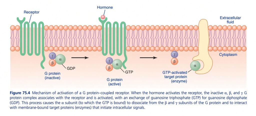
Figure 75.4 — ELI5: How a G Protein–Coupled Receptor (GPCR) Works
🧠 BIG IDEA FIRST
Think of a GPCR as a doorbell on the cell membrane.
- Hormone = person pressing the doorbell
- Receptor = doorbell
- G protein = messenger inside the house
- GDP = OFF signal
- GTP = ON signal
- Target enzyme = machine that produces the intracellular response
So the whole figure is:
Hormone binds receptor → G protein switches ON → α-subunit activates target enzyme → intracellular signal begins
1. 🧱 Cell membrane
The orange/pink double layer is the cell membrane.
It separates:
Outside
Extracellular fluid
from
Inside
Cytoplasm
The hormone is outside the cell.
The G protein and target enzyme are on the cytoplasmic side.
2. 🟢 Receptor — the “doorbell”
The green structure crossing the membrane is the:
G protein–coupled receptor (GPCR)
Notice that it passes through the membrane multiple times.
Most important concept:
The hormone cannot simply enter the cell.
Instead:
The hormone binds to the receptor on the outside of the membrane.
This changes the shape/function of the receptor.
That activated receptor then communicates with the G protein on the inside.3. 🔴 Hormone binds to receptor
The red circle labeled Hormone attaches to the receptor.
Think:
Before hormone:
Receptor = OFF
After hormone binds:
Receptor = ON
The receptor now activates the nearby G protein.
4. 🟣🟠🟢 What is the G protein?
The G protein has three subunits:
α (alpha)
β (beta)
γ (gamma)
Together:
α + β + γ = G protein
In the first part of the figure, all three are together.
5. 🔴 GDP means “OFF”
Look at the first G protein.
The α-subunit contains GDP.
GDP = OFF state
So:
α + GDP + β + γ
means:
🛑 G protein is inactive
This is why the figure labels it:
G protein (inactive)
6. 🔄 Hormone activates the receptor → GDP is replaced by GTP
This is the MOST IMPORTANT step.
When the hormone activates the receptor:
GDP leaves α
and
GTP binds α
So:
GDP OFF → GTP ON
Think of it like changing a battery switch:
GDP = OFF
GTP = ON
Now the G protein becomes:
G protein ACTIVE
7. 🟢 α-subunit separates from β + γ
Once GTP binds to α:
α-GTP separates from β and γ.
So instead of:
α + β + γ
we now have:
α-GTP ⟶ moves toward target protein
and
β + γ ⟶ remain together
Why does this matter?
Because α-GTP can now interact with another protein.
That other protein is the:
Target protein / enzyme
8. 🎯 Target protein — the next machine
The large yellow structure is the:
Target protein / enzyme
The activated α-GTP binds to it.
Think:
G protein = messenger
Target enzyme = machine
The messenger tells the machine:
“START!”
The enzyme then begins processes that produce intracellular signals.
9. ⚡ What happens after the target enzyme is activated?
The target enzyme can generate intracellular signaling molecules.
Depending on the type of G protein, this can lead to pathways involving things such as:
- cAMP
- IP₃
- DAG
- changes in Ca²⁺
These intracellular signals eventually produce the hormone’s effect.
Very important:
The hormone itself may be outside the cell, but its message is transmitted inside the cell.
🔥 The entire figure as a simple story
Imagine you are outside a house.
🧑 Hormone
You press the doorbell.
⬇️
🟢 GPCR
The doorbell receives the signal.
⬇️
🧬 G protein
The receptor activates the G protein.
⬇️
🔴 GDP leaves
The α-subunit loses its OFF signal.
⬇️
🟢 GTP binds
The α-subunit receives its ON signal.
⬇️
α-GTP separates
α separates from βγ.
⬇️
🎯 Target enzyme
α-GTP activates the target enzyme.
⬇️
⚡ Intracellular signals
The enzyme starts signaling inside the cell.
⬇️
🧠 Cellular response
The cell performs the hormone’s action.
⭐ The most important ON/OFF concept
GDP = OFF
GTP = ON
Memorize this:
GDP → inactive
GTP → active
This is one of the most important concepts in GPCR signaling.
🧩 Understand every label in the figure
| Label | Easiest meaning |
|---|---|
| Hormone | External messenger |
| Receptor | Detects the hormone |
| Extracellular fluid | Outside of cell |
| Cytoplasm | Inside of cell |
| G protein | Intracellular messenger |
| α subunit | Main signaling subunit shown |
| β + γ subunits | Other two G-protein subunits |
| GDP | OFF form |
| GTP | ON form |
| Target protein/enzyme | Activated by G protein |
| Intracellular signals | Messages produced inside cell |
🧠 10-SECOND EXAM REVISION
Inactive state:
GPCR + G protein (αβγ) + GDP
⬇️ Hormone binds
Activation:
GDP → GTP
⬇️
Separation:
α-GTP separates from βγ
⬇️
Action:
α-GTP → target enzyme
⬇️
Result:
Intracellular signaling → cellular response
🚨 One subtle but important point
The figure ends with α-GTP activating the target protein.
But the signal does not stay ON forever.
The α-subunit eventually hydrolyzes GTP → GDP.
Therefore:
GTP → GDP
and α becomes inactive again and can reassociate with βγ.
So the cycle is:
OFF → ON → OFF
GDP
⬇️ hormone/receptor activation
GTP
⬇️ target enzyme activation
GTP hydrolysis
⬇️
GDP
🔑 FINAL MASTER CONCEPT
Hormone outside the cell
↓
GPCR detects it
↓
GDP on α is replaced by GTP
↓
α-GTP becomes active
↓
α-GTP separates from βγ
↓
α-GTP activates target enzyme
↓
Intracellular signaling molecules are produced
↓
Cellular response
❤️ Remember it as:
“Hormone presses the receptor → receptor changes GDP to GTP → GTP turns G protein ON → α activates the enzyme → the message travels inside the cell.”
Figure 75.4 = GPCR is basically an OUTSIDE hormone signal being converted into an INSIDE cellular signal.
