


- Surgeons must be experts in fluid management because their patients depend on it.
- They care for patients who cannot eat or drink because they:
- Have severe bleeding.
- Have undergone surgery.
- Lose fluids from tubes, drains, or wounds.
- Surgeons must know how to manage these patients because their lives depend on proper fluid care.
- Fluid management may look simple.
- In reality, it is difficult because the body has complex fluid control systems.
- Managing a patient’s blood volume is one of the biggest challenges for surgeons.
- It requires careful control of:
- Fluid intake.
- Fluid output.
- Electrolytes.
- Blood loss must also be considered.
- Surgeons still do not completely understand the physiology of shock and resuscitation.
- Their knowledge is still incomplete.
- Because surgeons frequently treat bleeding patients, they continue to study these topics.
- Experience during wars has greatly improved knowledge of:
- Fluid management.
- Resuscitation.
- The wars in Iraq and Afghanistan also provided important new knowledge.
- Fluid treatment must be checked and adjusted continuously.
- This is because the human body is always changing.
- The first step is to know the patient’s starting condition.
- The surgeon must remember that the patient’s fluid status keeps changing.
- Many conditions can change fluid balance, including:
- Bleeding.
- Sepsis.
- Neuroendocrine disturbances.
- Problems in normal regulatory systems.
- These changes occur while the patient is ill and healing.
- Correct blood volume management depends greatly on timing.
- When blood volume is managed properly, surgeons can focus on other treatments such as:
- Nutrition.
- Antibiotics.
- Draining abscesses.
- Relieving obstruction.
- Relieving incarceration.
- Treating ischemia.
- Removing tumors.
- It is very important to recognize the difference between:
- Dehydration.
- Anemia.
- Hemorrhage.
- Overresuscitation.
- The human body is made mostly of water.
- Water is present in:
- Intravascular space.
- Intracellular space.
- Interstitial (third) space.
- Water moves between these spaces depending on many factors.
- This chapter mainly discusses the intravascular space.
- Surgeons can directly treat only the intravascular space.
- Managing the intravascular space also affects the other two fluid spaces.
- This chapter also explains the history of shock, fluids, and electrolytes.
- The purpose is not only to remember important discoveries.
- It is also to understand how medical knowledge developed over time.
- This helps explain:
- Why treatment methods changed in the past.
- Why they may change again in the future.
- Many old discoveries were important.
- However, some old ideas were later found to be incorrect.
- Future surgeons may also discover that some of today’s knowledge is incomplete or wrong.
- Studying history helps prevent repeating old mistakes.
- After the historical review, this chapter discusses:
- The fluids currently used.
- New fluids being developed.
- Finally, it explains the daily fluid care of perioperative patients.
KEY CONCEPT
- Fluid management is one of the surgeon’s most important responsibilities.
- Blood volume changes continuously, so fluid therapy must be adjusted continuously.
- Correct treatment depends on knowing the patient’s current fluid status.
- Surgeons mainly manage the intravascular space, which also influences the other body fluid compartments.
- Learning the history of fluid therapy helps understand present treatment and improve future care.
Conceptual Examples
- Patient with severe bleeding → Replace blood volume quickly → Maintain circulation.
- Patient losing fluid from a surgical drain → Monitor intake and output → Replace lost fluids appropriately.
- Patient with dehydration → Give fluids carefully → Restore normal blood volume without overresuscitation.
HISTORY
Resuscitation
- History may seem unimportant to people who only want the final answer.
- But learning history helps us know:
- Which treatments worked.
- Which treatments did not work.
- Medical ideas should always be questioned.
- We should ask whether today’s treatments are based on good science.
- Studying the history of shock is important for three reasons:
- Doctors and scientists had to understand blood loss because it was necessary.
- We need to know which experiments have already been done.
- We still need to learn more because our understanding of shock is still basic.
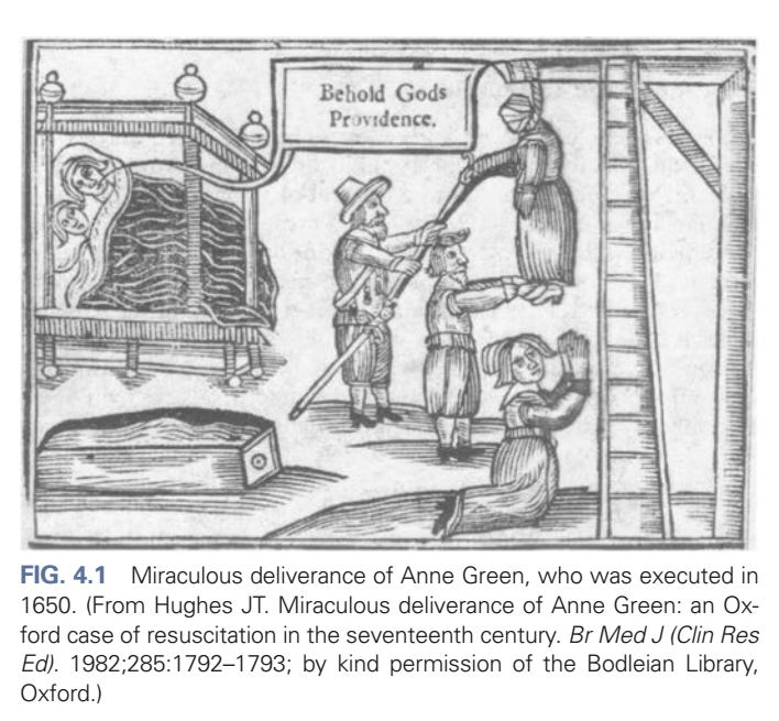
- One of the earliest recorded examples of resuscitation was the case of Anne Green.
- Anne Green was hanged on December 14, 1650.
- She was executed by hanging from a ladder.
- She remained hanging for about 30 minutes.
- During that time, some of her friends pulled on her legs, hoping to end her suffering more quickly (Fig. 4.1).
- Everyone believed she had died.
- Her body was placed in a coffin.
- The coffin was taken to Dr. William Petty.
- He had permission from the King to perform autopsies on executed prisoners.
- When the coffin was opened, Anne Green took a breath.
- A rattling sound was heard in her throat.
- Dr. Petty and Thomas Willis stopped preparing for the autopsy.
- They immediately tried to revive her.
- They sat her upright in the coffin.
- They forced her mouth open.
- They poured a hot cordial into her mouth.
- She coughed after drinking it.
- They rubbed her:
- Fingers.
- Hands.
- Arms.
- Feet.
- After about 15 minutes, they gave her more cordial.
- They gently tickled her throat with a feather.
- She briefly opened her eyes.
- They then opened a vein.
- About 5 ounces of blood were removed.
- They continued giving the cordial.
- They continued rubbing her arms and legs.
- They wrapped tight bandages around her arms and legs.
- They placed warming plasters on her chest.
- Another warming plaster was given as an enema to warm her bowels.
- They placed her in a warm bed.
- Another woman lay beside her to keep her warm.
- After 12 hours, Anne Green began to speak.
- After 24 hours, she answered questions normally.
- After 2 days, her memory had returned to normal.
- She remembered everything except:
- Her execution.
- The resuscitation.
KEY CONCEPT
- Studying history helps doctors understand which treatments worked and which did not.
- Our knowledge of shock is still incomplete, so learning from the past is important.
- The case of Anne Green is one of the earliest recorded examples of resuscitation.
- Doctors recognized signs of life, stopped the autopsy, and continued treatment until she recovered.
Conceptual Examples
- Patient appears dead but takes a breath → Doctors immediately stop other procedures and begin resuscitation.
- Patient becomes warmer and receives continuous care → Gradually regains consciousness, speech, and memory.
Shock
- Hemorrhagic shock has been studied for many centuries.
- Because injuries happen often, surgeons have learned most about shock while treating injured patients.
- Shock means the body’s tissues do not receive enough blood flow (perfusion).
- This definition is correct but does not explain everything.
- Cells need nutrients to survive.
- The exact nutrients needed are not completely known.
- Oxygen is the most important nutrient.
- But giving oxygen alone is not enough.
- Blood carries many important things, including:
- Nutrients.
- Buffers.
- Blood cells.
- Antibodies.
- Hormones.
- Chemicals.
- Electrolytes.
- Antitoxins.
- Blood delivery to tissues depends on:
- Blood volume.
- Anemia.
- Cardiac output (CO).
- How cells use nutrients is also affected by:
- Infection.
- Drugs.
- Blood vessel tone also affects shock.
- In neurogenic shock, sympathetic tone is lost.
- In sepsis, systemic vascular resistance decreases because normal regulation is disturbed.
- The word shock was first used in 1743.
- It appeared in a translation of Henri Francois Le Dran’s book about battlefield wounds.
- At that time, it meant the impact of injury, not the body’s response.
- In 1815, Guthrie used the word to describe physiologic instability after injury.
- Early theories about shock continued until the late 1800s.
- In 1830, Herman described one of the first uses of intravenous (IV) fluids.
- During a cholera outbreak, he injected 6 ounces of water into a vein to treat dehydration.
- In 1831, O’Shaughnessy treated cholera patients with large amounts of IV salt solution.
- He published these results in The Lancet.
- These were the first recorded attempts to replace intravascular fluid volume.
- However, treating cholera dehydration is not the same as treating hemorrhagic shock.
- In 1872, Gross described shock as “a manifestation of the rude unhinging of the machinery of life.”
- This definition became widely quoted.
- During the late 1800s, the cause of shock was still unclear.
- George Washington Crile believed shock happened because the autonomic nervous system failed.
- Between 1888 and 1918, ideas about shock changed greatly.
- Most surgeons then believed shock was mainly a nervous system problem.
- Today, this is known not to be the main cause.
- Even now, the exact mechanisms of hemorrhagic shock are still not fully understood.
- In 1899, Crile used the sphygmomanometer to measure blood pressure (BP).
- He suggested that very low BP explained the signs of shock.
- Before this, surgeons mainly judged shock by:
- Breathing.
- Pulse.
- Mental status.
- After Crile’s work, BP measurement became common.
- Harvey Cushing began measuring BP during all operations.
- This helped BP measurement become standard in medicine.
- Crile believed shock was the body’s attempt to survive, not simply the process of dying.
- Later, he concluded that low circulating blood volume was more important than low BP itself.
- In 1913, Crile proposed the kinetic system theory.
- He studied the role of thyroid hormone.
- He later recognized that epinephrine was very important in shock.
- His work was based partly on Walter Cannon’s experiments.
- Cannon showed that pain or emotion releases epinephrine.
- Epinephrine:
- Moves blood from the intestines to the brain and muscles.
- Causes the liver to release sugar from glycogen.
- Cannon believed these changes help the body defend itself.
- Crile added Cannon’s findings to his theory.
- He suggested that injury causes the brain to stimulate hormone release.
- These hormones produce changes throughout the body.
- His kinetic system included:
- Brain.
- Heart.
- Lungs.
- Blood vessels.
- Muscles.
- Thyroid gland.
- Liver.
- He believed severe stress could use up:
- Epinephrine.
- Glycogen.
- Thyroid hormone.
- Brain energy.
- When these energy stores were exhausted:
- BP fell.
- Shock developed.
- Henderson showed that reduced venous return lowers:
- Cardiac output.
- Arterial pressure.
- Better techniques helped measure heart chamber volume.
- Fat embolism also produced a shock-like condition.
- Its exact role remained uncertain because results were difficult to repeat.
- During the early 1900s, researchers also studied the vasomotor center.
- In 1914, Mann found that blood vessels supplied by nerves became more constricted during shock than denervated vessels.
- Experiences from World War I increased research on shock.
- Walter Cannon studied both soldiers and animal experiments.
- He suggested that:
- Toxins.
- Acidosis.
reduced blood vessel tone.
- Researchers then studied:
- Acidosis.
- Alkali treatment.
- They also studied the adrenal gland and adrenal hormones.
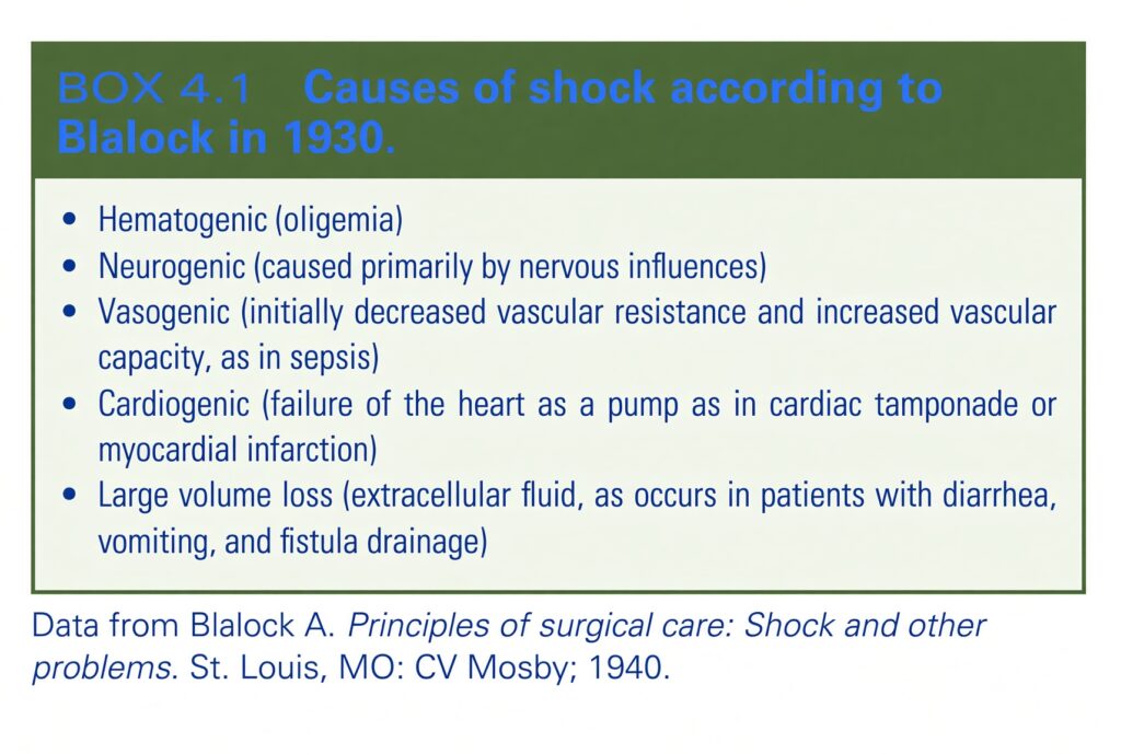
- In the 1930s, Blalock showed that almost all acute injuries change fluid and electrolyte balance.
- These changes mainly happen because effective circulating blood volume decreases.
- Blalock showed that reduced blood volume after injury can occur by several mechanisms (Box 4.1).
- He proved that injured tissues lose extracellular fluid (ECF).
- This fluid cannot help maintain blood circulation.
- From these studies, the idea of the “third space” developed.
- Third-space fluid is not available to the intravascular space.
- Carl John Wiggers introduced the idea of irreversible shock.
- His 1950 book Physiology of Shock summarized the knowledge of that time.
- He combined previous research with his own findings (Fig. 4.2).
- Wiggers used experiments now called the Wiggers preparation.
- He removed the spleens of dogs before the experiments.
- He inserted tubes into their arteries.
- This allowed him to measure arterial pressure.
- He lowered BP by removing blood.
- He usually reduced BP to about 40 mm Hg.
- After blood removal, BP rose by itself for a short time.
- This happened because fluid moved into the bloodstream.
- To keep BP at 40 mm Hg, Wiggers had to keep removing more blood.
- During this compensated phase, the dogs survived by using body fluid reserves.
- Water moved from:
- Intracellular space.
- Extracellular space.
- The body tried to keep enough blood flowing.
- Later, BP could no longer stay at 40 mm Hg without giving blood back.
- Wiggers called this uncompensated (irreversible) shock.
- If this stage continued, the dogs eventually died.
The best experimental model for studying shock is uncontrolled hemorrhage. Its main problem is that the amount of bleeding cannot be controlled. Because of this, results vary greatly. Even with this limitation, it is the most realistic model. Computer-assisted pressure models can copy the pressure changes seen in uncontrolled hemorrhage. These models reduce the artificial conditions found in pressure-controlled models. Smith and colleagues developed a hybrid hemorrhage model. In this model, a standard grade V liver injury is created in pigs. The pigs are allowed to bleed until:
- A fixed blood pressure is reached, or
- A fixed blood volume is lost.
The bleeding is then controlled by packing. This method reduces the variability seen in uncontrolled hemorrhage experiments.
KEY CONCEPT
- Shock = Inadequate tissue perfusion.
- Tissue perfusion depends on:
- Blood volume.
- Cardiac output.
- Blood vessel tone.
- Oxygen and other blood contents.
- Knowledge of shock has improved over centuries through:
- Clinical experience.
- War injuries.
- Animal experiments.
- Blalock introduced the idea of third-space fluid (Box 4.1).
- Wiggers described compensated and irreversible shock (Fig. 4.2).
History helps doctors understand which treatments worked and which did not. The case of Anne Green is one of the earliest recorded examples of successful resuscitation(Fig. 4.1). Uncontrolled hemorrhage is the most realistic way to study shock, but it produces variable results. A hybrid hemorrhage model makes bleeding more controlled while remaining close to real-life conditions.
Conceptual Examples
- Heavy bleeding → Blood volume falls → Tissue perfusion decreases → Hemorrhagic shock.
- Early shock → Body moves fluid into blood vessels → BP is temporarily maintained (compensated shock).
- Severe prolonged shock → Body can no longer compensate → BP continues to fall → Irreversible shock.
Patient appears dead but takes a breath → Doctors immediately stop the autopsy and begin resuscitation. Uncontrolled bleeding after trauma → Blood loss varies from patient to patient, making treatment and research more difficult. Hybrid experimental model → Bleeding is allowed first, then controlled with packing to produce more consistent results.
Fluids
- IV fluids like normal saline are used every day in medicine.
- Many people think these fluids were developed through strong scientific testing.
- Actually, that was not how they were introduced.
- Normal saline has been used for a long time.
- It is very useful.
- But today we know it can also be harmful.
- In 1882, Hartog Jakob Hamburger studied red blood cells in the laboratory.
- He incorrectly believed that 0.9% saline matched the salt concentration of human blood.
- He chose 0.9% saline because it had the same freezing point as human serum.
- This fluid is called normal (physiologic) saline.
- However, it is not truly physiologic and not truly normal.
- In 1831, O’Shaughnessy described treating cholera.
- He observed that cholera caused:
- Blood to move slowly in the veins.
- Poor oxygenation of blood.
- Blue skin.
- Loss of body heat.
- Stopped body secretions.
- Dark blood in arteries.
- Reduced carbon dioxide release from the lungs.
- Cold expired air.
- O’Shaughnessy wrote this after graduating from medical school at 22 years of age.
- He first tested IV fluid infusion in a dog.
- The dog showed no harmful effects.
- His goal was:
- To restore the normal thickness of blood.
- To replace lost salts.
- His experience also showed:
- Bloodletting reduced venous congestion.
- Nitrous oxide (laughing gas) did not improve oxygenation.
- In 1832, Robert Lewins reported seeing Thomas Latta inject large amounts of saline into veins.
- The treatment immediately:
- Improved blood flow.
- Improved blood color.
- Improved lung function.
- Latta’s solution contained:
- Sodium.
- Chloride.
- Bicarbonate.
- This solution was different from modern 0.9% normal saline.
- During the next 50 years, many different saline mixtures were used for cholera.
- None of them were the same as 0.9% normal saline.
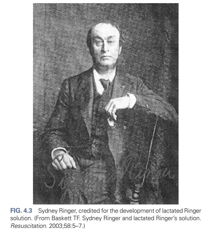
- In 1883, Sydney Ringer studied how blood components affected heart contraction (Fig. 4.3).
- He used:
- Frog hearts.
- 0.75% saline.
- Bullock blood.
- He first thought egg white improved heart contraction.
- He believed the benefit came from:
- Albumin.
- Potassium chloride.
- He performed many experiments by changing different ingredients.
- Later, Ringer found that his earlier results could not be repeated.
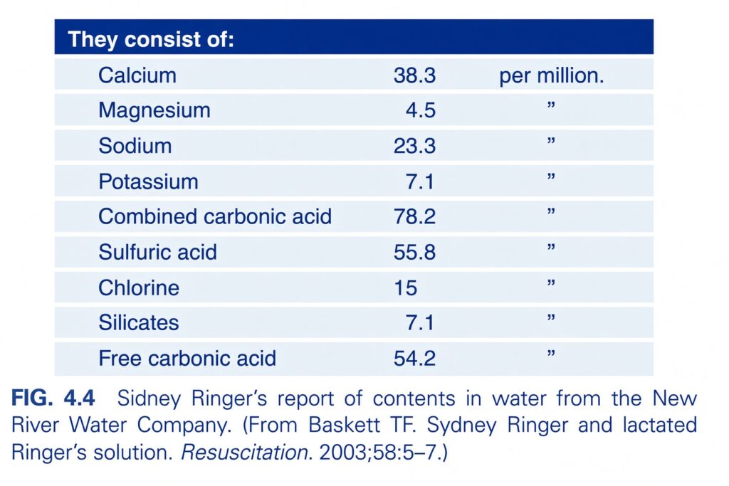
- He discovered that the water used was tap water, not distilled water as he had believed.
- The technician had used tap water instead.
- Ringer tested the tap water (Fig. 4.4).
- He found it contained many trace minerals.
- He discovered that:
- Calcium bicarbonate.
- Calcium chloride.
- restored good heart contractions.
- He also found sodium bicarbonate was necessary.
- Therefore, Ringer concluded that the important components were:
- Potassium.
- Calcium.
- Bicarbonate.
- Ringer solution soon became widely used in laboratory experiments.
- In the early 1900s, fluids were commonly given:
- Under the skin (hypodermoclysis).
- Into the rectum (proctoclysis).
- Hartwell and Hoguet used saline in dogs with intestinal obstruction.
- Their work became the basis for saline treatment in humans with intestinal obstruction.
- As IV crystalloid fluids developed, Ringer solution was modified.
- Alexis Hartmann made the most important change.
- In 1932, he wanted a fluid to treat acidosis.
- He added sodium lactate to Ringer solution.
- This created lactated Ringer (LR) or Hartmann solution.
- He used lactate instead of bicarbonate.
- Lactate changes into bicarbonate slowly.
- This reduced the risk of changing acidosis into alkalosis too quickly.
- In 1924, Rudolph Matas introduced the idea of a continuous IV drip.
- He also warned that normal saline could cause problems.
- He stated that metabolic problems from saline had been reported many times.
- However, these warnings were often ignored.
- Modern studies in healthy volunteers show that normal saline can cause:
- Abdominal discomfort.
- Pain.
- Nausea.
- Drowsiness.
- Reduced ability to perform complex mental tasks.
- Normal saline and lactated Ringer were not originally designed to replace blood.
- They were developed mainly for dehydration.
- When given in small amounts (1–3 L/day):
- They are generally well tolerated.
- They are usually safe.
- They replace water.
- The body can handle their electrolyte content.
- Over time, lactated Ringer became widely used for hemorrhagic shock.
- Both normal saline and lactated Ringer easily leave the blood vessels.
- They are poorly retained inside the bloodstream.
- After a few hours, only about 175–200 mL of a 1-liter infusion remains inside the blood vessels.
- Outside the United States:
- Lactated Ringer is often called Hartmann solution.
- Normal saline is often called physiologic (fisiologic) solution.
- Despite major scientific advances over the last 50 years, fluid resuscitation has changed very little.
KEY CONCEPT
- Normal saline became common because of historical practice, not because of perfect scientific evidence.
- Ringer solution was improved after discovering the importance of potassium, calcium, and bicarbonate (Fig. 4.3, Fig. 4.4).
- Hartmann added lactate to create lactated Ringer (LR).
- Normal saline and LR are good for dehydration, but they are not ideal blood replacements because most of the fluid leaves the blood vessels within a few hours.
Conceptual Examples
- Patient with dehydration → Normal saline or LR replaces lost water and electrolytes.
- Patient with severe bleeding → Giving 1 liter of saline does not mean all 1 liter stays in the bloodstream; after a few hours, only about 175–200 mL remains inside the blood vessels.
Blood Transfusions
- George Crile wanted to replace the blood lost by injured patients.
- He believed saline, adrenaline, and surgery could only replace lost blood up to a certain limit.
- At the beginning of the 20th century, blood transfusions were rare.
- Their use increased and decreased because:
- Transfusion reactions occurred.
- Blood often clotted before transfusion.
- From dog experiments, Crile believed blood could be exchanged without blood group matching.
- Alexis Carrel developed a method of sewing blood vessels together for transfusions.
- Crile thought this method was too slow for humans.
- He designed a short cannula to make transfusions easier.
- By World War II, shock was recognized as the most common treatable cause of illness and death.
- At the time of the Pearl Harbor attack (December 7, 1941):
- Blood banks were not available.
- Good blood transfusion facilities were not available.
- Most military hospitals also had very little dried plasma.
- Although wounded soldiers reached hospitals quickly, death rates remained high.
- IV fluids were almost unavailable.
- Only small amounts of saline were made in the operating room.
- Fluids were given using:
- A Salvesen flask.
- Reused rubber tubing.
- Reused tubing often caused high fever (febrile reactions).
- The first written report of resuscitation during World War II appeared in December 1942.
- It came from the 77th Evacuation Hospital in North Africa.
- E. D. Churchill reported that:
- Many wounded soldiers had already recovered from shock or died before reaching the hospital.
- Some patients arriving later were still in shock.
- Some patients urgently needed whole blood transfusion.
- Plenty of reconstituted plasma was available.
- However, whole blood was lacking.
- They also lacked:
- Blood transfusion sets.
- Sodium citrate.
- Sterile distilled water.
- Blood donors.
- At first, the Army mainly chose plasma instead of whole blood.
- This decision was supported by:
- The Office of the Surgeon General.
- Civilian researchers.
- They believed that shock caused:
- Thick blood.
- High hematocrit.
- On April 8, 1943, the Surgeon General decided not to send blood to combat areas.
- Seven months later, this decision remained unchanged because:
- Plasma was believed to be enough.
- Transporting blood was difficult.
- Shipping space was limited.
- Epinephrine and other vasoconstrictor drugs were not recommended.
- They were believed to reduce tissue blood flow by narrowing blood vessels.
- During World War II, the need for blood transfusion increased.
- This led to the development of blood banks.
- Better understanding of low blood volume (hypovolemia) improved shock treatment.
- Both plasma and whole blood became important for resuscitation.
- Treatment of traumatic shock improved greatly.
- Whole blood transfusion became widely used.
- Mixing whole blood with sodium citrate (6:1 ratio) prevented clotting by binding calcium.
- Doctors noticed that:
- Blood volume increased by only part of the amount transfused.
- This happened with:
- Blood.
- Colloids.
- Crystalloids.
- During the Korean War, doctors learned that more blood had to be given than the amount lost.
- The exact reason was not known.
- Possible reasons included:
- Hemolysis.
- Blood pooling in capillaries.
- Fluid moving into tissues.
- Raising the patient’s feet became an important part of shock treatment.
KEY CONCEPT
- Blood transfusion became important because IV fluids alone could not fully replace blood loss.
- World War II greatly improved the development of:
- Blood banks.
- Blood transfusion.
- Plasma therapy.
- Whole blood mixed with sodium citrate (6:1) prevented clotting during transfusion.
- During the Korean War, doctors learned that replacing blood loss often required giving more blood than the amount lost.
Conceptual Examples
- Patient with severe bleeding → Whole blood transfusion restores blood volume better than saline alone.
- Blood donation → Sodium citrate is added to prevent clotting before transfusion.
- Major trauma → Even after transfusion, additional blood may be needed because some blood is lost into tissues or trapped in small blood vessels.

MADE BY SELF LEARNING DR SHEEN
