

- The amount of food a person eats mainly depends on hunger.
- Hunger is the body’s natural (intrinsic) desire to eat food.
- The type of food a person chooses depends on appetite.
- Appetite is the desire for a particular type of food.
- Hunger and appetite are very important for maintaining an adequate nutritional supply for the body.
- The control of hunger and appetite is discussed in Chapter 72 in relation to body nutrition.
- This section focuses only on the mechanics of food ingestion.
- It mainly discusses:
- Mastication (chewing)
- Swallowing
KEY CONCEPT
- Food intake depends on two factors:
- Hunger → Determines how much food you eat.
- Appetite → Determines which type of food you want to eat.
- Both are important for maintaining proper nutrition.
- This chapter discusses only the mechanical process of eating, especially:
- Mastication (chewing)
- Swallowing
Conceptual Examples
- You have not eaten for many hours → Hunger increases → You eat a larger amount of food.
- You are craving sweet food → Appetite determines your food choice → You choose dessert ( SWEET DISH ) instead of vegetables.
- After choosing food → Chewing (mastication) begins → Swallowing moves the food into the digestive tract.
Easy Memory Trick
Hunger = How Much 🍽️
Appetite = What Type 🍕🍎
- Hunger → Amount of food
- Appetite → Choice of food
MASTICATION (CHEWING)
- The teeth are specially designed for chewing.
- The front teeth (incisors) are used for cutting food.
- The back teeth (molars) are used for grinding food.
- When all the jaw muscles work together, the teeth can produce:
- About 55 pounds of force on the incisors.
- About 200 pounds of force on the molars.
- Most chewing muscles are supplied by the motor branch of the fifth cranial nerve (trigeminal nerve).
- The chewing process is controlled by nuclei in the brainstem.
- Stimulation of specific reticular areas in the brainstem taste centers produces rhythmic chewing movements.
- Stimulation of the hypothalamus, amygdala, and cerebral cortex near the taste and smell sensory areas can also produce chewing.
- Chewing can begin voluntarily.
- After it begins, part of the chewing process occurs through a chewing reflex.
- When a bolus (food mass) enters the mouth, it first causes reflex relaxation (inhibition) of the chewing muscles.
- This relaxation allows the lower jaw to drop.
- The dropping jaw stretches the jaw muscles.
- This stretch triggers a stretch reflex.
- The stretch reflex causes the jaw muscles to contract again.
- This contraction raises the jaw and closes the teeth.
- The closing teeth compress the food bolus against the mouth lining.
- This again inhibits the jaw muscles.
- The jaw drops again.
- Another stretch reflex occurs.
- This cycle repeats again and again during chewing.
- Chewing is important for digestion of all foods.
- It is especially important for fruits and raw vegetables.
- These foods have cellulose coverings around their nutrients.
- These cellulose coverings must be broken before digestion can occur.
- Chewing also helps digestion because digestive enzymes act only on the surface of food particles.
- Therefore, the greater the surface area of the food, the faster digestion occurs.
- Chewing breaks food into very small particles.
- This prevents injury (excoriation) to the gastrointestinal tract.
- It also helps food leave the stomach more easily.
- Food then moves more easily through the small intestine and the rest of the gastrointestinal tract.
KEY CONCEPT
- Incisors cut food, while molars grind food.
- Jaw muscles can produce:
- 55 pounds of force on the incisors.
- 200 pounds of force on the molars.
- Most chewing muscles are supplied by the motor branch of the fifth cranial (trigeminal) nerve.
- Chewing is controlled by the brainstem and can also be influenced by the hypothalamus, amygdala, and cerebral cortex.
- The chewing reflex follows this sequence:
- Food enters the mouth
- Jaw muscles relax
- Jaw drops
- Stretch reflex occurs
- Jaw closes
- Food is compressed
- The cycle repeats
- Chewing:
- Breaks cellulose coverings of fruits and vegetables
- Increases the food surface area for digestive enzymes
- Protects the gastrointestinal tract
- Helps food move more easily through the digestive tract
Conceptual Examples
- Eating an apple → Incisors cut the apple → Molars grind it into small pieces.
- Food enters the mouth → Jaw drops → Stretch reflex closes the jaw → This cycle repeats automatically until the food is well chewed.
- Well-chewed food → Larger surface area → Digestive enzymes work faster → Food leaves the stomach more easily.
KEEP IN MIND
- Maximum chewing force:
- Incisors = 55 pounds
- Molars = 200 pounds
SWALLOWING (DEGLUTITION)
- Swallowing is a complex process.
- It is complex because the pharynx is used for both breathing and swallowing.
- During swallowing, the pharynx acts as a passage for food only for a few seconds.
- It is very important that breathing is not disturbed during swallowing.
- Swallowing is divided into three stages:
- Voluntary stage
- Pharyngeal stage
- Esophageal stage
- The voluntary stage starts the swallowing process.
- The pharyngeal stage is involuntary.
- During this stage, food passes through the pharynx into the esophagus.
- The esophageal stage is also involuntary.
- During this stage, food moves through the esophagus into the stomach.
KEY CONCEPT
- Swallowing (deglutition) is a complex process because the pharynx is shared by the respiratory and digestive systems.
- During swallowing, the pharynx temporarily becomes a food passage.
- Breathing must be protected during swallowing.
- Swallowing has three stages:
- Voluntary stage → Starts swallowing.
- Pharyngeal stage (involuntary) → Moves food from the pharynx to the esophagus.
- Esophageal stage (involuntary) → Moves food from the esophagus to the stomach.
Conceptual Examples
- You decide to swallow food → Voluntary stage begins.
- Food enters the pharynx → Pharyngeal stage automatically moves it into the esophagus.
- Food enters the esophagus → Esophageal stage automatically carries it to the stomach.
Easy Flow Chart
Food in Mouth
⬇️
Voluntary Stage → Swallowing begins
⬇️
Pharyngeal Stage (Involuntary) → Food passes through the pharynx into the esophagus
⬇️
Esophageal Stage (Involuntary) → Food moves into the stomach
Voluntary Stage of Swallowing
- When the food is ready to be swallowed, the tongue voluntarily pushes (squeezes or rolls) the food backward into the pharynx.
- The tongue does this by pressing the food upward and backward against the palate (roof of the mouth) (Fig. 64.1).
- After the food reaches the pharynx, swallowing becomes completely or almost completely automatic.
- Once this automatic process begins, it normally cannot be stopped.
KEY CONCEPT
- The voluntary stage is the first stage of swallowing.
- The tongue pushes the food upward and backward against the palate, moving it into the pharynx (Fig. 64.1).
- After the food enters the pharynx, swallowing becomes automatic.
- Once the automatic swallowing process starts, it usually cannot be stopped.
Conceptual Examples
- You decide to swallow food → Tongue pushes the food backward against the palate → Food enters the pharynx → Automatic swallowing begins.
- Food reaches the pharynx → The remaining stages of swallowing occur automatically without conscious control.
Easy Flow Chart
Food ready to swallow
⬇️
Tongue pushes food upward and backward against the palate
⬇️
Food enters the pharynx (Fig. 64.1)
⬇️
Swallowing becomes automatic
⬇️
Process usually cannot be stopped
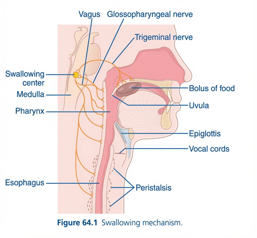
Figure 64.1: Swallowing Mechanism
Easiest and Most Conceptual Explanation
⭐ One-Line Concept
Swallowing is a coordinated reflex in which the brainstem (swallowing center in the medulla) safely moves a food bolus from the mouth to the stomach while protecting the airway from food entry.
First Understand the Whole Story
Swallowing occurs in a sequence:
- Food is pushed to the back of the mouth.
- The swallowing center in the medulla is activated.
- The airway closes temporarily.
- The food enters the esophagus, not the trachea.
- Peristaltic waves push the food toward the stomach.
Everything happens automatically within a few seconds.
Understanding Every Part of the Figure
1. Bolus of Food
The bolus is the soft, rounded mass of chewed food mixed with saliva.
Function
- It is the material being swallowed.
- It moves from the mouth to the pharynx and then into the esophagus.
Easy Concept
Think of the bolus as a small food ball ready to travel to the stomach.
2. Pharynx
The pharynx (throat) is the common passage for:
- Food
- Air
This is the place where the body decides:
- Food → Esophagus
- Air → Trachea
Easy Concept
The pharynx is the traffic intersection where food and air briefly share the same pathway.
3. Swallowing Center
Located in the medulla of the brainstem.
Function
It coordinates the entire swallowing reflex.
It:
- Receives sensory signals.
- Sends motor commands.
- Activates muscles in the correct order.
Easy Concept
The swallowing center is the control room that directs every step of swallowing.
4. Medulla
The medulla oblongata is part of the brainstem.
It contains:
- Swallowing center
- Respiratory center
- Cardiovascular center
Easy Concept
The medulla is the automatic control center for vital reflexes.
5. Trigeminal Nerve (Cranial Nerve V)
Function
Provides:
- Sensation from the mouth.
- Motor supply to muscles of chewing.
During swallowing, it helps prepare and move the food bolus.
Easy Concept
The trigeminal nerve mainly helps chew and position food.
6. Glossopharyngeal Nerve (Cranial Nerve IX)
This nerve carries sensory information from the:
- Pharynx
- Back of the tongue
When the food bolus touches the pharynx:
↓
This nerve sends signals to the swallowing center.
Easy Concept
It is the sensor that tells the brain:
“Food has reached the throat.”
7. Vagus Nerve (Cranial Nerve X)
This nerve sends motor signals from the swallowing center to:
- Pharynx
- Larynx
- Esophagus
It also initiates esophageal peristalsis.
Easy Concept
The vagus nerve is the main motor nerve of swallowing.
8. Uvula
The uvula is the small projection hanging from the soft palate.
During swallowing:
- It moves upward.
- It helps close the opening to the nasal cavity.
Why?
To prevent food from entering the nose.
Easy Concept
The uvula acts like a roof door that closes the path to the nose.
9. Epiglottis
The epiglottis is a flap of elastic cartilage.
During swallowing:
- It bends downward.
- It covers the entrance of the larynx.
Why?
To prevent food from entering the airway.
Easy Concept
The epiglottis is the safety lid over the windpipe.
10. Vocal Cords
The vocal cords also close tightly during swallowing.
This provides another protective barrier.
Why?
To stop food from entering the lungs.
Easy Concept
The vocal cords form the second safety gate protecting the airway.
11. Esophagus
The esophagus is the muscular tube connecting the pharynx to the stomach.
Its only function is transporting food.
Easy Concept
The esophagus is the food pipe.
12. Peristalsis
The dashed waves shown in the esophagus represent peristalsis.
Peristalsis is a coordinated wave of:
- Contraction behind the bolus.
- Relaxation in front of the bolus.
This pushes food toward the stomach.
Easy Concept
Think of squeezing toothpaste from the back of the tube.
The squeeze moves the paste forward.
That is exactly how peristalsis moves food.
Sequence of Swallowing (Step by Step)
Step 1
Food is chewed.
↓
Mixed with saliva.
↓
Forms a bolus.
Step 2
The tongue pushes the bolus backward.
↓
The bolus enters the pharynx.
Step 3
Stretch receptors in the pharynx are stimulated.
↓
Glossopharyngeal nerve carries sensory signals.
↓
Swallowing center in the medulla is activated.
Step 4
The swallowing center sends motor signals mainly through the vagus nerve.
↓
The swallowing reflex begins.Step 5
The uvula moves upward.
↓
The nasal cavity closes.
Step 6
The epiglottis folds downward.
↓
The laryngeal inlet closes.
Step 7
The vocal cords close.
↓
The airway is completely protected.
Step 8
The upper esophageal sphincter opens.
↓
Food enters the esophagus.
Step 9
Peristaltic waves push the bolus downward.
↓
Food reaches the stomach.
Why Doesn’t Food Normally Enter the Lungs?
Three protective mechanisms occur simultaneously:
- Uvula closes the nasal passage.
- Epiglottis covers the laryngeal opening.
- Vocal cords close tightly.
These mechanisms ensure that food enters the esophagus instead of the trachea.
Clinical Importance
If any part of this mechanism fails:
- Food may enter the airway (aspiration).
- This can cause choking or aspiration pneumonia.
Easy Story
Imagine food approaching a railway junction.
The swallowing center is the traffic controller.
Before allowing the train (food) to move:
- The road to the nose is closed by the uvula.
- The road to the lungs is closed by the epiglottis and vocal cords.
- The only open track is the esophagus.
Then, peristaltic waves gently push the food all the way to the stomach.
Understanding Every Label in the Figure
Bolus of Food
- Chewed food mixed with saliva.
- Ready to be swallowed.
Pharynx
- Common passage for food and air.
- Initiates the swallowing reflex.
Swallowing Center
- Located in the medulla.
- Coordinates the swallowing sequence.
Medulla
- Brainstem region containing the swallowing center.
Trigeminal Nerve (CN V)
- Helps with chewing and oral movements.
Glossopharyngeal Nerve (CN IX)
- Carries sensory signals from the pharynx to the swallowing center.
Vagus Nerve (CN X)
- Provides motor control to the pharynx and esophagus.
- Initiates esophageal peristalsis.
Uvula
- Elevates to close the nasopharynx.
Epiglottis
- Folds over the laryngeal inlet to prevent aspiration.
Vocal Cords
- Close during swallowing to protect the airway.
Esophagus
- Conducts food from the pharynx to the stomach.
Peristalsis
- Coordinated contraction behind and relaxation ahead of the bolus that propels food toward the stomach.
High-Yield Summary Table
| Structure | Main Function |
|---|---|
| Bolus of food | Food mass being swallowed |
| Pharynx | Common pathway where swallowing begins |
| Swallowing center | Coordinates the swallowing reflex |
| Medulla | Contains the swallowing center |
| Trigeminal nerve (CN V) | Assists chewing and oral phase of swallowing |
| Glossopharyngeal nerve (CN IX) | Carries sensory input from the pharynx |
| Vagus nerve (CN X) | Provides motor control to the pharynx and esophagus |
| Uvula | Closes the nasopharynx |
| Epiglottis | Covers the laryngeal inlet |
| Vocal cords | Close to protect the airway |
| Esophagus | Transports food to the stomach |
| Peristalsis | Propels the food bolus toward the stomach |
Key Concept
Swallowing is a highly coordinated reflex controlled by the swallowing center in the medulla. When the food bolus reaches the pharynx, sensory signals travel mainly through the glossopharyngeal nerve (CN IX) to the swallowing center. The center then sends motor signals primarily through the vagus nerve (CN X) to activate the muscles of swallowing. During this process, the uvula closes the nasopharynx, the epiglottis folds over the laryngeal inlet, and the vocal cords close to protect the airway. The upper esophageal sphincter opens, allowing the bolus to enter the esophagus, where peristaltic waves move it safely to the stomach.
Involuntary Pharyngeal Stage of Swallowing
- When the food bolus enters the back of the mouth and the pharynx, it stimulates swallowing receptors in the lining of the pharynx.
- These receptors are found especially around the tonsillar pillars.
- Nerve impulses from these receptors travel to the brainstem.
- The brainstem then starts a series of automatic contractions of the pharyngeal muscles.
- 1. The soft palate moves upward.
- This closes the posterior nares (back openings of the nose).
- This prevents food from entering the nasal cavity.
- 2. The palatopharyngeal folds on both sides move toward each other.
- They form a narrow vertical (sagittal) opening.
- The food passes through this opening into the posterior pharynx.
- This narrow opening allows only well-chewed food to pass easily.
- Because this stage lasts less than 1 second, large objects usually cannot enter the esophagus.
- 3. The vocal cords close tightly.
- The larynx moves upward and forward because of the neck muscles.
- At the same time, the epiglottis bends backward over the opening of the larynx.
- These actions prevent food from entering the nose and the trachea.
- The tight closure of the vocal cords is the most important protection.
- The epiglottis provides additional protection by helping keep food away from the vocal cords.
- If the vocal cords or their muscles are damaged, the risk of choking and airway blockage during swallowing increases.
- 4. As the larynx moves upward, it pulls open the entrance of the esophagus.
- At the same time, the upper 3–4 cm of the esophagus, called the upper esophageal sphincter (pharyngoesophageal sphincter), relaxes.
- This allows food to pass easily from the pharynx into the upper esophagus.
- Between swallows, this sphincter remains tightly closed.
- This prevents air from entering the esophagus during breathing.
- The upward movement of the larynx also moves the glottis away from the path of the food.
- Therefore, the food passes mainly on both sides of the epiglottis instead of over it.
- This provides additional protection against food entering the trachea.
- 5. After the larynx is elevated and the upper esophageal sphincter relaxes, the muscles of the pharynx contract.
- The contraction starts in the upper pharynx.
- It then moves downward through the middle and lower pharynx.
- This peristaltic wave pushes the food into the esophagus.
- In summary, during the pharyngeal stage of swallowing:
- The trachea closes.
- The esophagus opens.
- A rapid peristaltic wave pushes the food into the upper esophagus.
- The entire process is completed in less than 2 seconds.
KEY CONCEPT
- The pharyngeal stage of swallowing is involuntary.
- Food stimulates swallowing receptors in the pharynx.
- The brainstem automatically coordinates the swallowing reflex.
- During this stage:
- Soft palate closes the nose.
- Palatopharyngeal folds allow only well-chewed food to pass.
- Vocal cords close tightly.
- Epiglottis covers the laryngeal opening.
- Upper esophageal sphincter relaxes.
- Peristaltic contractions push food into the esophagus.
- The entire pharyngeal stage takes less than 2 seconds.
Conceptual Examples
- Food reaches the pharynx → Swallowing receptors are stimulated → Brainstem starts the swallowing reflex.
- Soft palate moves upward → Food cannot enter the nose.
- Vocal cords close and the epiglottis bends backward → Food cannot enter the trachea.
- Upper esophageal sphincter relaxes → Peristaltic wave pushes food into the esophagus.
Easy Flow Chart
Food enters the pharynx
⬇️
Swallowing receptors are stimulated
⬇️
Brainstem starts the swallowing reflex
⬇️
Soft palate closes the nose
⬇️
Palatopharyngeal folds form a narrow passage
⬇️
Vocal cords close + Epiglottis covers the larynx
⬇️
Upper esophageal sphincter relaxes
⬇️
Pharyngeal muscles contract (peristalsis)
⬇️
Food enters the esophagus
⬇️
Whole process completes in less than 2 seconds
Figure Number: None mentioned in the provided text.
KEEP IN MIND
- Duration of the pharyngeal stage: Less than 2 seconds.
- Length of the upper esophageal sphincter: 3–4 cm.
Nervous Initiation of the Pharyngeal Stage of Swallowing
- The most sensitive touch (tactile) areas for starting the pharyngeal stage of swallowing are located around the opening of the pharynx.
- The tonsillar pillars are the most sensitive areas.
- When food touches these areas, sensory nerve impulses are generated.
- These impulses travel through the sensory fibers of the trigeminal nerve (5th cranial nerve) and the glossopharyngeal nerve (9th cranial nerve).
- The impulses reach the medulla oblongata.
- They enter or pass very close to the tractus solitarius, which receives almost all sensory signals from the mouth.
- After this, the successive stages of swallowing begin automatically in the correct order.
- This automatic sequence is controlled by neuronal areas in the reticular substance of the medulla and the lower part of the pons.
- The sequence of the swallowing reflex is the same every time a person swallows.
- The timing of the swallowing cycle also remains the same with each swallow.
- The medulla and lower pons together form the deglutition (swallowing) center.
- The motor impulses from the swallowing center travel to the pharynx and upper esophagus.
- These impulses pass through:
- 5th cranial nerve (Trigeminal)
- 9th cranial nerve (Glossopharyngeal)
- 10th cranial nerve (Vagus)
- 12th cranial nerve (Hypoglossal)
- A few superior cervical nerves
- In summary, the pharyngeal stage of swallowing is mainly a reflex.
- It usually begins when food is voluntarily pushed to the back of the mouth.
- This stimulates the pharyngeal sensory receptors.
- The receptors then trigger the involuntary swallowing reflex.
KEY CONCEPT
- The tonsillar pillars are the most sensitive areas for starting the swallowing reflex.
- Sensory impulses travel through:
- 5th cranial nerve (Trigeminal)
- 9th cranial nerve (Glossopharyngeal)
- These impulses reach the medulla oblongata through the tractus solitarius.
- The deglutition (swallowing) center is located in the medulla and lower pons.
- Motor impulses leave the swallowing center through:
- 5th cranial nerve
- 9th cranial nerve
- 10th cranial nerve (Vagus)
- 12th cranial nerve (Hypoglossal)
- Some superior cervical nerves
- The pharyngeal stage is mainly an involuntary reflex, but it is usually triggered by the voluntary movement of food into the back of the mouth.
Conceptual Examples
- Tongue pushes food to the back of the mouth → Tonsillar pillars are stimulated → Sensory impulses travel to the medulla → Swallowing center starts the swallowing reflex.
- Swallowing center is activated → Motor impulses travel through cranial nerves V, IX, X, and XII → Pharyngeal muscles contract in the correct sequence.
- Every swallow → The same sequence and timing are repeated automatically.
Easy Flow Chart
Food reaches the back of the mouth
⬇️
Tonsillar pillars (most sensitive receptors) are stimulated
⬇️
Sensory impulses travel through CN V (Trigeminal) and CN IX (Glossopharyngeal)
⬇️
Signals reach the Medulla (Tractus Solitarius)
⬇️
Deglutition (Swallowing) Center in the Medulla + Lower Pons is activated
⬇️
Motor impulses travel through CN V, IX, X, XII (+ some superior cervical nerves)
⬇️
Automatic pharyngeal swallowing reflex occurs
The Pharyngeal Stage of Swallowing Momentarily Interrupts Respiration
- During the pharyngeal stage of swallowing, breathing stops for a short time.
- The entire pharyngeal stage usually finishes in less than 6 seconds.
- Therefore, breathing is interrupted for only a small part of one normal breathing cycle.
- During this time, the swallowing (deglutition) center temporarily inhibits the respiratory center in the medulla.
- This stops breathing, regardless of the stage of the breathing cycle.
- This temporary pause allows swallowing to occur safely.
- Even when a person is talking, swallowing interrupts breathing for such a short time that it is usually not noticed.
KEY CONCEPT
- The pharyngeal stage of swallowing temporarily stops breathing.
- This stage lasts less than 6 seconds.
- The swallowing center inhibits the respiratory center in the medulla.
- Breathing pauses briefly so that food can pass safely into the esophagus instead of the airway.
- The pause is so short that it is usually not noticeable, even during speaking.
Conceptual Examples
- Food enters the pharynx → Swallowing center becomes active → Respiratory center is temporarily inhibited → Breathing stops briefly → Food enters the esophagus safely → Breathing resumes.
- While talking, you swallow → Breathing pauses for only a moment → Most people do not notice the interruption.
Easy Flow Chart
Food enters the pharynx
⬇️
Swallowing center is activated
⬇️
Respiratory center in the medulla is temporarily inhibited
⬇️
Breathing stops briefly
⬇️
Food passes safely into the esophagus
⬇️
Breathing starts again
KEEP IN MIND
- Duration of the pharyngeal stage: Less than 6 seconds.
- Breathing interruption: Only a fraction of one normal respiratory cycle.
The Esophageal Stage of Swallowing Involves Two Types of Peristalsis
- The main function of the esophagus is to quickly carry food from the pharynx to the stomach.
- The movements of the esophagus are specially organized for this purpose.
- The esophagus normally has two types of peristaltic movements:
- Primary peristalsis
- Secondary peristalsis
- Primary peristalsis is the continuation of the peristaltic wave that starts in the pharynx during the pharyngeal stage of swallowing.
- This wave travels from the pharynx to the stomach in about 8–10 seconds.
- In a person who is standing upright, food usually reaches the lower end of the esophagus in about 5–8 seconds.
- This is because gravity also helps pull the food downward.
- If primary peristalsis does not move all the food into the stomach, secondary peristalsis begins.
- Secondary peristalsis occurs because the remaining food stretches (distends) the esophagus.
- These waves continue until all the food enters the stomach.
- Secondary peristalsis is started:
- Partly by intrinsic neural circuits in the myenteric nervous system.
- Partly by reflexes that:
- Begin in the pharynx.
- Travel to the medulla through vagal afferent fibers.
- Return to the esophagus through glossopharyngeal and vagal efferent fibers.
- The pharynx and upper one-third of the esophagus contain striated (skeletal) muscle.
- Therefore, peristalsis in these regions is controlled by skeletal motor nerve impulses through the glossopharyngeal and vagus nerves.
- The lower two-thirds of the esophagus contain smooth muscle.
- This part is strongly controlled by the vagus nerves through the esophageal myenteric nervous system.
- If the vagus nerves to the esophagus are cut, the esophageal myenteric plexus becomes more excitable after several days.
- It can then produce strong secondary peristaltic waves even without vagal reflexes.
- Therefore, even if the brainstem swallowing reflex is paralyzed, food placed into the esophagus by a tube or another method can still move into the stomach.
KEY CONCEPT
- The esophagus transports food from the pharynx to the stomach.
- It uses two types of peristalsis:
- Primary peristalsis → Continues the swallowing wave from the pharynx.
- Secondary peristalsis → Starts when food remains in the esophagus and stretches it.
- Primary peristalsis:
- 8–10 seconds from pharynx to stomach.
- 5–8 seconds in an upright person because gravity helps.
- Upper one-third of the esophagus contains striated muscle and is controlled by the glossopharyngeal and vagus nerves.
- Lower two-thirds contain smooth muscle and are controlled mainly through the vagus nerves and the myenteric plexus.
- Even if vagus nerves are cut, the myenteric plexus can eventually produce secondary peristalsis, allowing food to reach the stomach.
Conceptual Examples
- You swallow normally → Primary peristaltic wave moves food to the stomach.
- A small amount of food remains in the esophagus → Esophagus stretches → Secondary peristalsis pushes the remaining food into the stomach.
- Standing while swallowing water → Gravity helps the water reach the stomach faster (5–8 seconds).
- After vagus nerve injury → The myenteric plexus gradually takes over secondary peristalsis, allowing food in the esophagus to continue moving toward the stomach.
Easy Flow Chart
Food enters the esophagus
⬇️
Primary peristalsis starts (from the pharynx)
⬇️
Food moves toward the stomach (8–10 sec)
⬇️
Standing upright? → Gravity helps → Food reaches the lower esophagus in 5–8 sec
⬇️
Food remains?
⬇️
Yes → Esophagus stretches
⬇️
Secondary peristalsis begins
⬇️
Food enters the stomach
KEEP IN MIND
- Primary peristalsis: 8–10 seconds (pharynx → stomach).
- Food reaches the lower esophagus in an upright person: 5–8 seconds (because gravity helps).
Receptive Relaxation of the Stomach
- When the peristaltic wave in the esophagus approaches the stomach, a wave of relaxation occurs before it.
- This relaxation wave is transmitted through myenteric inhibitory neurons.
- As this relaxation wave reaches the lower end of the esophagus, the entire stomach relaxes.
- The duodenum also relaxes slightly.
- This relaxation prepares the stomach and duodenum to receive the incoming food.
- As a result, the food entering from the esophagus during swallowing can enter the stomach easily.
KEY CONCEPT
- Receptive relaxation means the stomach relaxes before food arrives.
- The relaxation wave travels ahead of the esophageal peristaltic wave.
- It is carried by myenteric inhibitory neurons.
- The whole stomach, and to a lesser extent the duodenum, relax before the food reaches them.
- This prepares the stomach to receive food smoothly without increasing pressure.
Conceptual ExamplesEasy Flow Chart
Food moves down the esophagus
⬇️
Esophageal peristaltic wave approaches the stomach
⬇️
Relaxation wave travels ahead through myenteric inhibitory neurons
⬇️
Stomach relaxes
⬇️
Duodenum relaxes slightly
⬇️
Food enters the stomach easily
Easy Memory Trick
“Relax First → Receive Food Later.”
- Peristalsis follows behind 🚶
- Relaxation goes ahead ➜
- Stomach opens first 🫙
- Food enters smoothly 🍞
Function of the Lower Esophageal Sphincter (Gastroesophageal Sphincter)
- The lower end of the esophagus has a broad lower esophageal sphincter (gastroesophageal sphincter).
- This sphincter extends about 3 cm above the junction of the esophagus and the stomach.
- It is formed by the circular muscle of the esophagus.
- Normally, the lower esophageal sphincter remains continuously (tonically) contracted.
- The pressure inside this sphincter is about 30 mm Hg.
- In contrast, the middle part of the esophagus normally remains relaxed.
- When a peristaltic swallowing wave travels down the esophagus, the lower esophageal sphincter undergoes receptive relaxation.
- This relaxation occurs before the peristaltic wave reaches the sphincter.
- This allows food to pass easily into the stomach.
- Sometimes, the lower esophageal sphincter does not relax properly.
- This condition is called achalasia.
- The stomach contains highly acidic secretions.
- These secretions also contain proteolytic (protein-digesting) enzymes.
- Most of the esophageal lining (except the lower one-eighth) cannot resist these gastric secretions for a long time.
- Normally, the tonic contraction of the lower esophageal sphincter prevents stomach contents from flowing back (refluxing) into the esophagus.
- Therefore, significant reflux usually occurs only in abnormal conditions.
KEY CONCEPT
- The lower esophageal sphincter (gastroesophageal sphincter) is a 3 cm long muscular region at the lower end of the esophagus.
- It normally remains tightly contracted with a pressure of about 30 mm Hg.
- During swallowing:
- Receptive relaxation occurs.
- Food passes easily into the stomach.
- If the sphincter fails to relax, the condition is called achalasia.
- The sphincter also prevents acidic stomach contents from refluxing into the esophagus.
- This protection is important because most of the esophageal mucosa cannot tolerate stomach acid and digestive enzymes.
Conceptual Examples
- Food reaches the lower esophagus → Lower esophageal sphincter relaxes → Food enters the stomach smoothly.
- After food enters the stomach → The sphincter closes again → Stomach acid cannot flow back into the esophagus.
- If the sphincter does not relax → Food cannot enter the stomach easily → Achalasia develops.
- If the sphincter becomes weak → Acid refluxes into the esophagus → The esophageal lining may be damaged.
Easy Flow Chart
Lower esophageal sphincter
⬇️
Normally closed (30 mm Hg pressure)
⬇️
Swallowing occurs
⬇️
Receptive relaxation
⬇️
Food enters the stomach
⬇️
Sphincter closes again
⬇️
Prevents acid reflux into the esophagus
Easy Memory Trick
“Close → Open → Close”
KEEP IN MIND
- Length of the lower esophageal sphincter: About 3 cm
- Normal sphincter pressure: About 30 mm Hg
Prevention of Esophageal Reflux By Valvelike Closure of the Distal End of the Esophagus
- Another mechanism that prevents esophageal reflux is the valvelike closure at the lower (distal) end of the esophagus.
- A short part of the esophagus extends slightly into the stomach.
- When intra-abdominal pressure increases, this part of the esophagus is pressed inward (collapsed).
- This creates a valvelike closure.
- The valvelike closure helps prevent stomach contents from flowing back into the esophagus.
- Therefore, high intra-abdominal pressure cannot easily force stomach acid into the esophagus.
- Without this mechanism, walking, coughing, or heavy breathing could push stomach acid back into the esophagus.
KEY CONCEPT
- A short part of the lower esophagus extends into the stomach.
- Increased intra-abdominal pressure presses this part inward, forming a valvelike closure.
- This valve prevents reflux of stomach contents into the esophagus.
- It protects the esophagus during activities that increase abdominal pressure, such as:
- Walking
- Coughing
- Heavy breathing
Conceptual Examples
- You cough → Abdominal pressure increases → The lower esophagus is compressed → The valve closes → Stomach acid does not reflux into the esophagus.
- You lift a heavy object → Intra-abdominal pressure rises → Valvelike closure prevents reflux.
- Without this valvelike mechanism → Every cough or deep breath could push stomach acid into the esophagus.
Easy Flow Chart
Increase in intra-abdominal pressure
⬇️
Lower end of the esophagus is compressed inward
⬇️
Valvelike closure forms
⬇️
Stomach contents cannot reflux into the esophagus
⬇️
Esophagus remains protected from stomach acidy Memory Trick
“Pressure Goes Up → Valve Closes → Acid Stays Down.”
- ↑ Abdominal pressure
- ➜ Valve closes
- ➜ No acid reflux
MADE BY SELF LEARNING DR SHEEN
