


- Besides the circulating renin–angiotensin system, many tissues have their own local renin–angiotensin systems.
- These local systems produce angiotensin II for use within the same tissue.
- Components of the renin–angiotensin system are present in the walls of blood vessels.
- They are also present in:
- Uterus
- Placenta
- Fetal membranes
- Amniotic fluid contains a high concentration of prorenin.
- Components of the renin–angiotensin system are also found in:
- Eyes
- Exocrine pancreas
- Heart
- Fat (adipose tissue)
- Adrenal cortex
- Testis
- Ovary
- Anterior pituitary
- Intermediate pituitary
- Pineal gland
- Brain
- Renin produced by these tissues contributes very little to the renin circulating in the blood.
- After removal of the kidneys, Plasma Renin Activity (PRA) falls to undetectable levels.
- This shows that the kidneys are the main source of circulating renin.
- The exact functions of these tissue renin–angiotensin systems are still not completely understood.
- Evidence suggests that angiotensin II acts as an important growth factor in the heart and blood vessels.
- ACE inhibitors and AT₁ receptor blockers are the preferred treatments for heart failure.
- Part of their benefit may be due to blocking the growth-promoting effects of angiotensin II.
KEY CONCEPT
- Many organs have their own local renin–angiotensin systems that produce angiotensin II for local actions. Although these tissue systems contribute very little to circulating renin, angiotensin II appears to play an important role in the growth and remodeling of the heart and blood vessels. Blocking angiotensin II with ACE inhibitors or AT₁ receptor blockers is beneficial in heart failure.
Conceptual Examples
- Example 1: In heart failure, excessive local angiotensin II promotes enlargement and remodeling of the heart. ACE inhibitors reduce these harmful effects by decreasing angiotensin II production.
- Example 2: Even though the heart contains its own renin–angiotensin system, removal of the kidneys causes Plasma Renin Activity (PRA) to become almost undetectable, showing that the kidneys are the main source of circulating renin.
ANGIOTENSIN II RECEPTORS
- There are at least two main types of angiotensin II receptors:
- AT₁ receptors
- AT₂ receptors
- AT₁ receptors are G-protein-coupled receptors (GPCRs).
- They are linked to the Gq protein.
- Activation of AT₁ receptors stimulates phospholipase C (PLC).
- This increases the level of intracellular calcium (Ca²⁺).
- AT₁ receptor activation also stimulates many tyrosine kinases.
- In vascular smooth muscle, AT₁ receptors are associated with caveolae.
- Angiotensin II increases the production of caveolin-1, an important protein of caveolae.
- In rodents, there are two closely related AT₁ receptor subtypes:
- AT₁A
- AT₁B
- These two subtypes are produced by two separate genes.
- AT₁A receptors are found in:
- Blood vessel walls
- Brain
- Many other organs
- AT₁A receptors produce most of the known effects of angiotensin II.
- AT₁B receptors are mainly found in:
- Anterior pituitary
- Adrenal cortex
- In humans, the AT₁ receptor gene is located on chromosome 3.
- It is still uncertain whether humans have separate AT₁A and AT₁B receptor subtypes.
- AT₂ receptors are also present in the body.
- In humans, the AT₂ receptor gene is located on the X chromosome.
- Like AT₁ receptors, AT₂ receptors have seven transmembrane domains.
- However, AT₂ receptors produce different effects from AT₁ receptors.
- AT₂ receptors activate phosphatase enzymes through a G-protein.
- These phosphatases oppose (reduce) growth-promoting effects.
- AT₂ receptor activation also opens potassium (K⁺) channels.
- AT₂ receptors increase the production of nitric oxide (NO).
- Nitric oxide increases intracellular cyclic guanosine monophosphate (cGMP).
- The overall physiological effects of these signaling pathways are not yet fully understood.
- AT₂ receptors are more abundant during fetal and newborn life.
- They remain present in the brain and some other organs in adults.
- AT₁ receptors in blood vessels and the adrenal cortex are regulated differently.
- High levels of angiotensin II decrease (down-regulate) AT₁ receptors in blood vessels.
- This reduces the response of blood vessels to angiotensin II.
- High levels of angiotensin II increase (up-regulate) AT₁ receptors in the adrenal cortex.
- This makes the adrenal cortex more sensitive to the aldosterone-stimulating effect of angiotensin II.
KEY CONCEPT
- Angiotensin II acts through two main receptors. AT₁ receptors mediate most of the known actions of angiotensin II, including increased intracellular Ca²⁺, vasoconstriction, and aldosterone secretion. AT₂ receptors have different signaling pathways that oppose growth, increase nitric oxide (NO) and cGMP production, and open K⁺ channels. High angiotensin II levels down-regulate vascular AT₁ receptors but up-regulate adrenal AT₁ receptors, increasing aldosterone secretion.
Conceptual Examples
- Example 1: During dehydration, angiotensin II binds to AT₁ receptors in blood vessels, causing vasoconstriction, while simultaneously stimulating AT₁ receptors in the adrenal cortex to release aldosterone, which helps retain sodium and water.
- Example 2: In a patient with chronically elevated angiotensin II, blood vessels become less responsive because vascular AT₁ receptors are down-regulated, but the adrenal cortex becomes more responsive because its AT₁ receptors are up-regulated, allowing continued aldosterone secretion.
THE JUXTAGLOMERULAR APPARATUS
- Renin is produced by specialized kidney cells called juxtaglomerular (JG) cells.
- JG cells are modified smooth muscle (epitheloid) cells.
- They are located in the wall (media) of the afferent arteriole just before it enters the glomerulus.
- JG cells contain membrane-bound secretory granules.
- These secretory granules store renin.
- Renin is also present in agranular lacis cells.
- Lacis cells are located between the afferent arteriole and the efferent arteriole.
- The exact function of renin in lacis cells is still unknown.
- At the vascular pole of the glomerulus, the nephron tubule comes into contact with its own afferent and efferent arterioles.
- This point marks the beginning of the distal convoluted tubule (DCT).
- At this location, the tubular epithelial cells become modified to form the macula densa.
- The macula densa lies very close to the juxtaglomerular (JG) cells.
- Together, three structures form the juxtaglomerular apparatus (JGA):
- Macula densa
- Juxtaglomerular (JG) cells
- Lacis cells (extraglomerular mesangial cells)
Figure: Figure 38–8
KEY CONCEPT
- The juxtaglomerular apparatus (JGA) is a specialized structure located at the vascular pole of the nephron. It is made of three components: the macula densa, juxtaglomerular (JG) cells, and lacis cells. JG cells produce and store renin, the macula densa monitors tubular fluid near the start of the distal convoluted tubule, and the function of lacis cells is not fully understood. Together, these structures help regulate renin release and maintain blood pressure and kidney function.
Conceptual Examples
- Example 1: A patient has low blood pressure. The JG cells detect this and release renin, which activates the renin–angiotensin system to increase blood pressure.
- Example 2: When less sodium chloride reaches the macula densa, it signals the nearby JG cells to release more renin, helping increase blood pressure and improve kidney perfusion.
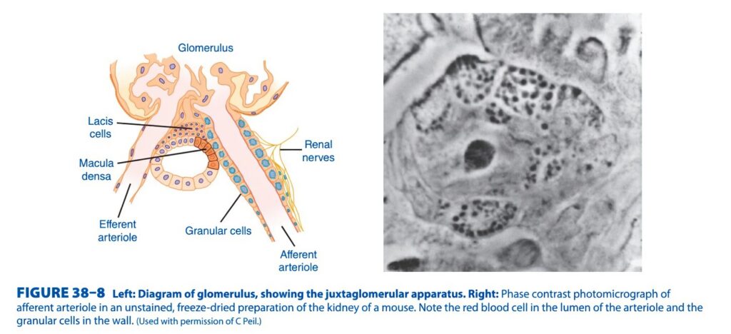
Figure 38-8: Juxtaglomerular Apparatus (JGA)
Easiest & Most Conceptual Explanation for SELF LEARNERS
This figure shows the Juxtaglomerular Apparatus (JGA), which is the kidney’s “blood pressure and salt sensor.”
Its main job is to monitor blood pressure and sodium (NaCl) delivery and regulate:
- Renin secretion
- Glomerular filtration rate (GFR)
- Blood pressure
- Body fluid volume
⭐ One-Line Concept
The Juxtaglomerular Apparatus is a specialized region where the distal tubule touches the afferent arteriole, allowing the kidney to sense blood pressure and sodium levels and regulate renin release and GFR.
First Understand the Whole Picture
Imagine a water filtration plant.
- A pipe brings water in → Afferent arteriole
- The filter → Glomerulus
- The cleaning tube checks the filtered fluid → Macula densa
- A pressure sensor sits on the incoming pipe → Granular (Juxtaglomerular) cells
- A communication centre connects everything → Lacis cells
All four structures together form the Juxtaglomerular Apparatus (JGA).
Components of the Figure
1. Glomerulus
Located at the top of the figure.
Function
- Filters blood
- Produces the initial filtrate
Easy Concept
Think of it as a coffee filter.
Blood enters → filtration occurs → filtrate enters the nephron.
2. Afferent Arteriole
This is the vessel bringing blood into the glomerulus.
Function
- Delivers blood to be filtered
- Contains granular (juxtaglomerular) cells
Easy Concept
It is the incoming water pipe supplying the filter.
3. Efferent Arteriole
This vessel carries blood away from the glomerulus.
Function
- Removes blood after filtration
- Helps maintain glomerular filtration pressure
Easy Concept
It is the outgoing pipe after the filter.4. Granular Cells (Juxtaglomerular Cells)
These blue-stained cells lie in the wall of the afferent arteriole.
Most Important Cells in This Figure
Function
They:
- Sense blood pressure inside the afferent arteriole
- Store renin in secretory granules
- Release renin when required
When do they release renin?
They release renin when:
- Blood pressure falls
- Blood volume decreases
- Sympathetic stimulation increases
- Macula densa senses low NaCl delivery
Easy Memory
Granular cells = Renin factory
5. Macula Densa
This is the dark patch of specialized cells in the distal tubule.
Function
It measures:
- Sodium chloride (NaCl)
- Tubular flow
It continuously checks how much NaCl reaches the distal tubule.
If NaCl is LOW
Macula densa tells granular cells:
“Not enough filtration—release renin!”
↓
Renin increases.
If NaCl is HIGH
Macula densa signals:
“Too much filtration—reduce GFR.”
This is called tubuloglomerular feedback.
Easy Concept
Macula densa is the kidney’s salt sensor.
6. Lacis Cells (Extraglomerular Mesangial Cells)
These cells lie between:
- Macula densa
- Afferent arteriole
- Efferent arteriole
Function
They are believed to:
- Support the JGA structurally
- Help transmit signals between the macula densa and granular cells
Easy Concept
Think of them as the communication bridge within the JGA.
7. Renal Nerves
These sympathetic nerve fibres reach the afferent arteriole.
Function
During:
- Blood loss
- Stress
- Shock
they stimulate granular cells to release renin.
Easy Concept
The renal nerves act as an emergency alarm system.
How Does the Entire JGA Work?
Step 1
Blood enters through the afferent arteriole.
↓
Step 2
The glomerulus filters blood.
↓
Step 3
The filtrate reaches the macula densa.
↓
Step 4
Macula densa measures NaCl concentration.
↓
Step 5
If NaCl delivery is low,
↓
Macula densa signals the granular cells.
↓
Step 6
Granular cells release renin.
↓
Step 7
Renin activates the renin–angiotensin–aldosterone system (RAAS).
↓
Blood pressure rises.
↓
Kidney filtration is maintained.
Easy Flow Diagram
Blood enters
Afferent arteriole
        │
        ▼
Granular cells monitor pressure
        │
        ▼
Glomerulus filters blood
        │
        ▼
Filtrate reaches macula densa
        │
        ▼
Macula densa checks NaCl
        │
        ├── High NaCl
        │      ↓
        │ Reduce GFR
        │
        └── Low NaCl
               ↓
      Signal granular cells
               ↓
         Renin release
               ↓
             RAAS
               ↓
      Blood pressure increases
Understanding the Right-Side Photomicrograph
The right panel is a phase-contrast photomicrograph of the afferent arteriole.
It demonstrates the real microscopic appearance of the granular (juxtaglomerular) cells.
What are the dark dots?
They are secretory granules containing renin.
These granules confirm that the granular cells are endocrine cells capable of storing and releasing renin.
Everyday Analogy
Imagine a water treatment plant.
- Afferent arteriole = Water inlet pipe
- Glomerulus = Water filter
- Macula densa = Water-quality sensor
- Granular cells = Pressure-control office
- Lacis cells = Communication cables
- Renal nerves = Emergency control line
If water pressure falls or the water-quality sensor detects reduced salt delivery, the control office releases renin to restore the system.
Easy Memory Trick
Remember the letters G-M-L-R:
- G = Granular cells → Release Renin
- M = Macula densa → Measures NaCl
- L = Lacis cells → Link/communication
- R = Renal nerves → Regulate renin release
High-Yield Exam Points
- The Juxtaglomerular Apparatus (JGA) is formed where the distal tubule (macula densa) contacts the vascular pole of the glomerulus.
- It consists of macula densa cells, granular (juxtaglomerular) cells, and lacis (extraglomerular mesangial) cells.
- Granular cells are modified smooth muscle cells in the wall of the afferent arteriole.
- Granular cells synthesize, store, and secrete renin.
- Macula densa senses the NaCl concentration in the distal tubular fluid.
- Low NaCl delivery or reduced renal perfusion stimulates renin release through macula densa and sympathetic mechanisms.
- The JGA regulates both glomerular filtration rate (GFR) and systemic blood pressure through tubuloglomerular feedback and activation of the renin–angiotensin–aldosterone system (RAAS).
- The photomicrograph demonstrates granular cells filled with renin-containing secretory granules.
KEY CONCEPT (Figure 38-8)
Figure 38-8 illustrates the Juxtaglomerular Apparatus (JGA), the kidney’s specialized regulatory unit that coordinates blood pressure control, renin secretion, and glomerular filtration. The macula densa monitors sodium chloride delivery in the distal tubule, while the granular (juxtaglomerular) cells of the afferent arteriole sense perfusion pressure and store renin. Lacis cells facilitate communication within the JGA. When renal perfusion pressure or tubular NaCl delivery falls, the JGA stimulates renin release, activates the renin–angiotensin–aldosterone system (RAAS), increases blood pressure, and helps preserve the glomerular filtration rate (GFR). The photomicrograph highlights the renin-containing granules within the granular cells.
REGULATION OF RENIN SECRETION
- Renin secretion is controlled by several factors.
- The amount of renin released at any time depends on the combined effect of all these factors.
Intra-Renal Baroreceptor Mechanism
- One important regulator is the intra-renal baroreceptor mechanism.
- The juxtaglomerular (JG) cells act as pressure sensors in the afferent arteriole.
- When pressure in the afferent arteriole increases, renin secretion decreases.
- When pressure in the afferent arteriole decreases, renin secretion increases.
Macula Densa Mechanism
- Another important regulator is the macula densa.
- Renin secretion is inversely related to the amount of sodium (Na⁺) and chloride (Cl⁻) reaching the distal renal tubule from the loop of Henle.
- High Na⁺ and Cl⁻ delivery to the macula densa decreases renin secretion.
- Low Na⁺ and Cl⁻ delivery to the macula densa increases renin secretion.
- Na⁺ and Cl⁻ enter the macula densa cells through Na–K–2Cl⁻ transporters in the apical membrane.
- This triggers a signal that acts on nearby JG cells to regulate renin release.
- Nitric oxide (NO) may act as this signaling molecule, but this has not been confirmed.
Effect of Potassium (K⁺)
- Renin secretion is also inversely related to plasma potassium (K⁺) concentration.
- High plasma K⁺ decreases renin secretion.
- This effect occurs mainly because K⁺ changes the amount of Na⁺ and Cl⁻ delivered to the macula densa.
Negative Feedback by Angiotensin II
- Angiotensin II directly inhibits renin secretion from JG cells.
- This is a negative feedback mechanism that prevents excessive activation of the renin–angiotensin system.
Effect of Vasopressin (ADH)
- Vasopressin (ADH) also inhibits renin secretion.
- This inhibitory effect has been demonstrated both in laboratory studies (in vitro) and in living organisms (in vivo).
- It is still uncertain whether the effect in the body is direct or indirect.
Sympathetic Nervous System
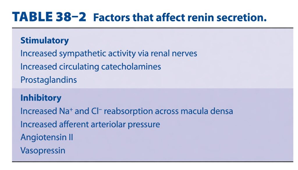
- Increased sympathetic nervous activity increases renin secretion.
- This occurs through:
- Circulating catecholamines
- Norepinephrine released from renal sympathetic nerves
- These catecholamines mainly stimulate β₁-adrenergic receptors on JG cells.
- β₁ receptor stimulation increases intracellular cyclic AMP (cAMP).
- The increase in cAMP stimulates renin release.
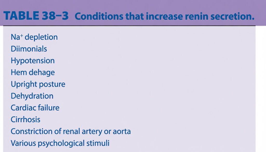
Conditions That Increase Renin Secretion
- Several conditions increase renin secretion in humans.
- Most of these conditions decrease central venous pressure.
- Reduced central venous pressure increases sympathetic nervous activity.
- Some conditions also reduce pressure in the renal arterioles.
- Constriction of the renal artery decreases renal arteriolar pressure and increases renin secretion.
- Constriction of the aorta above the renal arteries also decreases renal blood flow and stimulates renin release.
- Psychological stress increases renal sympathetic nerve activity.
- This also increases renin secretion.
Table: Table 38–2
Table: Table 38–3
Clinical Box: Clinical Box 38–3
KEY CONCEPT
- Renin secretion is mainly regulated by four mechanisms: the intra-renal baroreceptor, the macula densa, the sympathetic nervous system, and negative feedback by angiotensin II. Low renal perfusion pressure, low Na⁺ and Cl⁻ delivery to the macula densa, and increased sympathetic stimulation all increase renin secretion, whereas high blood pressure, high Na⁺ and Cl⁻ delivery, angiotensin II, and vasopressin decrease renin secretion.
Conceptual Examples
- Example 1: A patient with severe dehydration has low renal blood pressure and low Na⁺ delivery to the macula densa. Both signals stimulate JG cells to release renin, activating the renin–angiotensin–aldosterone system to restore blood pressure.
- Example 2: During acute stress or haemorrhage, the sympathetic nervous system releases norepinephrine, which stimulates β₁ receptors on JG cells, increases cAMP, and promotes renin secretion.
CLINICAL BOX 38–3
Role of Renin in Clinical Hypertension
- Narrowing (constriction) of one renal artery reduces blood flow to that kidney.
- The affected kidney interprets the reduced blood flow as low blood pressure.
- As a result, the juxtaglomerular (JG) cells release more renin.
- Increased renin activates the renin–angiotensin–aldosterone system (RAAS).
- This increases angiotensin II and aldosterone levels.
- Blood pressure rises and remains elevated, producing sustained hypertension.
- This type of hypertension is called:
- Renal hypertension
- Goldblatt hypertension
- If the narrowed renal artery is corrected or the ischemic (poorly perfused) kidney is removed early, the hypertension can be cured.
- This is true only if the hypertension has not been present for a long time.
One-Clip, Two-Kidney Goldblatt Hypertension
- One renal artery is narrowed (one clip).
- The other kidney remains normal.
- The affected kidney releases excessive renin.
- Circulating renin levels become increased.
- This causes hypertension.
- A similar condition occurs in humans when one renal artery becomes narrowed by atherosclerosis or other diseases of the renal circulation.
One-Clip, One-Kidney Goldblatt Hypertension
- Only one kidney is present, and its renal artery is narrowed.
- In this condition, Plasma Renin Activity (PRA) is usually normal.
- The exact reason for hypertension in this situation is still not fully understood.
Response to Treatment
- Many patients with hypertension improve after treatment with:
- ACE inhibitors
- Losartan (AT₁ receptor blocker)
- This benefit can occur even when:
- The renal circulation appears normal.
- Plasma Renin Activity (PRA) is normal.
- Plasma Renin Activity (PRA) is low.
- This shows that blocking the renin–angiotensin system can effectively reduce blood pressure in many patients, even when circulating renin levels are not elevated.
Clinical Box: Clinical Box 38–3
KEY CONCEPT
- Reduced blood flow to a kidney stimulates renin release, activating the renin–angiotensin–aldosterone system and causing hypertension (Goldblatt hypertension). Early correction of the reduced blood supply can reverse the hypertension. ACE inhibitors and AT₁ receptor blockers are effective treatments for many patients with hypertension, even when Plasma Renin Activity (PRA) is normal or low.
Conceptual Examples
- Example 1: A patient has narrowing of the right renal artery due to atherosclerosis. The right kidney senses low blood flow, releases renin, activates the RAAS, and causes persistent hypertension, even though the left kidney is normal.
- Example 2: A patient with essential hypertension has normal Plasma Renin Activity (PRA) but responds well to captopril or losartan because blocking the renin–angiotensin system still lowers blood pressure effectively.
MADE BY EASIEST SELF LEARNING CEO AND FOUNDER DR SHEEN
