

Physiologic Anatomy of the Gastrointestinal Wall
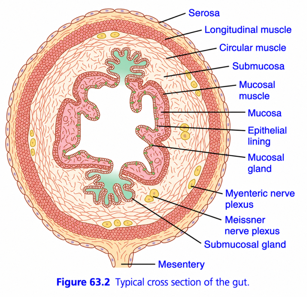
- Figure 63.2 shows a typical cross-section of the intestinal wall.
- From the outer surface to the inner surface, the intestinal wall has these layers:
- Serosa
- Longitudinal smooth muscle layer
- Circular smooth muscle layer
- Submucosa
- Mucosa
- Small bundles of smooth muscle fibers, called the mucosal muscle, are present in the deeper part of the mucosa.
- The different smooth muscle layers perform the motor (movement) functions of the gastrointestinal tract.
- The general structure and function of smooth muscle are explained in Chapter 8.
- Review Chapter 8 first because it provides the background needed to understand the following parts of this chapter.
KEY CONCEPT
- The gastrointestinal wall has five main layers from outside to inside.
- Mucosal muscle consists of small smooth muscle bundles in the deeper mucosa.
- Smooth muscle layers are responsible for gastrointestinal movements.
- Figure 63.2 illustrates the layers of the intestinal wall.
- Chapter 8 provides the basic concepts of smooth muscle needed for this topic.
GENERAL PRINCIPLES OF GASTROINTESTINAL MOTILITY
Gastrointestinal Smooth Muscle Functions as a Syncytium
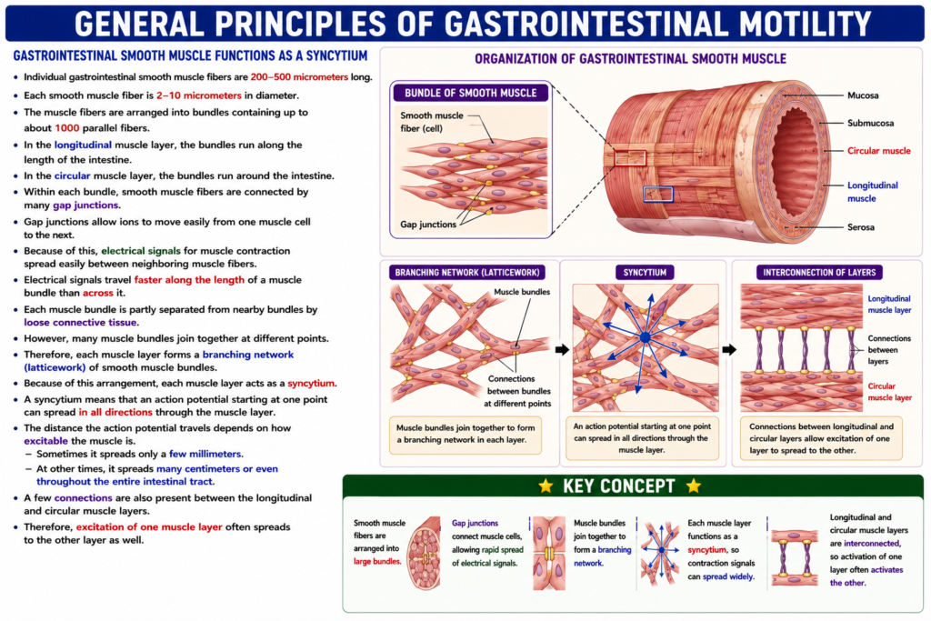
- Individual gastrointestinal smooth muscle fibers are 200–500 micrometers long.
- Each smooth muscle fiber is 2–10 micrometers in diameter.
- The muscle fibers are arranged into bundles containing up to about 1000 parallel fibers.
- In the longitudinal muscle layer, the bundles run along the length of the intestine.
- In the circular muscle layer, the bundles run around the intestine.
- Within each bundle, smooth muscle fibers are connected by many gap junctions.
- Gap junctions allow ions to move easily from one muscle cell to the next.
- Because of this, electrical signals for muscle contraction spread easily between neighboring muscle fibers.
- Electrical signals travel faster along the length of a muscle bundle than across it.
- Each muscle bundle is partly separated from nearby bundles by loose connective tissue.
- However, many muscle bundles join together at different points.
- Therefore, each muscle layer forms a branching network (latticework) of smooth muscle bundles.
- Because of this arrangement, each muscle layer acts as a syncytium.
- A syncytium means that an action potential starting at one point can spread in all directions through the muscle layer.
- The distance the action potential travels depends on how excitable the muscle is.
- Sometimes it spreads only a few millimeters.
- At other times, it spreads many centimeters or even throughout the entire intestinal tract.
- A few connections are also present between the longitudinal and circular muscle layers.
- Therefore, excitation of one muscle layer often spreads to the other layer as well.
KEY CONCEPT
- Gastrointestinal smooth muscle fibers are arranged into large bundles.
- Gap junctions electrically connect muscle cells, allowing rapid spread of electrical signals.
- Muscle bundles join together to form a branching network.
- Each muscle layer functions as a syncytium, so contraction signals can spread widely.
- Longitudinal and circular muscle layers are interconnected, so activation of one layer often activates the other.
GENERAL PRINCIPLES OF GASTROINTESTINAL MOTILITY
Electrical Activity of Gastrointestinal Smooth Muscle
- Gastrointestinal smooth muscle is continuously stimulated by slow, natural (intrinsic) electrical activity in the muscle cell membranes.
- This electrical activity has two basic types of electrical waves:
- Slow waves
- Spikes (Figure 63.3)
- The resting membrane potential of gastrointestinal smooth muscle can also change to different levels.
- Changes in the resting membrane potential play an important role in controlling gastrointestinal motor activity.
KEY CONCEPT
- Gastrointestinal smooth muscle has continuous intrinsic electrical activity.
- There are two main electrical waves:
- Slow waves
- Spikes (Figure 63.3)
- Changes in resting membrane potential also help regulate gastrointestinal movements.
GENERAL PRINCIPLES OF GASTROINTESTINAL MOTILITY
“Slow Waves” Caused by Undulating Changes in Resting Membrane Potential
- Most gastrointestinal contractions occur rhythmically.
- This rhythm is mainly determined by the frequency of slow waves in the smooth muscle membrane.
- Slow waves are shown in Figure 63.3.
- Slow waves are not action potentials.
- They are slow, wave-like (undulating) changes in the resting membrane potential.
- The amplitude (intensity) of slow waves is usually 5–15 mV.
- The frequency of slow waves varies in different parts of the gastrointestinal tract:
- Stomach (body): about 3/min
- Duodenum: up to 12/min
- Terminal ileum: about 8–9/min
- The exact cause of slow waves is not completely understood.
- Slow waves are believed to result from complex interactions between smooth muscle cells and interstitial cells of Cajal.
- Interstitial cells of Cajal act as the electrical pacemakers of gastrointestinal smooth muscle.
- These cells form a network with one another.
- They are located between the smooth muscle layers.
- They make synapse-like contacts with smooth muscle cells.
- Interstitial cells of Cajal undergo cyclic changes in membrane potential.
- These changes occur because special ion channels open periodically.
- Opening of these ion channels produces inward (pacemaker) currents.
- These pacemaker currents help generate slow wave activity.
- In most parts of the gastrointestinal tract, slow waves alone do not cause muscle contraction.
- The stomach may be an exception, where slow waves can sometimes contribute directly to contraction.
- Instead, slow waves mainly trigger intermittent spike potentials.
- Spike potentials then produce the actual smooth muscle contraction.
KEY CONCEPT
- Slow waves are rhythmic changes in resting membrane potential, not action potentials (Figure 63.3).
- Slow wave amplitude is 5–15 mV.
- Slow wave frequency varies by region:
- Stomach: ~3/min
- Duodenum: ~12/min
- Terminal ileum: ~8–9/min
- Interstitial cells of Cajal act as the electrical pacemakers of the gastrointestinal tract.
- Slow waves mainly trigger spike potentials, and spike potentials cause gastrointestinal smooth muscle contraction.
GENERAL PRINCIPLES OF GASTROINTESTINAL MOTILITY
Spike Potentials Are True Action Potentials
- Spike potentials are true action potentials.
- They occur automatically when the resting membrane potential becomes more positive than about −40 mV.
- The normal resting membrane potential of gastrointestinal smooth muscle is between −50 and −60 mV.
- In Figure 63.3, spike potentials appear whenever the peak of a slow wave becomes more positive than −40 mV.
- The higher the slow wave rises, the greater the frequency of spike potentials.
- Spike frequency usually ranges from 1–10 spikes per second.
- Gastrointestinal spike potentials last 10–40 times longer than action potentials in large nerve fibers.
- Each gastrointestinal spike potential lasts about 10–20 milliseconds.
- Gastrointestinal smooth muscle action potentials are generated differently from nerve fiber action potentials.
- In nerve fibers, action potentials are produced mainly by the rapid entry of sodium (Na⁺) ions through sodium channels.
- In gastrointestinal smooth muscle, action potentials are produced through calcium–sodium (Ca²⁺–Na⁺) channels.
- These channels allow large amounts of calcium ions and smaller amounts of sodium ions to enter the muscle cell.
- Calcium–sodium channels open and close much more slowly than sodium channels in nerve fibers.
- This slow opening and closing causes the longer duration of gastrointestinal action potentials.
- The entry of large amounts of calcium ions during the action potential also helps produce contraction of intestinal smooth muscle.
KEY CONCEPT
- Spike potentials are true action potentials of gastrointestinal smooth muscle.
- They occur when the membrane potential becomes more positive than −40 mV (Figure 63.3).
- Normal resting membrane potential is −50 to −60 mV.
- Higher slow waves produce more spike potentials (about 1–10 spikes/second).
- Gastrointestinal spike potentials last 10–20 ms, much longer than nerve action potentials.
- Gastrointestinal action potentials are produced by calcium–sodium channels, not mainly by sodium channels.
- Calcium entry is essential for intestinal smooth muscle contraction.
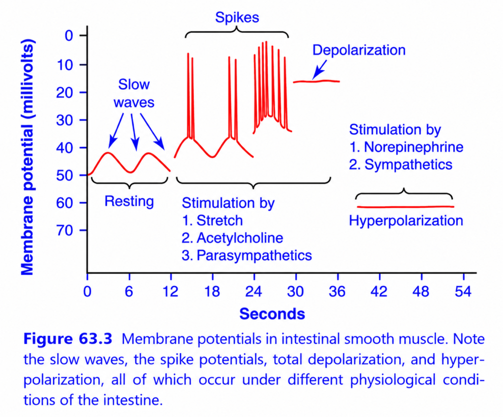
Membrane Potentials in Intestinal Smooth Muscle (Figure 63.3)
This figure explains the electrical activity of gastrointestinal (GI) smooth muscle under different physiological conditions.
It shows four important electrical events:
- Slow waves (Resting rhythm)
- Spike potentials (Action potentials)
- Depolarization
- Hyperpolarization
The figure demonstrates one major principle:
Slow waves set the rhythm of the intestine, but only spike potentials produce strong muscle contraction.
Basic Concept
Unlike skeletal muscle,
intestinal smooth muscle does not remain electrically silent.
Instead,
its membrane potential continuously rises and falls, producing rhythmic electrical activity.
This rhythmic activity controls:
- Intestinal contractions
- Mixing of food
- Movement of food (peristalsis)
Understanding the Axes
X-axis (Seconds)
Shows time.
Moving to the right means:
➡️ More time has passed.
Y-axis (Membrane Potential, mV)
Shows the electrical charge across the smooth muscle membrane.
Normal resting membrane potential is approximately:
−50 to −60 mV
When the membrane becomes less negative, it is called:
➡️ Depolarization
When it becomes more negative, it is called:
➡️ Hyperpolarization
1. Resting State – Slow Waves
This is the first part of the graph.
What are Slow Waves?
Slow waves are rhythmic fluctuations of the membrane potential.
They are not true action potentials.
What happens?
The membrane potential slowly rises and falls between approximately:
- −50 mV
- −40 mV
No spikes are produced.
Who generates slow waves?
They are produced by the:
Interstitial Cells of Cajal
These cells act as the pacemaker cells of the gastrointestinal tract.
What is their function?
They set the basic electrical rhythm of the intestine.
However,
slow waves alone usually do not cause significant muscle contraction.
Easy Concept
Think of slow waves as the heartbeat of the intestine.
They create a regular rhythm,
but they do not produce strong contractions by themselves.
Key Point
Slow waves establish the rhythm but are not true action potentials.
2. Spike Potentials
This is the second part of the graph.
What happens?
When the peak of a slow wave reaches the threshold (about −40 mV),
multiple sharp spikes appear.
These spikes are:
True Action Potentials
Why do spikes occur?
The membrane becomes sufficiently depolarized.
Voltage-gated:
- Calcium channels
- Calcium–sodium channels
open.
Calcium enters the cell.
The muscle contracts.
What is the result?
More spikes
➡️ More calcium enters
➡️ Stronger contraction
Easy Concept
Imagine a gun.
The slow wave is like pulling the trigger halfway.
Nothing happens.
Once the trigger reaches the firing point,
the gun fires.
Similarly,
once threshold is reached,
spike potentials occur.
Key Point
Spike potentials produce intestinal contraction.
Why Are There More Spikes Later?
Notice that later in the graph,
many spike potentials occur together.
Why?
The membrane remains depolarized for a longer period.
Therefore,
multiple action potentials are generated.
This allows:
- More calcium entry.
- Stronger contractions.
Easy Concept
More spikes mean:
➡️ More calcium
➡️ Stronger muscle contraction.
3. Depolarization
This is the third part of the graph.
What is Depolarization?
Depolarization means:
The membrane becomes less negative.
What causes depolarization?
The figure lists three important stimulatory factors:
1. Stretch
When food enters the intestine,
the intestinal wall stretches.
Stretch causes depolarization.
2. Acetylcholine (ACh)
Released by parasympathetic nerves.
ACh makes the membrane less negative.
3. Parasympathetic Stimulation
The parasympathetic nervous system increases intestinal activity.
It causes depolarization.
What happens after depolarization?
The membrane moves closer to threshold.
Spike potentials become more frequent.
Contractions become stronger.
Easy Concept
Imagine pushing a ball up a hill.
Depolarization pushes the membrane closer to the top (threshold),
making it much easier to trigger action potentials.
Key Point
Depolarization increases intestinal contractions.
4. Hyperpolarization
This is the final part of the graph.
What is Hyperpolarization?
Hyperpolarization means:
The membrane becomes more negative than normal.
What causes hyperpolarization?
The figure lists:
1. Norepinephrine
Released from sympathetic nerves.
2. Sympathetic Stimulation
The sympathetic nervous system inhibits intestinal activity.
What happens?
The membrane moves farther away from threshold.
Spike potentials cannot occur.
Therefore,
intestinal contractions decrease.
Easy Concept
Imagine pushing a ball farther away from the top of a hill.
Now,
it becomes much harder to reach the firing point.
Similarly,
hyperpolarization makes action potentials less likely.
Key Point
Hyperpolarization decreases intestinal contractions.
Role of the Autonomic Nervous System
Parasympathetic Nervous System
Releases:
Acetylcholine
Effects:
- Depolarization
- More spike potentials
- Increased calcium entry
- Stronger intestinal contractions
- Increased motility
Sympathetic Nervous System
Releases:
Norepinephrine
Effects:
- Hyperpolarization
- Fewer spike potentials
- Less calcium entry
- Weaker contractions
- Decreased motility
Comparison of the Four Electrical Events
| Electrical Event | What Happens? | Muscle Contraction |
|---|---|---|
| Slow waves | Rhythmic membrane oscillations | Little or no contraction by themselves |
| Spike potentials | True action potentials when threshold is reached | Strong contraction |
| Depolarization | Membrane becomes less negative | Increases spikes and contractions |
| Hyperpolarization | Membrane becomes more negative | Decreases spikes and contractions |
Clinical Importance
After a Meal
The stomach and intestine stretch.
Parasympathetic activity increases.
Acetylcholine is released.
More spike potentials occur.
Intestinal contractions increase.
Food moves forward.
During Fear or Stress
Sympathetic activity increases.
Norepinephrine is released.
Hyperpolarization occurs.
Contractions decrease.
Digestion slows.
Quick Memory Table
| Factor | Effect on Membrane | Effect on Motility |
|---|---|---|
| Stretch | Depolarization | ↑ Increases |
| Acetylcholine | Depolarization | ↑ Increases |
| Parasympathetic stimulation | Depolarization | ↑ Increases |
| Norepinephrine | Hyperpolarization | ↓ Decreases |
| Sympathetic stimulation | Hyperpolarization | ↓ Decreases |
Easy Memory Trick
Slow Waves = “Rhythm” 🥁
- Set the pace.
- Do not produce strong contractions alone.
Spike Potentials = “Strength” 💪
- Calcium enters.
- Muscle contracts.
Parasympathetic = “Push” 🚀
- Depolarization.
- More spikes.
- More movement.
Sympathetic = “Stop” 🛑
- Hyperpolarization.
- Fewer spikes.
- Less movement.
Key Concept
The intestinal smooth muscle membrane exhibits spontaneous rhythmic electrical activity called slow waves, which are generated by the interstitial cells of Cajal and establish the basic electrical rhythm of the gastrointestinal tract. Slow waves are not true action potentials and usually do not produce significant contraction by themselves. When the peak of a slow wave reaches the threshold (about −40 mV), spike potentials (true action potentials) are generated through the opening of voltage-gated calcium–sodium channels, allowing calcium to enter the cell and initiate smooth muscle contraction. Stretch, acetylcholine, and parasympathetic stimulation cause depolarization, bringing the membrane closer to threshold, increasing spike frequency, and enhancing gastrointestinal motility. In contrast, norepinephrine and sympathetic stimulation produce hyperpolarization, moving the membrane farther from threshold, suppressing spike potentials, reducing calcium entry, and decreasing intestinal contractions. Thus, slow waves determine the rhythm of intestinal activity, whereas spike potentials determine the strength of intestinal contraction.
Changes in Voltage of the Resting Membrane Potential
- In addition to slow waves and spike potentials, the resting membrane potential of gastrointestinal smooth muscle can also change.
- The average resting membrane potential is about −56 mV.
- Many factors can change this resting membrane potential.
- When the resting membrane potential becomes less negative, it is called depolarization.
- Depolarization makes the smooth muscle fibers more excitable.
- When the resting membrane potential becomes more negative, it is called hyperpolarization.
- Hyperpolarization makes the smooth muscle fibers less excitable.
- Factors that cause depolarization (increase excitability) are:
- Stretching of the muscle
- Acetylcholine released from parasympathetic nerve endings
- Several specific gastrointestinal hormones
- Factors that cause hyperpolarization (decrease excitability) are:
- Norepinephrine or epinephrine acting on the muscle fiber membrane
- Sympathetic nerve stimulation, which mainly releases norepinephrine
KEY CONCEPT
- The average resting membrane potential of gastrointestinal smooth muscle is −56 mV.
- Depolarization (less negative membrane potential) → Muscle becomes more excitable.
- Hyperpolarization (more negative membrane potential) → Muscle becomes less excitable.
- Stretching, acetylcholine, and gastrointestinal hormones cause depolarization.
- Norepinephrine, epinephrine, and sympathetic stimulation cause hyperpolarization.
Entry of Calcium Ions Causes Smooth Muscle Contraction
- Smooth muscle contraction occurs when calcium ions enter the muscle fiber.
- Calcium ions act through the calmodulin control mechanism.
- The calmodulin mechanism activates myosin filaments.
- Activated myosin filaments develop attractive forces with actin filaments.
- This interaction between myosin and actin causes the smooth muscle to contract.
- The detailed mechanism is explained in Chapter 8.
- Slow waves do not allow calcium ions to enter the smooth muscle fiber.
- Slow waves mainly allow sodium ions to enter.
- Therefore, slow waves alone usually do not produce muscle contraction.
- During spike potentials, which occur at the peaks of slow waves, large amounts of calcium ions enter the muscle fibers.
- The entry of calcium ions during spike potentials causes most gastrointestinal smooth muscle contractions.
KEY CONCEPT
- Calcium ion entry is essential for gastrointestinal smooth muscle contraction.
- Calcium works through the calmodulin mechanism to activate myosin, which interacts with actin to produce contraction.
- Slow waves mainly allow sodium entry, so they usually do not cause contraction.
- Spike potentials allow calcium entry and are responsible for most smooth muscle contractions.
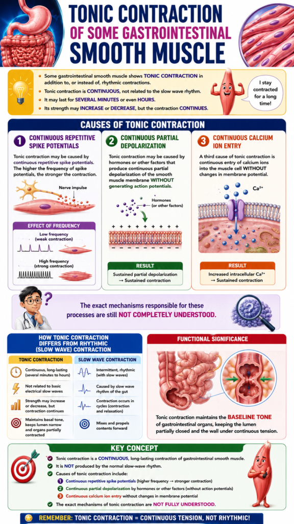
Tonic Contraction of Some Gastrointestinal Smooth Muscle
- Some gastrointestinal smooth muscle shows tonic contraction in addition to, or instead of, rhythmic contractions.
- Tonic contraction is continuous.
- It is not related to the basic electrical rhythm of slow waves.
- Tonic contraction may last for several minutes or even hours.
- Its strength may increase or decrease, but the contraction continues.
- Tonic contraction may be caused by continuous repetitive spike potentials.
- The higher the frequency of spike potentials, the stronger the contraction.
- Tonic contraction may also be caused by hormones or other factors.
- These factors produce continuous partial depolarization of the smooth muscle membrane.
- This occurs without generating action potentials.
- A third cause of tonic contraction is continuous entry of calcium ions into the muscle cell.
- This calcium entry occurs without changes in membrane potential.
- The exact mechanisms responsible for these processes are still not completely understood.
KEY CONCEPT
- Tonic contraction is a continuous, long-lasting contraction of gastrointestinal smooth muscle.
- It is not produced by the normal slow-wave rhythm.
- Causes of tonic contraction include:
- Continuous repetitive spike potentials
- Continuous partial depolarization by hormones or other factors
- Continuous calcium ion entry without membrane potential changes
- The exact mechanisms of tonic contraction are not fully understood.
MADE BY SELF LEARNING DR SHEEN
