


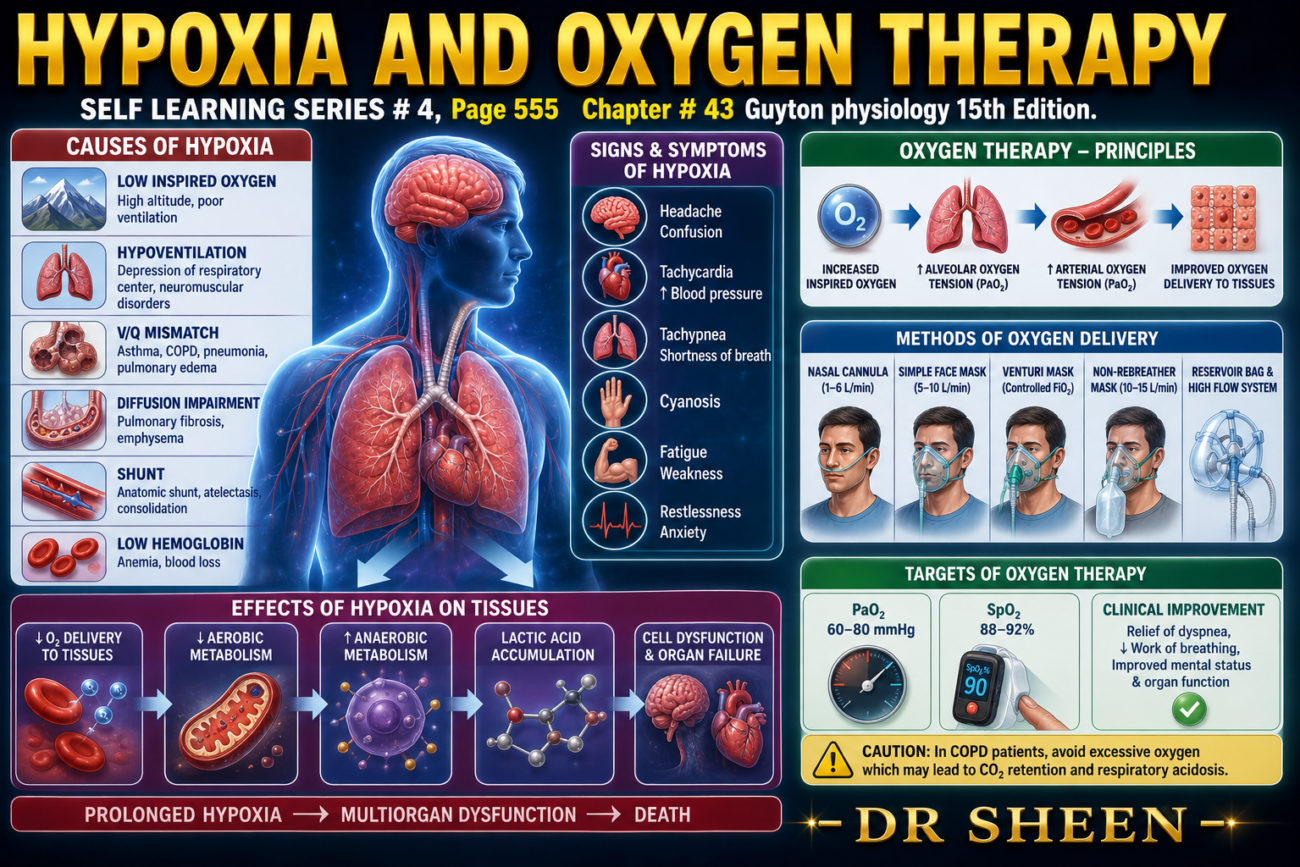
- Almost any condition discussed earlier in this chapter can cause serious hypoxia (low oxygen) in the body’s cells.
- Oxygen (O₂) therapy may be:
- Very effective in some conditions.
- Moderately effective in others.
- Almost ineffective in some situations.
- Therefore, it is important to understand the different types of hypoxia before understanding oxygen therapy.
- The causes of hypoxia are classified as follows:
1. Inadequate Oxygenation of the Blood in the Lungs Because of Extrinsic Reasons
- Low oxygen (O₂) in the atmosphere.
- Hypoventilation caused by neuromuscular disorders.
2. Pulmonary Disease
- Hypoventilation due to increased airway resistance or decreased lung (pulmonary) compliance.
- Abnormal alveolar ventilation-perfusion (V/Q) ratio, including:
- Increased physiological dead space.
- Increased physiological shunt.
- Reduced diffusion across the respiratory membrane.
3. Venous-to-Arterial Shunts
- Right-to-left cardiac shunts, where venous blood enters the arterial circulation without adequate oxygenation.
4. Inadequate Oxygen (O₂) Transport to the Tissues by the Blood
- Anemia or abnormal hemoglobin.
- General circulatory deficiency.
- Localized circulatory deficiency, such as in:
- Peripheral blood vessels
- Cerebral blood vessels
- Coronary blood vessels
- Tissue edema.
5. Inadequate Tissue Capability of Using Oxygen (O₂)
- Poisoning of cellular oxidation enzymes.
- Reduced cellular ability to use oxygen because of toxicity, vitamin deficiency, or other factors.
- Most types of hypoxia listed above have already been explained earlier in the chapter.
- The type of hypoxia caused by the inability of tissue cells to use oxygen requires further explanation.
KEY CONCEPT
- Hypoxia is a condition in which body tissues receive or utilize insufficient oxygen.
- The effectiveness of oxygen therapy depends on the underlying cause of hypoxia.
- The five major causes of hypoxia are:
- Inadequate oxygenation of blood due to extrinsic causes.
- Pulmonary disease.
- Venous-to-arterial (right-to-left) shunts.
- Inadequate oxygen transport by the blood.
- Inability of tissues to utilize oxygen.
- The last type—impaired tissue utilization of oxygen—requires further discussion.
Inadequate Tissue Capability to Use Oxygen
- This type of hypoxia occurs when body tissues cannot use oxygen (O₂), even if enough oxygen is available in the blood.
- The classic cause is cyanide poisoning.
- In cyanide poisoning, cyanide blocks the enzyme cytochrome oxidase.
- When cytochrome oxidase is blocked, the tissues cannot use oxygen, even though oxygen is present in adequate amounts.
- Deficiency of tissue oxidative enzymes can also reduce the ability of cells to use oxygen.
- Defects in other parts of the tissue oxidative system can produce the same type of hypoxia.
- A special example is beriberi.
- In beriberi, vitamin B deficiency interferes with several important steps involved in tissue utilization of oxygen and the formation of carbon dioxide (CO₂).
Effects of Hypoxia on the Body
- If hypoxia is severe, it can cause death of cells throughout the body.
- In less severe hypoxia, the main effects are:
- Depressed mental activity, which may progress to coma.
- Reduced ability of muscles to perform work.
- These effects are discussed further in Chapter 44 in relation to high-altitude physiology.
KEY CONCEPT
- Inadequate tissue capability to use oxygen is a type of hypoxia in which cells cannot utilize O₂ despite an adequate oxygen supply.
- The classic cause is cyanide poisoning, which blocks the enzyme cytochrome oxidase.
- Deficiency of oxidative enzymes or defects in the tissue oxidative system can also impair oxygen utilization.
- Beriberi, caused by vitamin B deficiency, reduces tissue utilization of oxygen and CO₂ formation.
- Severe hypoxia can cause widespread cell death, while milder hypoxia causes depressed mental activity, coma, and reduced muscle work capacity.
OXYGEN THERAPY IN DIFFERENT TYPES OF HYPOXIA
- Oxygen (O₂) therapy is used to increase the amount of oxygen available to the body.
- Oxygen can be given by:
- An oxygen tent around the patient’s head.
- An oxygen mask delivering pure or high-concentration oxygen.
- An intranasal tube (nasal cannula).
- The benefit of oxygen therapy depends on the cause of hypoxia.
1. Atmospheric Hypoxia
- Atmospheric hypoxia occurs when the air contains too little oxygen.
- Oxygen therapy raises the oxygen level in the inspired air to normal.
- Therefore, oxygen therapy can completely correct this type of hypoxia.
- It is almost 100% effective.
2. Hypoventilation Hypoxia
- Hypoventilation means inadequate air reaches the alveoli.
- When the patient breathes 100% oxygen, about five times more oxygen enters the alveoli with each breath than when breathing normal air.
- Therefore, oxygen therapy is highly effective.
- However, oxygen therapy does not remove the excess carbon dioxide (CO₂) that accumulates because of hypoventilation.
3. Diffusion Hypoxia (Impaired Alveolar Membrane Diffusion)
- This type of hypoxia occurs when oxygen cannot diffuse efficiently across the respiratory membrane.
- Oxygen therapy raises alveolar PO₂ from about 100 mm Hg to about 600 mm Hg.
- As a result, the oxygen pressure gradient from the alveoli to the blood increases from about 60 mm Hg to about 560 mm Hg.
- This is an increase of more than 800%.
- The much larger pressure gradient greatly speeds the diffusion of oxygen into the blood.
- Therefore, oxygen therapy is highly beneficial in diffusion hypoxia. (Fig. 43.8)
- For example, in pulmonary edema, blood absorbs oxygen three to four times faster during oxygen therapy than without treatment. (Fig. 43.8)
4. Hypoxia Due to Impaired Oxygen Transport
This includes:
- Anemia
- Abnormal hemoglobin
- General circulatory deficiency
- Physiological shunt
- In these conditions, oxygen levels in the alveoli are already normal.
- The problem is not oxygen entering the lungs, but poor transport of oxygen from the lungs to the tissues.
- Therefore, oxygen therapy is much less effective.
- However, increasing alveolar oxygen raises the amount of oxygen dissolved directly in plasma.
- This dissolved oxygen can increase by about 7%–30%.
- Although the oxygen carried by hemoglobin changes very little, the extra dissolved oxygen may be enough to save a patient’s life.
5. Hypoxia Due to Inadequate Tissue Use of Oxygen
- In this type of hypoxia, oxygen reaches the lungs normally.
- Oxygen is transported to the tissues normally.
- The problem is that the tissue metabolic enzymes cannot use the delivered oxygen.
- Therefore, oxygen therapy provides no measurable benefit.
Summary of Oxygen Therapy in Different Types of Hypoxia
| Type of Hypoxia | Benefit of Oxygen Therapy | Reason |
|---|---|---|
| Atmospheric hypoxia | Completely effective | Corrects low oxygen in inspired air |
| Hypoventilation hypoxia | Highly effective | Greatly increases alveolar oxygen, but does not remove excess CO₂ |
| Diffusion hypoxia | Highly effective | Greatly increases oxygen diffusion across the respiratory membrane |
| Anemia / Abnormal hemoglobin / Circulatory deficiency / Physiological shunt | Limited benefit | Only increases dissolved oxygen because oxygen transport is impaired |
| Inadequate tissue utilization of oxygen | No benefit | Cells cannot use oxygen despite adequate delivery |
KEY CONCEPT
- The effectiveness of oxygen therapy depends on the underlying cause of hypoxia.
- It is completely effective in atmospheric hypoxia and highly effective in hypoventilation and diffusion hypoxia.
- It has limited benefit in anemia, abnormal hemoglobin, circulatory deficiency, and physiological shunts because oxygen transport—not oxygen uptake—is impaired.
- It provides no measurable benefit when tissues are unable to utilize oxygen, such as in cyanide poisoning, because oxygen delivery is normal but cellular oxygen use is defective.
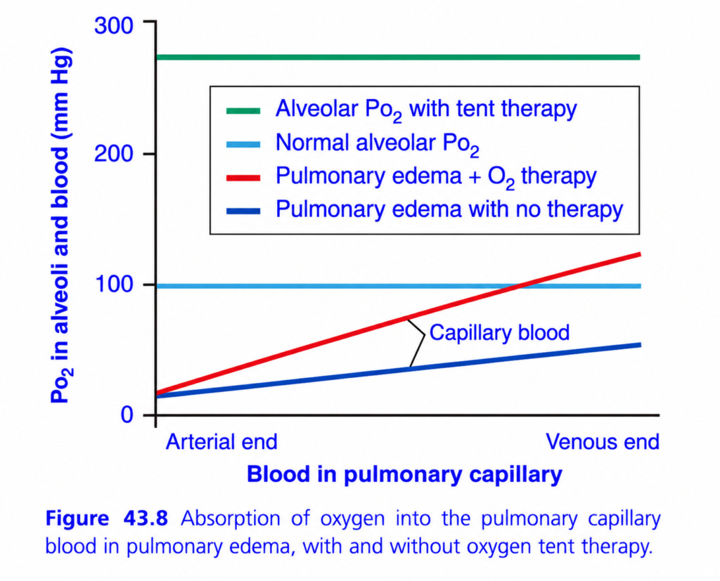
bsorption of Oxygen into Pulmonary Capillary Blood in Pulmonary Edema (Figure 43.8) – Easy Conceptual Summary
This figure explains how pulmonary edema affects oxygen diffusion from the alveoli into pulmonary capillary blood and why oxygen (O₂) therapy is helpful.
It compares three situations:
- Normal lungs
- Pulmonary edema without oxygen therapy
- Pulmonary edema with oxygen therapy (oxygen tent)
The main message is:
Pulmonary edema slows oxygen diffusion, but oxygen therapy increases alveolar PO₂ and greatly improves oxygen transfer into the blood.
Basic Concept
Normally,
Oxygen moves:
Alveolus → Pulmonary capillary blood
because:
Alveolar PO₂ > Blood PO₂
The larger this pressure difference (diffusion gradient),
➡️ the faster oxygen diffuses.
Understanding the Axes
X-axis (Blood in Pulmonary Capillary)
Shows blood flowing through the pulmonary capillary:
- Left side = Arterial end
- Right side = Venous end
As blood moves from left to right,
it spends about 0.75 seconds in the pulmonary capillary.
Y-axis (PO₂ in Alveoli and Blood, mm Hg)
Shows the partial pressure of oxygen (PO₂).
Higher PO₂
➡️ More oxygen available.
Lower PO₂
➡️ Less oxygen available.
Understanding the Horizontal Lines
These lines represent alveolar PO₂.
Because alveolar air remains relatively constant,
these lines are horizontal.
1. Light Blue Horizontal Line – Normal Alveolar PO₂
Value
Approximately:
100 mm Hg
What does it mean?
This is the normal oxygen pressure inside healthy alveoli.
Normally,
oxygen diffuses until blood PO₂ also reaches about 100 mm Hg.
Key Point
Normal alveolar PO₂ provides an adequate oxygen diffusion gradient.
2. Green Horizontal Line – Alveolar PO₂ with Oxygen Tent Therapy
Value
Approximately:
270–280 mm Hg
What happens?
The patient breathes oxygen-rich air.
Alveolar PO₂ increases dramatically.
The diffusion gradient becomes much larger.
Therefore,
oxygen enters the blood much more rapidly.
Easy Concept
Imagine water flowing down a hill.
A steeper hill allows water to flow faster.
Similarly,
a larger PO₂ difference causes oxygen to diffuse faster.
Key Point
Higher alveolar PO₂ greatly increases oxygen diffusion.
Understanding the Sloping Lines
These lines represent the PO₂ of pulmonary capillary blood as it travels through the lung.
3. Blue Sloping Line – Pulmonary Edema Without Oxygen Therapy
What happens?
Blood enters the pulmonary capillary with a PO₂ of about:
20–25 mm Hg
Because pulmonary edema thickens the diffusion barrier,
oxygen enters the blood very slowly.
Even at the venous end,
blood PO₂ reaches only about:
50–60 mm Hg
Why?
Pulmonary edema causes fluid to accumulate between:
- Alveolus
- Capillary
The diffusion distance becomes much greater.
According to Fick’s law:
Increased diffusion distance → Decreased diffusion rate.
Therefore,
blood cannot fully oxygenate before leaving the lungs.
Easy Concept
Imagine trying to smell perfume through:
- One sheet of paper (easy)
- A thick blanket (very difficult)
Pulmonary edema acts like the thick blanket.
Oxygen diffuses much more slowly.
Key Point
Without oxygen therapy,
blood remains poorly oxygenated.
4. Red Sloping Line – Pulmonary Edema with Oxygen Therapy
What happens?
Oxygen therapy increases alveolar PO₂.
Now,
even though the diffusion barrier is still thick,
the pressure difference becomes much larger.
Therefore,
oxygen diffuses more rapidly.
Blood PO₂ rises much higher,
reaching about:
120 mm Hg
Why?
The increased pressure gradient compensates for the thickened diffusion barrier.
Even though diffusion is slow,
the stronger driving force pushes more oxygen into the blood.
Easy Concept
Imagine pushing a heavy door.
If you push gently,
it barely moves.
If you push much harder,
it opens despite the resistance.
Similarly,
high alveolar PO₂ overcomes the resistance created by pulmonary edema.
Key Point
Oxygen therapy improves oxygenation despite pulmonary edema.
Why Does Oxygen Therapy Help?
According to Fick’s Law of Diffusion:
Diffusion increases when:
- Surface area increases.
- Membrane thickness decreases.
- Pressure difference increases.
In pulmonary edema,
the membrane becomes thicker,
which decreases diffusion.
Oxygen therapy cannot reduce membrane thickness immediately,
but it greatly increases the pressure gradient, allowing more oxygen to diffuse.
Comparison of the Three Conditions
| Condition | Alveolar PO₂ | Blood PO₂ at End of Capillary | Oxygen Diffusion |
|---|---|---|---|
| Normal lung | ~100 mm Hg | ~100 mm Hg | Normal |
| Pulmonary edema (No therapy) | ~100 mm Hg | ~50–60 mm Hg | Greatly reduced |
| Pulmonary edema (O₂ therapy) | ~270–280 mm Hg | ~120 mm Hg | Markedly improved |
Clinical Importance
Pulmonary Edema
Occurs in conditions such as:
- Left-sided heart failure
- Acute respiratory distress syndrome (ARDS)
- Fluid overload
- High-altitude pulmonary edema
Fluid accumulates around the alveoli,
making oxygen diffusion difficult.
Oxygen Therapy
Increases:
- Alveolar PO₂
- Diffusion gradient
- Oxygen transfer to blood
Therefore,
oxygen therapy is one of the first treatments for pulmonary edema.
Quick Memory Table
| Line | Represents | Meaning |
|---|---|---|
| Light Blue Horizontal | Normal alveolar PO₂ | Normal oxygen pressure (~100 mm Hg) |
| Green Horizontal | Alveolar PO₂ during oxygen therapy | Much higher oxygen pressure (~270–280 mm Hg) |
| Blue Sloping | Capillary blood in pulmonary edema without therapy | Poor oxygen uptake |
| Red Sloping | Capillary blood in pulmonary edema with oxygen therapy | Improved oxygen uptake |
Easy Memory Trick
Normal Lung = Thin Wall 🫁
- Oxygen crosses easily.
- Blood reaches normal PO₂ quickly.
Pulmonary Edema = Thick Wall 💧
- Oxygen crosses slowly.
- Blood remains poorly oxygenated.
Oxygen Therapy = Stronger Push 🌬️
- Higher alveolar PO₂.
- Larger diffusion gradient.
- More oxygen enters the blood.
Key Concept
This figure demonstrates that pulmonary edema impairs oxygen diffusion by increasing the thickness of the alveolar–capillary membrane, thereby slowing the movement of oxygen from the alveoli into the pulmonary capillary blood. In normal lungs, alveolar PO₂ is approximately 100 mm Hg, allowing blood PO₂ to rapidly equilibrate with alveolar PO₂ before reaching the end of the pulmonary capillary. In pulmonary edema without oxygen therapy, the thickened diffusion barrier markedly reduces oxygen transfer, so blood leaves the lungs with a much lower PO₂. When oxygen therapy is administered, alveolar PO₂ rises substantially (to about 270–280 mm Hg), creating a much larger diffusion gradient. Although the membrane remains thick, the increased pressure difference drives more oxygen across the barrier, significantly improving arterial oxygenation. Thus, oxygen therapy does not remove the edema but improves oxygen diffusion by increasing the alveolar PO₂ and the diffusion gradient across the alveolar–capillary membrane.
Made by self learning CEO and founder Dr sheen
