


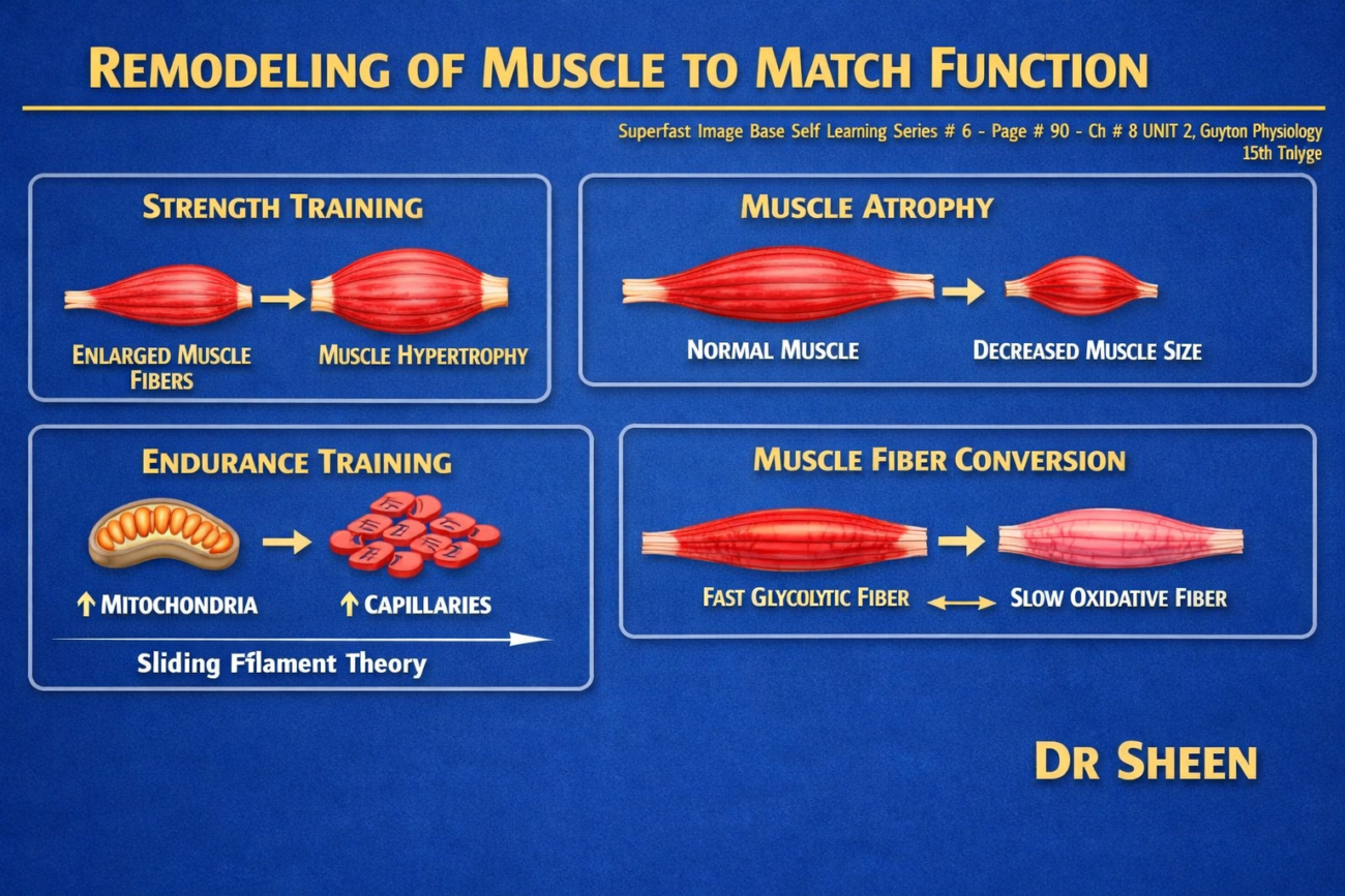
- Skeletal muscles continuously remodel themselves to meet the functional demands placed on them.
- This remodeling helps the muscles adapt to changing levels of activity and workload.
- During remodeling, several muscle properties can change:
- Muscle diameter (thickness) can increase or decrease.
- Muscle length can change.
- Muscle strength can increase or decrease.
- Blood (vascular) supply to the muscle can be altered.
- The types of muscle fibers can also change slightly.
- These changes allow the muscle to perform its required function more effectively.
- Muscle remodeling is often a rapid process.
- Significant changes can occur within a few weeks.
- Experiments in animals have shown that the contractile proteins of some small, highly active muscles can be completely replaced in as little as 2 weeks.
- This demonstrates that skeletal muscle is a highly adaptable tissue with continuous protein turnover.
KEY CONCEPT
- Skeletal muscles continuously remodel to match their functional demands.
- Remodeling can change muscle diameter, length, strength, blood supply, and even muscle fiber type.
- These adaptations help muscles perform their required functions more efficiently.
- Muscle remodeling occurs rapidly, often within a few weeks.
- In some small, active muscles, contractile proteins can be replaced in as little as 2 weeks.
Muscle Hypertrophy and Muscle Atrophy
- Muscle hypertrophy means an increase in the total mass (size) of a muscle.
- Muscle atrophy means a decrease in the total mass (size) of a muscle.
Muscle Hypertrophy
- Almost all muscle hypertrophy occurs because the number of actin and myosin filaments increases within each muscle fiber.
- As a result, each individual muscle fiber becomes larger.
- This enlargement of a muscle fiber is called fiber hypertrophy.
- Hypertrophy occurs much more when the muscle contracts against a load (resistance).
- Only a few strong muscle contractions each day are enough to produce significant hypertrophy within 6–10 weeks.
- The exact mechanism by which forceful contraction causes hypertrophy is not completely understood.
- During hypertrophy:
- The rate of synthesis of muscle contractile proteins increases greatly.
- More actin and myosin filaments are formed inside the myofibrils.
- The number of these filaments may increase by as much as 50%.
- In some hypertrophying muscles:
- Existing myofibrils have been observed to split and form new myofibrils.
- However, the importance of this process in normal muscle enlargement is still uncertain.
- Along with the increase in myofibril size:
- The muscle’s energy-producing enzyme systems also increase.
- Glycolytic enzymes increase especially, allowing the muscle to produce ATP rapidly during short, forceful contractions.
Muscle Atrophy
- When a muscle is not used for many weeks, it gradually becomes smaller.
- This decrease in muscle size is called muscle atrophy.
- During atrophy:
- Contractile proteins are broken down faster than they are replaced.
- As a result, muscle mass decreases.
- Much of this protein breakdown occurs through the ATP-dependent ubiquitin–proteasome pathway.
- Proteasomes are large protein complexes that:
- Break down damaged or unnecessary proteins.
- They do this by proteolysis, which is the chemical breakdown of peptide bonds.
- Ubiquitin is a regulatory protein that:
- Tags (labels) proteins for destruction.
- These tagged proteins are then degraded by proteasomes.
KEY CONCEPT
- Muscle hypertrophy is an increase in muscle size, while muscle atrophy is a decrease in muscle size.
- Hypertrophy occurs mainly because more actin and myosin filaments are added to each muscle fiber, producing fiber hypertrophy.
- Resistance (loaded) contractions stimulate hypertrophy, and noticeable changes can occur within 6–10 weeks.
- During hypertrophy, contractile protein synthesis increases, myofibrils enlarge, glycolytic enzymes increase, and myofibrils may occasionally split to form new ones.
- Muscle atrophy develops when muscles remain unused for weeks because protein degradation exceeds protein replacement.
- The ATP-dependent ubiquitin–proteasome pathway is a major mechanism responsible for protein breakdown during muscle atrophy.
Adjustment of Muscle Length
- Muscles can remodel their length to match the functional demands placed on them.
When a Muscle Is Stretched
- When a muscle is stretched beyond its normal length, another type of muscle hypertrophy occurs.
- This stretching stimulates the formation of new sarcomeres.
- New sarcomeres are added at the ends of the muscle fibers, where the fibers attach to the tendons.
- In newly developing muscle, new sarcomeres can be added as rapidly as several per minute.
- This demonstrates that muscle length can adapt very quickly.
When a Muscle Remains Shortened
- When a muscle remains continuously shorter than its normal length, the opposite change occurs.
- Sarcomeres at the ends of the muscle fibers gradually disappear.
- This reduces the overall length of the muscle fiber.
Purpose of Muscle Length Adjustment
- By adding sarcomeres during stretching and removing sarcomeres during prolonged shortening, muscles are continuously remodeled.
- This remodeling maintains the appropriate muscle length.
- Maintaining the proper length allows the muscle to contract efficiently and produce normal force.
KEY CONCEPT
- Muscles continuously adjust their length to meet functional requirements.
- Stretching a muscle beyond its normal length stimulates the addition of new sarcomeres at the ends of muscle fibers where they attach to tendons.
- In developing muscle, several new sarcomeres can be added each minute.
- When a muscle remains shortened for a long time, sarcomeres at the ends of the muscle fibers disappear.
- The addition and removal of sarcomeres help maintain the optimal muscle length for effective contraction.
Satellite Cells and Muscle Repair
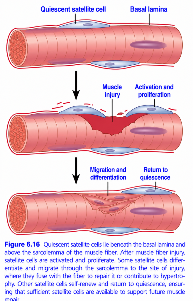
- Skeletal muscle contains special adult stem cells called satellite cells.
- Under normal conditions, satellite cells remain inactive (quiescent).
- When muscle fibers are damaged by:
- Normal daily activity, or
- Muscle injury,
- After activation, satellite cells begin to multiply (proliferate).
- Following several cycles of proliferation, they differentiate into muscle cells.
- These differentiated cells then fuse with the damaged muscle fibers at the site of injury.
- This process repairs the injured muscle fibers.
- Satellite cells are essential for:
- Maintaining normal muscle homeostasis.
- Muscle hypertrophy (increase in muscle size).
- Muscle regeneration after injury.
Satellite Cells and Aging
- As people age, the number of satellite cells decreases.
- Their ability to repair damaged muscle fibers also declines.
- This reduction may contribute to the age-related loss of muscle mass, known as sarcopenia.
- Exercise activates satellite cells.
- Therefore, regular exercise may help prevent or reverse some of the age-related loss of muscle mass.
KEY CONCEPT
- Satellite cells are adult stem cells found in skeletal muscle.
- They normally remain inactive but become activated after muscle damage (Fig. 6.16).
- Activated satellite cells proliferate, differentiate, and fuse with damaged muscle fibers to repair them.
- Satellite cells are essential for normal muscle maintenance, muscle hypertrophy, and muscle regeneration after injury.
- Aging reduces the number and repair capacity of satellite cells, contributing to sarcopenia.
- Exercise stimulates satellite cells and may reduce or reverse some age-related muscle loss.
Hyperplasia of Muscle Fibers
- Under rare conditions of extremely high muscle force generation, the number of muscle fibers may increase slightly.
- This increase occurs in addition to the enlargement (hypertrophy) of existing muscle fibers.
- The increase in the number of muscle fibers is called muscle fiber hyperplasia.
- Fiber hyperplasia is uncommon.
- Even when it occurs, the increase in muscle fiber number is usually only a few percent.
- The main mechanism of fiber hyperplasia is the linear splitting of previously enlarged (hypertrophied) muscle fibers.
- As an enlarged muscle fiber splits, new muscle fibers are formed, leading to a small increase in the total number of muscle fibers.
KEY CONCEPT
- Muscle fiber hyperplasia is the increase in the number of muscle fibers.
- It occurs only under rare conditions of extreme muscle force generation.
- The increase in fiber number is usually small (only a few percent).
- Hyperplasia occurs in addition to muscle hypertrophy.
- The primary mechanism is the linear splitting of previously enlarged (hypertrophied) muscle fibers.
Muscle Denervation Causes Rapid Atrophy
- Denervation means loss of the nerve supply to a muscle.
- When a muscle loses its nerve supply, it no longer receives the nerve impulses (contractile signals) needed to maintain its normal size.
- As a result, muscle atrophy begins almost immediately.
Early Changes After Denervation
- Within about 2 months, degenerative changes begin to appear in the muscle fibers.
- If the nerve grows back quickly, the muscle can recover completely.
- Full return of muscle function may occur within about 3 months.
- After this period:
- The chance of functional recovery gradually decreases.
- After 1–2 years, no further functional recovery usually occurs, even if the nerve regrows.
Late Changes in Denervation Atrophy
- In the final stage of denervation atrophy:
- Most muscle fibers are destroyed.
- They are replaced by fibrous tissue and fatty tissue.
- The remaining muscle fibers:
- Consist mainly of a long cell membrane with a row of muscle cell nuclei.
- Have few or no contractile properties.
- Have little or no ability to regenerate myofibrils, even if the nerve later regrows.
Contracture
- The fibrous tissue that replaces muscle fibers continues to shorten for many months.
- This progressive shortening is called contracture.
- Contracture can cause severe disability and deformity if it is not prevented.
Prevention of Contracture
- One of the major goals of physical therapy is to prevent contractures in muscles undergoing denervation atrophy.
- This is achieved by:
- Daily stretching of the affected muscles.
- Using splints or other appliances that keep the muscles stretched during the atrophying process.
KEY CONCEPT
- Denervation causes rapid muscle atrophy because the muscle no longer receives the nerve impulses required to maintain its size.
- Degenerative changes appear after about 2 months.
- If the nerve regrows early, complete recovery may occur within about 3 months, but the chance of recovery declines with time and is usually absent after 1–2 years.
- In advanced denervation, muscle fibers are replaced by fibrous and fatty tissue, and the remaining fibers have little or no contractile or regenerative ability.
- Fibrous tissue gradually shortens, producing contracture.
- Daily stretching and supportive devices are essential to prevent contractures during denervation atrophy.
Recovery of Muscle Contraction in Poliomyelitis: Development of Macromotor Units
- In poliomyelitis (polio), some nerve fibers supplying a muscle are destroyed, while others remain intact.
- The remaining healthy nerve fibers produce new branches (axons).
- These new axons reinnervate many of the previously paralyzed muscle fibers.
- As a result, very large motor units are formed, called macromotor units.
- A macromotor unit may contain up to five times the normal number of muscle fibers supplied by one spinal motoneuron.
- Formation of macromotor units:
- Restores part of the muscle’s strength.
- Reduces the precision (fine control) of muscle movements, because one nerve now controls many more muscle fibers.
KEY CONCEPT
- After partial nerve damage in poliomyelitis, surviving nerve fibers sprout new axons to reinnervate paralyzed muscle fibers.
- This forms macromotor units that are much larger than normal.
- Macromotor units improve muscle strength but reduce fine motor control.
Rigor Mortis
- Within a few hours after death, all the skeletal muscles become stiff and contracted.
- This condition is called rigor mortis.
- During rigor mortis:
- Muscles contract and become rigid even without action potentials.
- The main cause is the complete depletion of ATP.
- ATP is required to detach myosin cross-bridges from actin during muscle relaxation.
- Without ATP, the cross-bridges remain attached, so the muscles stay contracted.
- Rigor mortis lasts until the muscle proteins begin to break down, usually 15–25 hours after death.
- This breakdown is believed to occur because of autolysis, in which lysosomal enzymes digest the muscle proteins.
- Higher temperatures accelerate all of these processes, causing rigor mortis to develop and disappear more quickly.
KEY CONCEPT
- Rigor mortis is the stiffness of muscles that develops a few hours after death.
- It occurs because ATP is depleted, preventing separation of actin and myosin cross-bridges.
- Rigor ends after 15–25 hours when muscle proteins are digested by lysosomal enzymes.
- Higher temperatures speed up both the onset and disappearance of rigor mortis.
Muscular Dystrophy
- Muscular dystrophies are inherited disorders that cause:
- Progressive muscle weakness.
- Degeneration of muscle fibers.
- Replacement of muscle tissue by fatty tissue and collagen.
Duchenne Muscular Dystrophy (DMD)
- One of the most common muscular dystrophies is Duchenne muscular dystrophy (DMD).
- DMD affects only males because it is inherited as an X-linked recessive disorder.
- DMD is caused by a mutation in the gene that produces dystrophin.
- Dystrophin is a protein that:
- Links actin filaments to proteins in the muscle cell membrane.
- Forms a connection between the intracellular contractile apparatus and the extracellular connective tissue matrix.
Effects of Dystrophin Deficiency
- Absence or abnormal dystrophin destabilizes the muscle cell membrane.
- It also activates several pathophysiological changes, including:
- Abnormal intracellular Ca²⁺ handling.
- Reduced ability to repair the muscle cell membrane after injury.
- One major effect is increased membrane permeability to Ca²⁺.
- As a result:
- Extracellular Ca²⁺ enters the muscle fiber.
- This activates intracellular enzymes.
- These enzymes cause proteolysis (protein breakdown).
- Eventually, muscle fibers degenerate and are destroyed.
Clinical Features of DMD
- Muscle weakness begins in early childhood.
- The weakness progressively worsens.
- Most patients require a wheelchair by about 12 years of age.
- Many patients die from respiratory failure before the age of 30 years.
Becker Muscular Dystrophy (BMD)
- Becker muscular dystrophy (BMD) is also caused by mutations in the dystrophin gene.
- Compared with DMD:
- Symptoms begin later.
- Disease progression is slower.
- Survival is longer.
- DMD and BMD together affect approximately 1 in every 5600–7700 males between 5 and 24 years of age.
- Currently, there is no effective treatment for DMD or BMD.
- However, understanding their genetic basis has created the potential for future gene therapy.
KEY CONCEPT
- Muscular dystrophies are inherited diseases that cause progressive muscle weakness and replacement of muscle fibers by fat and collagen.
- Duchenne muscular dystrophy (DMD) is an X-linked recessive disorder caused by mutations in the dystrophin gene and affects only males.
- Dystrophin connects the contractile apparatus to the muscle cell membrane and helps maintain membrane stability.
- Loss of dystrophin increases Ca²⁺ entry into muscle fibers, leading to enzyme activation, proteolysis, and muscle fiber destruction.
- DMD begins in early childhood, progresses rapidly, often requires wheelchair use by about 12 years of age, and commonly leads to respiratory failure before 30 years of age.
- Becker muscular dystrophy (BMD) is a milder form with later onset and longer survival.
- Although no effective treatment currently exists, advances in genetics offer the possibility of future gene therapy.
prepare and made by Dr sheen
