


Motor Unit—All the Muscle Fibers Innervated By a Single Nerve Fiber
- Each motor neuron (motoneuron) leaving the spinal cord supplies multiple muscle fibers.
- The number of muscle fibers supplied by one motor neuron varies depending on the type of muscle.
- All the muscle fibers supplied by a single motor neuron are called a motor unit.
- Fig. 6.13 shows a motor unit consisting of one motor neuron and all the muscle fibers it innervates.
- In general, small muscles that contract rapidly and require precise control have fewer muscle fibers in each motor unit.
- Therefore, these muscles have more motor neurons relative to the number of muscle fibers.
- For example, some laryngeal muscles have only 2–3 muscle fibers in each motor unit.
- In contrast, large muscles that do not require fine control have many muscle fibers in each motor unit.
- For example, the soleus muscle may have several hundred muscle fibers in one motor unit.
- The average motor unit in the human body contains about 80–100 muscle fibers.
- The muscle fibers belonging to one motor unit are not grouped together in one place.
- Instead, they are scattered throughout the muscle.
- They overlap with muscle fibers of other motor units in small bundles (microbundles) of about 3–15 muscle fibers.
- This interdigitation allows different motor units to contract together and support one another, rather than contracting as completely separate groups.
KEY CONCEPT
- A motor unit consists of one motor neuron and all the muscle fibers it innervates (Fig. 6.13).
- The number of muscle fibers in a motor unit depends on the function of the muscle.
- Small muscles requiring precise movements have few muscle fibers per motor unit (e.g., laryngeal muscles).
- Large muscles requiring less precise control have many muscle fibers per motor unit (e.g., soleus muscle).
- On average, one motor unit contains about 80–100 muscle fibers.
- Muscle fibers of different motor units overlap within the muscle, allowing coordinated and efficient muscle contraction.
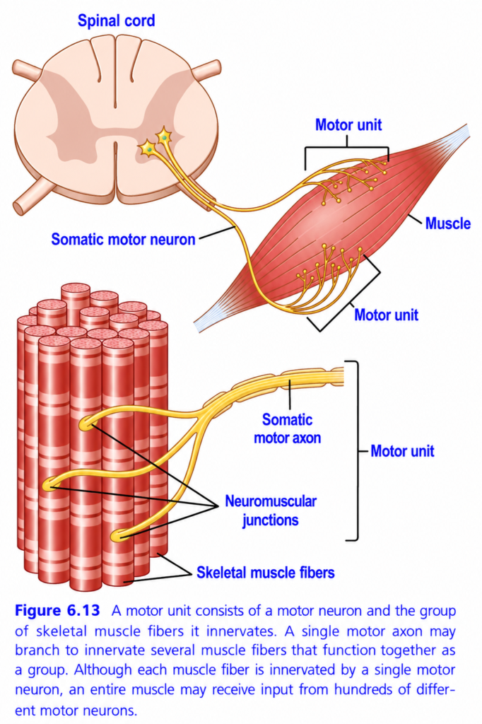
Motor Unit (Figure 6.13) — Easiest Conceptual Summary
This figure explains how the brain/spinal cord controls skeletal muscle contraction through a motor unit.
Main Idea
A motor unit is the smallest functional unit of muscle contraction.
Motor Unit = One Somatic Motor Neuron + All the Skeletal Muscle Fibers Supplied by That Neuron
Whenever the motor neuron fires, every muscle fiber belonging to that motor unit contracts together.
Understanding the Figure Step by Step
1. Spinal Cord (Control Center)
- The motor neuron cell body is located in the anterior (ventral) horn of the spinal cord.
- It receives commands from the brain.
- It generates nerve impulses that travel to the muscle.
Concept
Brain → Spinal cord → Motor neuron → Muscle contraction
2. Somatic Motor Neuron
- The yellow nerve leaving the spinal cord is the somatic motor neuron.
- This is the only nerve that directly stimulates skeletal muscle.
- It carries electrical impulses from the spinal cord to the muscle.
Think of it as
An electrical wire carrying current from the control room to a machine.
3. Motor Axon Travels Toward Muscle
The long yellow fiber is called the motor axon.
Its job is to
- carry action potentials
- travel all the way to the muscle
- branch into many smaller endings near the muscle
4. Branching of the Axon
Near the muscle, the single axon divides into many branches.
Each branch goes to a different muscle fiber.
So
One neuron → Many muscle fibers
This branching forms a motor unit.
5. Neuromuscular Junctions
Each branch ends at a neuromuscular junction (NMJ).
Here,
- the nerve communicates with the muscle fiber
- acetylcholine is released
- the muscle fiber receives the signal to contract
Every muscle fiber has only one neuromuscular junction from one motor neuron.
6. Skeletal Muscle Fibers
The red cylinders represent individual skeletal muscle fibers.
Each fiber
- is an independent muscle cell
- receives nerve supply from only one motor neuron
- contracts only when its motor neuron sends an impulse
7. What Is a Motor Unit?
The bracket in the figure highlights one motor unit.
It consists of
- one motor neuron
- its axon
- all muscle fibers supplied by that neuron
Formula
Motor Unit = One Motor Neuron + All Muscle Fibers It Innervates
8. Why Does One Neuron Supply Many Fibers?
Instead of controlling each muscle fiber separately,
the nervous system saves energy by controlling many fibers together.
When the neuron fires,
- Fiber 1 contracts
- Fiber 2 contracts
- Fiber 3 contracts
- Fiber 4 contracts
—all at exactly the same time.
This produces smooth and coordinated muscle contraction.
9. One Muscle Fiber Has Only One Motor Neuron
This is a very important concept.
Each skeletal muscle fiber receives input from only one motor neuron.
It cannot receive signals from two different motor neurons.
This prevents conflicting commands.
10. One Muscle Has Many Motor Units
Although each muscle fiber belongs to only one motor neuron,
an entire muscle contains hundreds of different motor units.
For example
A large muscle may contain
- Motor Unit 1
- Motor Unit 2
- Motor Unit 3
- Motor Unit 4
- …
- Hundreds more
The brain activates different motor units depending on how much force is needed.
How Muscle Force Is Controlled
Light Work
Example
- Holding a pen
- Writing
- Typing
Only a few motor units are activated.
↓
Small force is produced.
Moderate Work
Example
- Lifting a book
More motor units are recruited.
↓
Greater force is produced.
Heavy Work
Example
- Lifting a heavy suitcase
Almost all motor units are activated.
↓
Maximum muscle force is produced.
Simple Flow of Events
Brain
      ↓
Spinal cord
      ↓
Motor neuron
      ↓
Motor axon
      ↓
Branches
      ↓
Neuromuscular junctions
      ↓
Muscle fibers
      ↓
Muscle contraction
Easy Real-Life Analogy
Imagine a teacher and a classroom.
- Teacher = Motor neuron
- Students = Muscle fibers
- Teacher’s instruction = Nerve impulse
When the teacher says,
“Stand up!”
All students in that classroom stand together.
Similarly,
when one motor neuron fires,
all muscle fibers belonging to that motor unit contract together.
Important Points from the Figure
- Motor neuron cell body lies in the spinal cord.
- A single motor axon leaves the spinal cord and travels to the muscle.
- The axon branches into many terminals.
- Each terminal forms one neuromuscular junction with one muscle fiber.
- One motor neuron supplies many muscle fibers.
- One muscle fiber is supplied by only one motor neuron.
- All muscle fibers supplied by one motor neuron form a motor unit.
- When the motor neuron fires, every muscle fiber in that motor unit contracts simultaneously.
- A whole skeletal muscle contains many different motor units, allowing precise control of muscle force.
KEY CONCEPT (Figure 6.13)
A motor unit is one somatic motor neuron and all the skeletal muscle fibers it innervates. A single motor axon branches to supply multiple muscle fibers, and all fibers within that motor unit contract together whenever the motor neuron is activated. Although each muscle fiber is innervated by only one motor neuron, an entire skeletal muscle contains many motor units, allowing the nervous system to produce anything from delicate, precise movements to powerful contractions by recruiting different numbers of motor units.
Muscle Contractions of Different Force—Force Summation
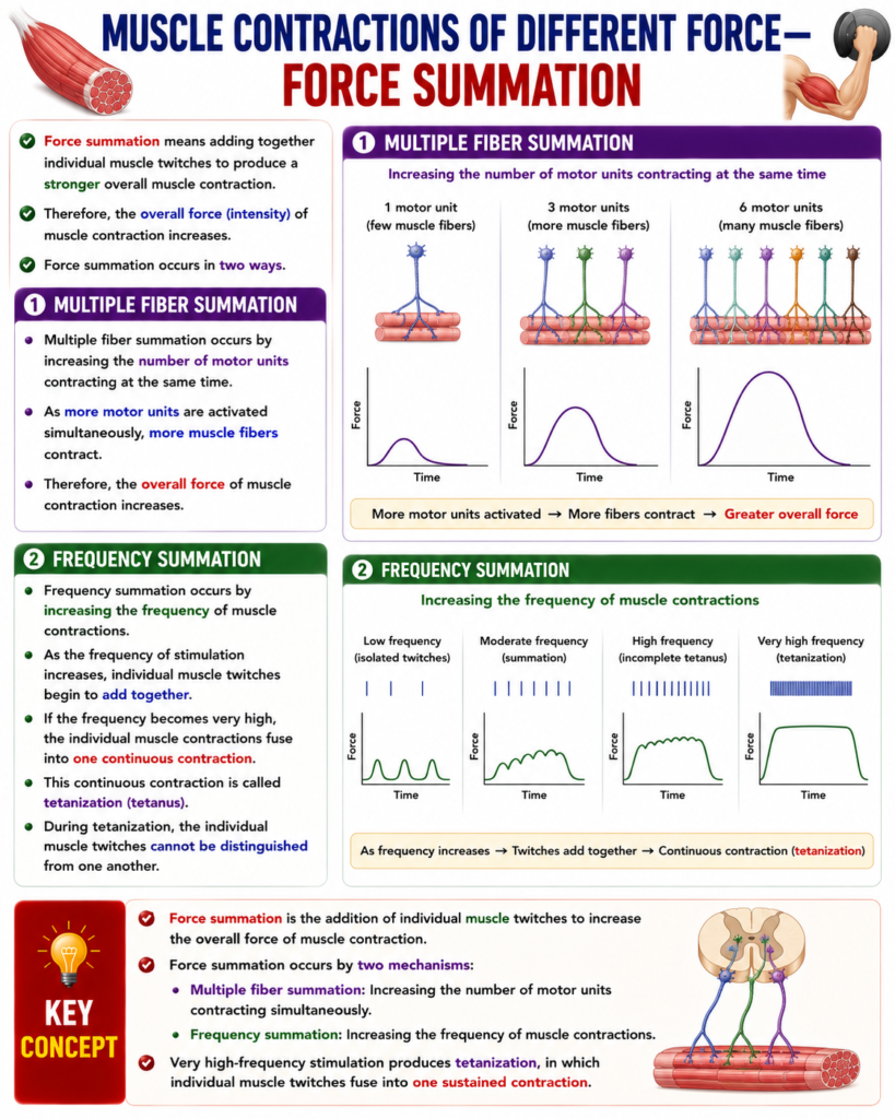
- Force summation means adding together individual muscle twitches to produce a stronger overall muscle contraction.
- Therefore, the overall force (intensity) of muscle contraction increases.
- Force summation occurs in two ways.
1. Multiple Fiber Summation
- Multiple fiber summation occurs by increasing the number of motor units contracting at the same time.
- As more motor units are activated simultaneously, more muscle fibers contract.
- Therefore, the overall force of muscle contraction increases.
2. Frequency Summation
- Frequency summation occurs by increasing the frequency of muscle contractions.
- As the frequency of stimulation increases, individual muscle twitches begin to add together.
- If the frequency becomes very high, the individual muscle contractions fuse into one continuous contraction.
- This continuous contraction is called tetanization (tetanus).
- During tetanization, the individual muscle twitches cannot be distinguished from one another.
KEY CONCEPT
- Force summation is the addition of individual muscle twitches to increase the overall force of muscle contraction.
- Force summation occurs by two mechanisms:
- Multiple fiber summation: Increasing the number of motor units contracting simultaneously.
- Frequency summation: Increasing the frequency of muscle contractions.
- Very high-frequency stimulation produces tetanization, in which individual muscle twitches fuse into one sustained contraction.
Multiple Fiber Summation
- When the central nervous system sends a weak signal to a muscle, the small motor units are activated first.
- The larger motor units are not activated during weak stimulation.
- As the strength of the nerve signal increases, larger and larger motor units are gradually recruited.
- The largest motor units may produce up to 50 times more contractile force than the smallest motor units.
- This pattern of recruiting motor units from small to large is called the size principle.
- The size principle allows muscle force to increase gradually.
- During weak muscle contractions, the force increases in small, precise steps.
- When greater muscle force is required, larger motor units are recruited, producing larger increases in force.
- The size principle occurs because:
- Small motor units are supplied by small motor nerve fibers.
- Small motor neurons in the spinal cord are more excitable than large motor neurons.
- Therefore, small motor neurons are activated before large motor neurons.
- Another important feature of multiple fiber summation is that different motor units are activated asynchronously by the spinal cord.
- This means that motor units do not all contract at the same time.
- Instead, different motor units contract one after another in an alternating pattern.
- This alternating activation produces smooth muscle contraction, even when the frequency of nerve impulses is low.
KEY CONCEPT
- Multiple fiber summation increases muscle force by recruiting more motor units.
- Small motor units are activated first, followed by larger motor units as the strength of the nerve signal increases.
- The largest motor units can generate up to 50 times more force than the smallest motor units.
- This orderly recruitment is called the size principle.
- The size principle occurs because small motor neurons are more excitable than large motor neurons.
- Motor units are activated asynchronously, allowing smooth and coordinated muscle contraction even at low frequencies of nerve stimulation.
Frequency Summation and Tetanization
- Fig. 6.14 shows the principles of frequency summation and tetanization.
- At a low frequency of stimulation, each muscle twitch occurs separately.
- Each twitch contraction is completed before the next one begins.
- As the frequency of stimulation increases, the next muscle contraction begins before the previous contraction has completely ended.
- Therefore, the new contraction is added to the previous contraction.
- This process is called frequency summation.
- As the frequency continues to increase, the total force of muscle contraction increases progressively.
- When the frequency reaches a critical level, the individual muscle contractions occur so rapidly that they fuse together.
- The separate twitches can no longer be distinguished.
- The muscle produces one smooth, continuous contraction.
- This sustained contraction is called tetanization (tetanus).
- At a slightly higher stimulation frequency, the muscle reaches its maximum force of contraction.
- Further increases in stimulation frequency do not produce any additional increase in muscle force.
- Tetany occurs because sufficient Ca²⁺ remains in the sarcoplasm between successive action potentials.
- Therefore, the muscle remains in a fully contracted state without relaxing between action potentials.
KEY CONCEPT
- Fig. 6.14 demonstrates frequency summation and tetanization.
- At low stimulation frequencies, individual muscle twitches occur separately.
- As stimulation frequency increases, successive twitches overlap, producing frequency summation and increasing muscle force.
- At a critical frequency, the twitches fuse into one smooth, continuous contraction called tetanization.
- At slightly higher frequencies, the muscle reaches its maximum force, and further increases in frequency do not increase contraction strength.
- Tetany occurs because Ca²⁺ remains in the sarcoplasm between action potentials, preventing muscle relaxation.
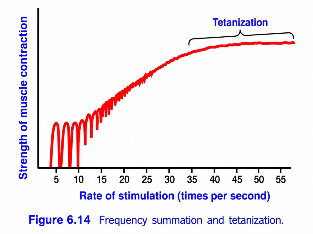
Frequency Summation and Tetanization (Figure 6.14) – Easy Conceptual Summary for MBBS Students
This figure explains how the strength of muscle contraction changes when the frequency (rate) of stimulation increases.
It demonstrates two very important concepts in muscle physiology:
- Frequency (Wave) Summation
- Tetanization (Tetanic Contraction)
The graph shows that:
The faster a muscle is stimulated, the stronger its contraction becomes until it reaches a maximum sustained contraction called tetanus.
Basic Concept
A single stimulus produces one muscle twitch.
Normally,
The muscle contracts,
then completely relaxes before the next stimulus arrives.
However,
If another stimulus arrives before the muscle has completely relaxed, the contractions begin to add together.
This is called:
Frequency (Wave) Summation
If the stimulation becomes even faster,
The muscle no longer has time to relax at all.
It remains in a sustained contraction called:
Tetanic Contraction (Tetanization)
Understanding the Axes
X-axis (Rate of Stimulation)
Shows how many electrical stimuli are given per second.
Moving to the right means:
➡️ The frequency of stimulation increases.
Y-axis (Strength of Muscle Contraction)
Shows the force produced by the muscle.
Higher curve
➡️ Stronger contraction
Lower curve
➡️ Weaker contraction
Understanding the Graph
The graph can be divided into four stages.
Stage 1 – Low Frequency (About 5–10 Stimuli/Second)
What happens?
Each stimulus produces a separate muscle twitch.
The muscle contracts,
then completely relaxes,
before the next stimulus arrives.
Therefore,
The graph shows separate peaks.
Why?There is enough time between stimuli for:
- Calcium to return to the sarcoplasmic reticulum.
- Cross-bridges to detach.
- The muscle to relax completely.
Easy Concept
Imagine clapping your hands once every few seconds.
Each clap is completely separate.
Key Point
One stimulus → One twitch → Complete relaxation
Stage 2 – Moderate Frequency (About 10–25 Stimuli/Second)
What happens?
The next stimulus arrives before complete relaxation occurs.
Each new contraction begins while some tension from the previous twitch still remains.
The contractions add together.
This is called:
Frequency (Wave) SummationWhy?
Some calcium is still present inside the muscle cell.
The next stimulus releases even more calcium.
More calcium means:
- More actin binding sites exposed.
- More cross-bridges form.
- Greater force develops.
Easy Concept
Imagine pushing a swing.
Instead of waiting until it completely stops,
you push it again while it is still moving.
Each push adds more movement.
The swing goes higher and higher.
Exactly the same happens with muscle contraction.
Key Point
More frequent stimulation produces stronger contractions.
Stage 3 – High Frequency (About 25–35 Stimuli/Second)
What happens?
The muscle has very little time to relax.
The contractions almost fuse together.
The graph becomes much smoother.
Force continues to increase.
Why?
Calcium remains elevated in the muscle cell.
Cross-bridges continue cycling without complete relaxation.
Therefore,
The muscle develops nearly continuous tension.
Easy Concept
Imagine pushing the swing continuously without allowing it to slow down.
Key Point
Relaxation becomes incomplete.
Stage 4 – Very High Frequency (About 35–55 Stimuli/Second)
The bracket labeled “Tetanization” represents this stage.
What happens?
The muscle receives stimuli so rapidly that:
- It never relaxes.
- Individual twitches disappear.
- The muscle develops one smooth, sustained contraction.
This is called:
Tetanic Contraction (Tetanization)
Why?
Calcium remains continuously high inside the muscle cell.
The sarcoplasmic reticulum has no time to remove calcium between stimuli.
Therefore,
Cross-bridges remain active continuously.
Maximum force is maintained.
Easy Concept
Imagine keeping a light switch permanently ON.
Instead of flashing on and off,
the light remains continuously illuminated.
Similarly,
The muscle remains continuously contracted.
Key Point
Tetanus produces the maximum force that a muscle can generate.
Why Does Muscle Strength Increase?
Each new stimulus adds to the previous contraction because:
- Calcium has not been completely removed.
- More calcium accumulates in the sarcoplasm.
- More troponin molecules bind calcium.
- More actin binding sites become available.
- More actin-myosin cross-bridges form.
Therefore,
Muscle tension progressively increases.
Why Does the Curve Become Flat During Tetanization?
Eventually,
Every available cross-bridge is already active.
The muscle has reached its maximum force.
Giving more stimuli cannot increase force further.
Therefore,
The graph reaches a plateau.
Clinical Importance
Normal Skeletal Muscle Movement
Most normal movements are produced by repeated nerve impulses, not by single twitches.
Therefore,
Our muscles usually contract by summation and partial tetanus.
Complete Tetanus
Occurs during powerful voluntary contractions such as:
- Lifting heavy weights
- Jumping
- Sprinting
The nervous system sends rapid impulses,
allowing the muscle to generate maximum force.
Fatigue
If tetanic stimulation continues for a long time,
ATP and energy stores decrease,
and muscle force eventually falls.
Comparison of the Stages
| Stage | Stimulation Frequency | Muscle Response |
|---|---|---|
| Low | 5–10/sec | Separate twitches with complete relaxation |
| Moderate | 10–25/sec | Frequency (wave) summation with incomplete relaxation |
| High | 25–35/sec | Twitches nearly fuse; force increases further |
| Very High | 35–55/sec | Complete tetanus (tetanization); sustained maximum contraction |
Quick Memory Table
| Term | Meaning |
|---|---|
| Single Twitch | One stimulus produces one contraction |
| Frequency (Wave) Summation | Repeated stimuli before complete relaxation increase muscle force |
| Incomplete (Unfused) Tetanus | Small relaxation occurs between contractions |
| Complete (Fused) Tetanus | No relaxation; sustained maximum contraction |
Easy Memory Trick
Slow Stimulation = Separate Twitches 👏
- Stimulus
- Relax
- Stimulus
- Relax
Faster Stimulation = Summation 🌊
Very Fast Stimulation = Tetanus 💪
- No relaxation.
- Continuous contraction.
- Maximum force.
Key Concept
This figure demonstrates that the strength of skeletal muscle contraction depends on the frequency of stimulation. At low stimulation frequencies, each stimulus produces a separate muscle twitch because the muscle has enough time to relax completely before the next stimulus arrives. As the stimulation frequency increases, the next stimulus reaches the muscle before relaxation is complete, causing frequency (wave) summation, in which the force of successive contractions adds together. This occurs because calcium remains in the sarcoplasm, allowing more actin-myosin cross-bridges to form with each new stimulus. At very high stimulation frequencies, the muscle has no time to relax, and the individual twitches merge into a smooth, sustained contraction called tetanization (tetanic contraction). During tetanus, calcium remains continuously elevated, cross-bridge cycling continues without interruption, and the muscle develops its maximum possible force.
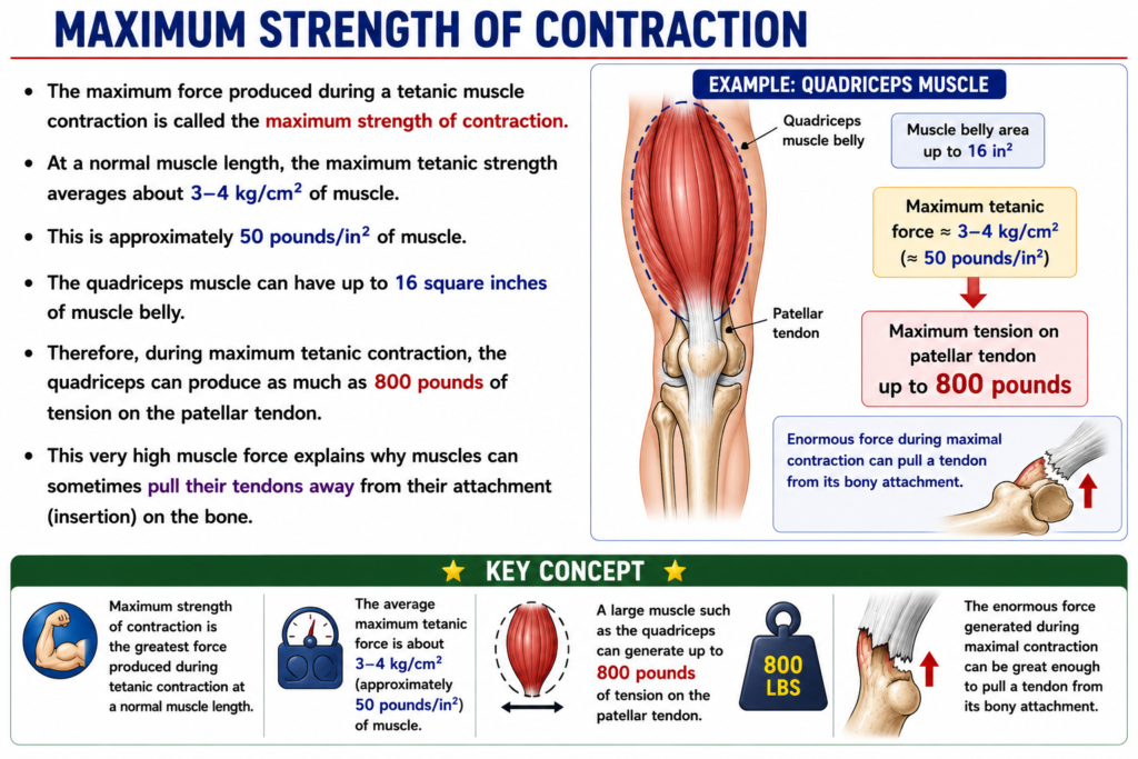
Maximum Strength of Contraction
- The maximum force produced during a tetanic muscle contraction is called the maximum strength of contraction.
- At a normal muscle length, the maximum tetanic strength averages about 3–4 kg/cm² of muscle.
- This is approximately 50 pounds/in² of muscle.
- The quadriceps muscle can have up to 16 square inches of muscle belly.
- Therefore, during maximum tetanic contraction, the quadriceps can produce as much as 800 pounds of tension on the patellar tendon.
- This very high muscle force explains why muscles can sometimes pull their tendons away from their attachment (insertion) on the bone.
KEY CONCEPT
- Maximum strength of contraction is the greatest force produced during tetanic contraction at a normal muscle length.
- The average maximum tetanic force is about 3–4 kg/cm² (approximately 50 pounds/in²) of muscle.
- A large muscle such as the quadriceps can generate up to 800 pounds of tension on the patellar tendon.
- The enormous force generated during maximal contraction can be great enough to pull a tendon from its bony attachment.
Increases in Muscle Strength After the Onset of Contraction—The Staircase Effect (Treppe)
- When a muscle begins to contract after a long period of rest, its first few contractions are relatively weak.
- The initial strength of contraction may be only about one-half of the strength reached after 10–50 muscle twitches.
- With each successive muscle twitch, the strength of contraction gradually increases.
- Eventually, the strength reaches a maximum stable level (plateau).
- This gradual increase in contraction strength is called the staircase effect (treppe).
- The exact cause of the staircase effect is not completely known.
- It is believed to occur mainly because the amount of Ca²⁺ in the cytosol gradually increases.
- With each successive muscle action potential, more Ca²⁺ is released from the sarcoplasmic reticulum.
- At the same time, the sarcoplasmic reticulum does not immediately recapture all of the released Ca²⁺.
- As a result, more Ca²⁺ remains in the cytosol.
- The increased cytosolic Ca²⁺ allows more actin–myosin cross-bridges to form, producing stronger muscle contractions.
- Therefore, the force of contraction increases gradually until it reaches a plateau.
KEY CONCEPT
- The staircase effect (treppe) is the gradual increase in muscle contraction strength after a muscle begins contracting following a period of rest.
- The initial contraction may be only about half as strong as the contractions that occur after 10–50 twitches.
- The contraction strength increases progressively until it reaches a plateau.
- The staircase effect is believed to result mainly from a gradual increase in cytosolic Ca²⁺ due to repeated Ca²⁺ release from the sarcoplasmic reticulum and incomplete immediate reuptake of Ca²⁺.
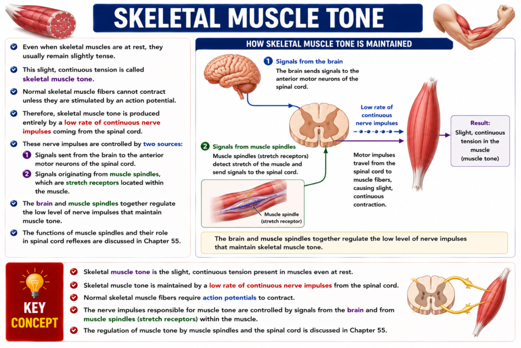
Skeletal Muscle Tone
- Even when skeletal muscles are at rest, they usually remain slightly tense.
- This slight, continuous tension is called skeletal muscle tone.
- Normal skeletal muscle fibers cannot contract unless they are stimulated by an action potential.
- Therefore, skeletal muscle tone is produced entirely by a low rate of continuous nerve impulses coming from the spinal cord.
- These nerve impulses are controlled by two sources:
- Signals sent from the brain to the anterior motor neurons of the spinal cord.
- Signals originating from muscle spindles, which are stretch receptors located within the muscle.
- The brain and muscle spindles together regulate the low level of nerve impulses that maintain muscle tone.
- The functions of muscle spindles and their role in spinal cord reflexes are discussed in Chapter 55.
KEY CONCEPT
- Skeletal muscle tone is the slight, continuous tension present in muscles even at rest.
- Skeletal muscle tone is maintained by a low rate of continuous nerve impulses from the spinal cord.
- Normal skeletal muscle fibers require action potentials to contract.
- The nerve impulses responsible for muscle tone are controlled by signals from the brain and from muscle spindles (stretch receptors) within the muscle.
- The regulation of muscle tone by muscle spindles and the spinal cord is discussed in Chapter 55.
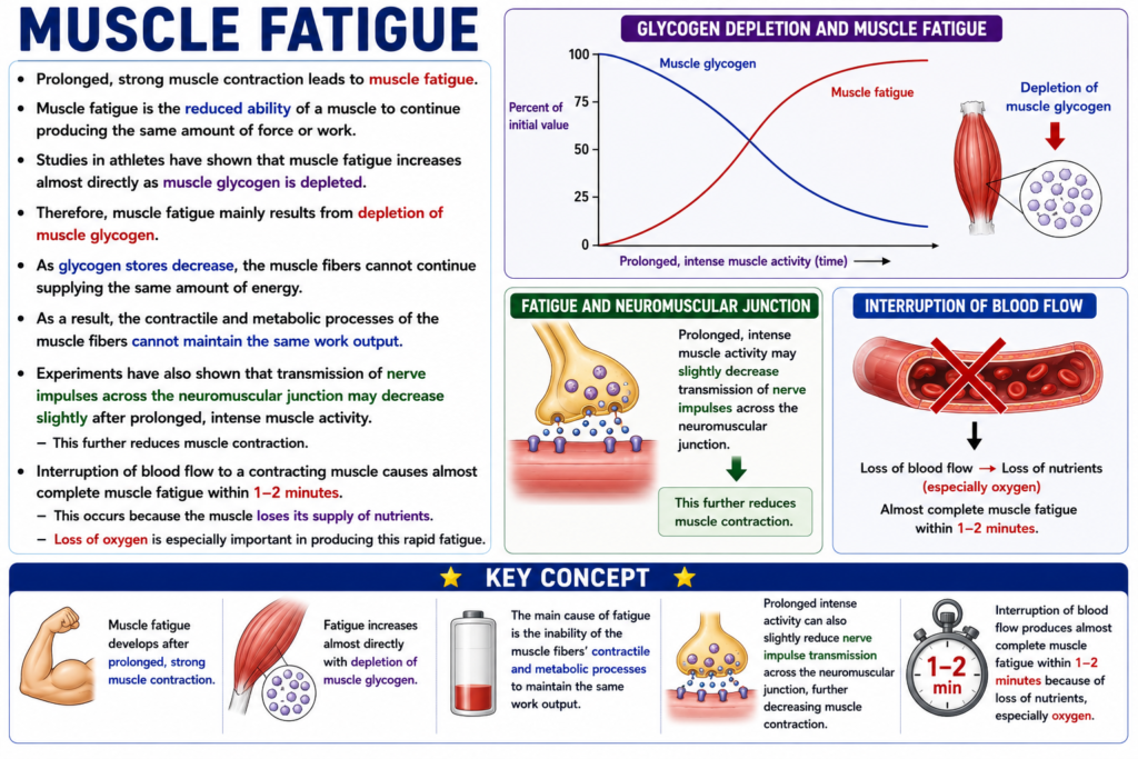
Muscle Fatigue
- Prolonged, strong muscle contraction leads to muscle fatigue.
- Muscle fatigue is the reduced ability of a muscle to continue producing the same amount of force or work.
- Studies in athletes have shown that muscle fatigue increases almost directly as muscle glycogen is depleted.
- Therefore, muscle fatigue mainly results from depletion of muscle glycogen.
- As glycogen stores decrease, the muscle fibers cannot continue supplying the same amount of energy.
- As a result, the contractile and metabolic processes of the muscle fibers cannot maintain the same work output.
- Experiments have also shown that transmission of nerve impulses across the neuromuscular junction may decrease slightly after prolonged, intense muscle activity.
- This further reduces muscle contraction.
- Interruption of blood flow to a contracting muscle causes almost complete muscle fatigue within 1–2 minutes.
- This occurs because the muscle loses its supply of nutrients.
- Loss of oxygen is especially important in producing this rapid fatigue.
KEY CONCEPT
- Muscle fatigue develops after prolonged, strong muscle contraction.
- Fatigue increases almost directly with depletion of muscle glycogen.
- The main cause of fatigue is the inability of the muscle fibers’ contractile and metabolic processes to maintain the same work output.
- Prolonged intense activity can also slightly reduce nerve impulse transmission across the neuromuscular junction, further decreasing muscle contraction.
- Interruption of blood flow produces almost complete muscle fatigue within 1–2 minutes because of loss of nutrients, especially oxygen.
Lever Systems of the Body
- Muscles produce movement by pulling on their points of attachment (insertions) on bones.
- The bones act as lever systems to produce body movements.
- Fig. 6.15 shows the lever system formed by the biceps muscle to lift the forearm against a load.
- A large biceps muscle with a cross-sectional area of 6 square inches can produce a maximum contraction force of about 300 pounds.
- When the forearm is at a right angle (90°) to the upper arm:
- The biceps tendon is attached about 2 inches in front of the elbow joint (fulcrum).
- The total length of the forearm lever is about 14 inches.
- Therefore, the lifting force at the hand is only one-seventh of the muscle force.
Calculation
- Muscle force = 300 pounds
- Mechanical advantage = 2 inches / 14 inches = 1/7
Lifting force at the hand=7300≈43 pounds
- Therefore, the biceps can lift about 43 pounds at the hand.
- When the arm is fully extended:
- The biceps tendon lies much closer to the elbow joint (fulcrum).
- Therefore, the lifting force at the hand becomes much less than 43 pounds.
- To understand any lever system in the body, four factors must be known:
- The point of muscle insertion.
- The distance between the muscle insertion and the fulcrum.
- The length of the lever arm.
- The position of the lever.
- The body performs many different types of movements.
- Some movements require great strength.
- Other movements require a large range of movement.
- Therefore, the body contains many different types of muscles.
- Some muscles are long and shorten over a long distance.
- Other muscles are short but have a large cross-sectional area, allowing them to produce very strong contractions over short distances.
- The study of muscles, lever systems, and body movements is called kinesiology.
- Kinesiology is an important part of human physiology.
KEY CONCEPT
- Muscles move bones by pulling on their insertions, and bones function as lever systems (Fig. 6.15).
- A large biceps can generate about 300 pounds of force, but because of the lever arrangement, only about 43 pounds of lifting force is produced at the hand when the elbow is flexed to 90°.
- When the arm is fully extended, the lifting force decreases because the tendon is closer to the fulcrum.
- Analysis of a body lever system depends on the muscle insertion, its distance from the fulcrum, the length of the lever arm, and the position of the lever.
- Different muscles are specialized for either powerful contractions or large ranges of movement.
- The study of muscles, lever systems, and body movements is called kinesiology.
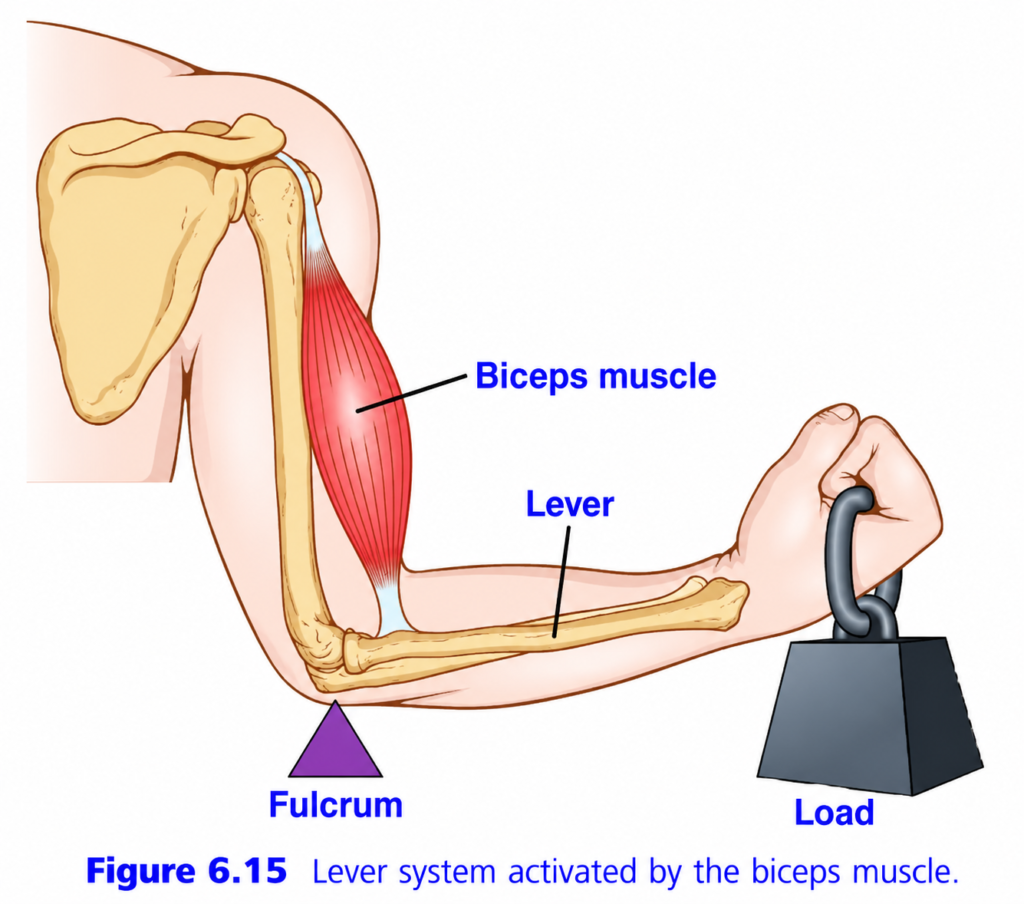
Lever System Activated by the Biceps Muscle (Figure 6.15) — Summary
This figure explains how the biceps muscle lifts an object using the bones of the forearm as a lever.
The body uses the same principles of mechanics (lever system) that are used in machines like crowbars, scissors, and seesaws.
Main Idea
When the biceps muscle contracts, it pulls on the forearm bone, causing the forearm to rotate upward around the elbow joint and lift the load.
Muscle pulls → Bone acts as a lever → Elbow acts as a pivot → Hand lifts the load
Understanding the Figure Step by Step
1. Biceps Muscle (Force Producer)
The red structure is the biceps muscle.
Function
- Contracts (shortens)
- Pulls the forearm upward
- Produces the force needed for movement
Simple Concept
The biceps is the engine that generates the pulling force.
2. Tendon (Force Transmitter)
The lower end of the biceps is attached to the radius by a tendon.
Its job is to
- transfer the force produced by the muscle
- pull the forearm bone
Without the tendon, the muscle could not move the bone.
3. Lever (Forearm Bone)
The forearm bones act as a lever.
A lever is simply a rigid bar that rotates around a fixed point.
Here,
- Radius and ulna together function as the lever.
Simple Concept
The bone is not producing force.
It only transmits the force generated by the muscle.
4. Fulcrum (Pivot Point)
The elbow joint is labeled as the fulcrum.
A fulcrum is the point around which movement occurs.
Here,
- the elbow remains fixed
- the forearm rotates around it
Simple Concept
Think of the elbow as the hinge of a door.
The door swings around the hinge.
Similarly,
the forearm swings around the elbow.
5. Load (Weight in the Hand)
The weight held in the hand is the load.
The load is the resistance that the muscle must overcome.
Examples
- Dumbbell
- Grocery bag
- Suitcase
- Bucket
The heavier the load,
the harder the biceps must contract.
6. How Movement Happens
Step 1
Brain sends a signal.
↓
Step 2
Motor neurons stimulate the biceps.
↓
Step 3
Biceps contracts (shortens).
↓
Step 4
The tendon pulls on the forearm.
↓
Step 5
The forearm rotates around the elbow.
↓
Step 6
The hand and weight move upward.
Easy Flow Diagram
Brain
   ↓
Motor neuron
   ↓
Biceps contracts
   ↓
Tendon pulls radius
   ↓
Forearm rotates around elbow
   ↓
Hand lifts the load
What Is a Lever?
A lever has three basic parts.
1. Fulcrum (F)
- Fixed point
- Pivot
- Elbow joint
2. Effort (E)
- Force applied
- Produced by the biceps muscle
3. Load (L)
- Object being lifted
- Weight in the hand
In This Figure
| Part | Structure in the Body |
|---|---|
| Fulcrum | Elbow joint |
| Lever | Forearm bones (radius and ulna) |
| Effort | Pull of the biceps muscle |
| Load | Weight held in the hand |
Why Is the Biceps Attached Close to the Elbow?
Notice that the tendon attaches very close to the elbow, while the load is held far away in the hand.
This means
- the muscle pulls over a short distance
- the hand moves through a much larger distance
Result
A small shortening of the muscle produces a large movement of the hand.
This allows
- fast movements
- wide range of motion
- precise control
What Is the Disadvantage?
Because the tendon is close to the fulcrum,
the biceps must generate a force much greater than the weight being lifted.
For example,
to lift a 10-kg weight, the biceps may need to produce much more than 10 kg of force because of the mechanical disadvantage of this lever arrangement.
Concept
The body sacrifices force efficiency to gain speed and a large range of movement.
What Type of Lever Is This?
This is a Third-Class Lever (Class III Lever).
In a third-class lever,
Effort lies between the fulcrum and the load.
Arrangement:
Fulcrum → Effort → LoadIn the figure:
Elbow → Biceps insertion → Weight in handWhy Does the Human Body Prefer Third-Class Levers?
Although third-class levers require more muscle force, they provide several important advantages:
- Large range of movement
- Faster limb movement
- Better precision
- Smooth and controlled actions
- Ideal for activities like writing, eating, throwing, lifting, and sports
Real-Life Analogy
Imagine using a fishing rod.
- One hand near the handle acts as the fulcrum.
- Your other hand applies the effort in the middle.
- The fish at the end of the rod is the load.
Even though you apply a large force, the tip of the rod moves through a much greater distance and speed.
The biceps and forearm work in the same way.
Important Points from the Figure
- The biceps muscle is the source of pulling force.
- The tendon transfers this force to the forearm.
- The forearm bones act as the lever.
- The elbow joint acts as the fulcrum (pivot).
- The weight in the hand is the load.
- When the biceps contracts, the forearm rotates upward around the elbow.
- This arrangement is a third-class lever, where the effort is applied between the fulcrum and the load.
- Third-class levers provide speed, precision, and a wide range of movement, even though they require the muscle to generate a force greater than the external load.
KEY CONCEPT (Figure 6.15)
The biceps muscle lifts the forearm using a third-class lever system. The elbow joint is the fulcrum, the forearm bones act as the lever, the biceps provides the effort, and the weight in the hand is the load. When the biceps contracts, it pulls on the forearm, causing it to rotate around the elbow and lift the load. Although this arrangement requires the biceps to produce a force greater than the weight being lifted, it provides the body with rapid, precise, and wide-ranging movements, making it ideal for everyday activities and skilled motor tasks.
Positioning of a Body Part By Contraction of Agonist and Antagonist Muscles on Opposite Sides of a Joint
- Almost all body movements occur by the simultaneous contraction of agonist and antagonist muscles on opposite sides of a joint.
- This simultaneous contraction is called coactivation of the agonist and antagonist muscles.
- Coactivation is controlled by the motor control centers of the brain and spinal cord.
- The position of each body part, such as an arm or a leg, depends on the relative degree of contraction of the agonist and antagonist muscles.
- For example, if an arm or leg is to be held in a midrange position:
- The agonist muscles are activated to about the same degree as the antagonist muscles.
- This balanced contraction holds the limb in the desired position.
- An elongated (stretched) muscle contracts with greater force than a shortened muscle.
- Fig. 6.9 showed that:
- Maximum force is produced when the muscle is at its normal functional length.
- Very little force is produced when the muscle is shortened to about half of its normal length.
- Therefore, the elongated muscle on one side of a joint initially produces greater force than the shortened muscle on the opposite side.
- As the arm or leg moves toward the midposition:
- The force produced by the elongated muscle gradually decreases.
- The force produced by the shortened muscle gradually increases.
- Eventually, the forces produced by the agonist and antagonist muscles become equal.
- At this point, movement stops, and the limb remains in the desired position.
- Thus, the nervous system controls the position of the arm or leg by adjusting the degree of activation of the agonist and antagonist muscles.
- The motor nervous system also has additional mechanisms to compensate for different muscle loads during body positioning, which are discussed in Chapter 55.
KEY CONCEPT
- Most body movements result from the simultaneous contraction (coactivation) of agonist and antagonist muscles.
- Coactivation is controlled by the brain and spinal cord.
- The position of a limb depends on the relative strength of contraction of the agonist and antagonist muscles.
- An elongated muscle generates greater force than a shortened muscle (Fig. 6.9).
- As the limb moves, the force of the elongated muscle decreases while the force of the shortened muscle increases until both become equal.
- When the forces are equal, movement stops and the limb is maintained in the desired position.
- The nervous system continuously adjusts agonist and antagonist activity to accurately position body parts.
PREPARE AND MADE SELF LEARNING BY DR SHEEN
